Review
. 2022 Feb 14;14(2):386.
doi: 10.3390/v14020386.

Vav Proteins in Development of the Brain: A Potential Relationship to the Pathogenesis of Congenital Zika Syndrome?

Affiliations
Review

Vav Proteins in Development of the Brain: A Potential Relationship to the Pathogenesis of Congenital Zika Syndrome?

Aidan J Norbury et al. Viruses. .

Abstract

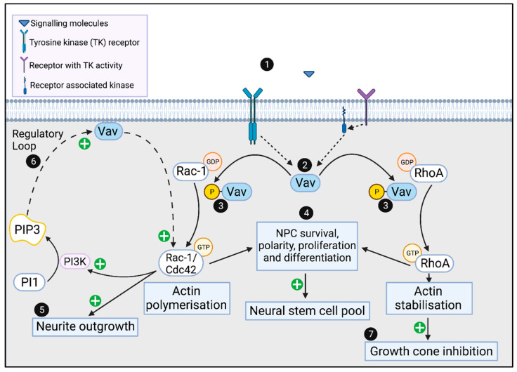
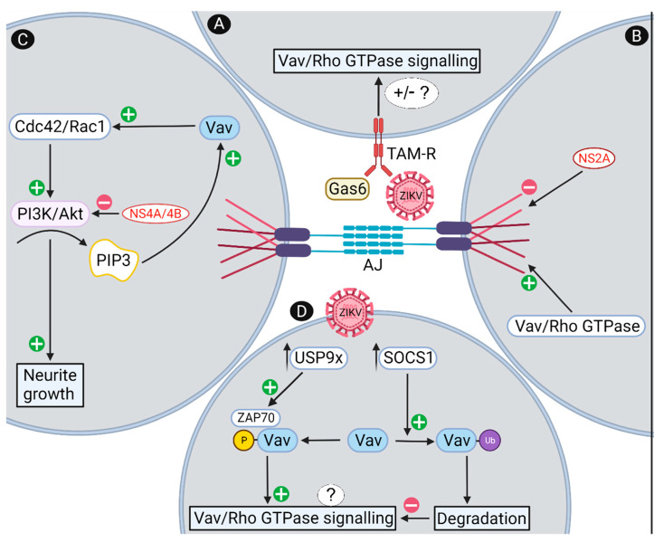
Zika virus (ZIKV) infection during pregnancy can result in a significant impact on the brain and eye of the developing fetus, termed congenital zika syndrome (CZS). At a morphological level, the main serious presentations of CZS are microcephaly and retinal scarring. At a cellular level, many cell types of the brain may be involved, but primarily neuronal progenitor cells (NPC) and developing neurons. Vav proteins have guanine exchange activity in converting GDP to GTP on proteins such as Rac1, Cdc42 and RhoA to stimulate intracellular signaling pathways. These signaling pathways are known to play important roles in maintaining the polarity and self-renewal of NPC pools by coordinating the formation of adherens junctions with cytoskeletal rearrangements. In developing neurons, these same pathways are adopted to control the formation and growth of neurites and mediate axonal guidance and targeting in the brain and retina. This review describes the role of Vavs in these processes and highlights the points of potential ZIKV interaction, such as (i) the binding and entry of ZIKV in cells via TAM receptors, which may activate Vav/Rac/RhoA signaling; (ii) the functional convergence of ZIKV NS2A with Vav in modulating adherens junctions; (iii) ZIKV NS4A/4B protein effects on PI3K/AKT in a regulatory loop via PPI3 to influence Vav/Rac1 signaling in neurite outgrowth; and (iv) the induction of SOCS1 and USP9X following ZIKV infection to regulate Vav protein degradation or activation, respectively, and impact Vav/Rac/RhoA signaling in NPC and neurons. Experiments to define these interactions will further our understanding of the molecular basis of CZS and potentially other developmental disorders stemming from in utero infections. Additionally, Vav/Rac/RhoA signaling pathways may present tractable targets for therapeutic intervention or molecular rationale for disease severity in CZS.

Keywords: Rac1; RhoA; Vav; brain development; congenital zika syndrome; guanosine nucleotide exchange factor; neuronal development; neuronal progenitor cell; zika virus.


Conflict of interest statement

The authors declare no conflict of interest.

Figures

Figure 1
Figure 1
Transcriptional profile of VAV in the developing human brain. VAV expression was analyzed using the developmental transcriptome provided by the Allen Institute for Brain Science, Allen Brain Atlas, Brain Span, Atlas of the developing human brain https://www.brainspan.org (accessed on 4 January 2022). VAV1, -2 and -3 mRNA intensity is assigned in accordance with the color map derived from data for donor tissue from 8–37 pcw, as indicated by the arrows and represented by the blue-green donors, and 4mth–40 yrs represented by the yellow-dark red donor bars. Data is grouped by brain region with 1 = dorsolateral prefrontal cortex; 2 = ventrolateral prefrontal cortex; 3 = anterior cingulate cortex; 4 = orbital cortex; 5 = primary motor cortex; 6/7 = primary motor-sensory cortex; 8 = primary somatosensory cortex; 9 = posteroventral parietal cortex; 10 = primary auditory cortex; 11 = temporal neocortex; 12 = posterior superior temporal cortex; 13 = inferolateral temporal cortex; 14 = occipital neocortex; 15 = primary visual cortex; 16 = hippocampus; 17 = amygdaloid complex; 18 = medial ganglionic eminence; 19 = striatum; 20 = dorsal thalamus; 21 = medial dorsal nucleus of the thalamus; 22 = upper rhombic lip; 23 = cerebellar cortex.
Figure 2
Figure 2
Vav and Rho family GTPase signaling in NPC and neurons. (1) signaling molecules such as NGF, BDNF and Ephrins bind to tyrosine kinase receptors or receptors with associated tyrosine kinases, such as Src/Syk, which can (2) stimulate phosphorylation of Vav. Phosphorylated Vav acts as a guanosine exchange factor to (3) activate Rho family GTPases (Rac1, Cdc42 and RhoA). In neurons, Rac1, Cdc42 and RhoA are involved in maintaining NPC polarity, regulating survival, proliferation and differentiation, which is important for (4) maintenance of the neural stem cell pool. GTP-Rac1/Cdc42 can mediate neurite outgrowth via actin polymerization within the growth cone (5) and activate PI3K, which generates PIP3 that in a regulatory loop, activates Vav to further activate Cdc42/Rac1 (6). GTP-RhoA inhibits the growth cone/neurite outgrowth through actin stabilization (7). Created with BioRender.com (https://biorender.com (accessed on 7 January 2022).
Figure 3
Figure 3
Proposed mechanisms by which ZIKV may affect Vav and Rho GTPase signaling. (A) ZIKV can bind and enter cells via TAM receptors, including through an interaction with the TAM ligand Gas6, which can signal via Vav and Rho GTPases with unknown outcomes for this signaling pathway; (B) ZIKV NS2A binds to and disrupts adherens junctions, a structure that is regulated by Vav and Rho GTPases, with Vav itself a potential adherens junction component; (C) ZIKV NS4A/B inhibits the PI3K/Akt pathway, which is part of a positive feedback loop via PIP3 activation of Vav to drive neurite outgrowth; (D) ZIKV induces SOCS1 and USP9X expression which can affect Vav ubiquitination (Ub) and proteosomal degradation, or ZAP70-induced Vav phosphorylation, which would be predicted to influence Vav activation of Rho GTPases. Predicted outcomes are reduced adherens junctions’ formation and alterations in the actin cytoskeleton that affect the polarization and NPC environment leading to a reduced NPC pool. In neurons, reduced Vav/RhoGTPase signaling is predicted to lead to reduced neurite outgrowth or growth cone collapse, affecting the dynamic changes that influence development of the complex brain architecture. AJ = adherens junctions. Created with BioRender.com https://biorender.com (accessed on 7 January 2022).

Similar articles

Cited by

References

    1. Baud D., Gubler D.J., Schaub B., Lanteri M.C., Musso D. An update on Zika virus infection. Lancet. 2017;390:2099–2109. doi: 10.1016/S0140-6736(17)31450-2. - DOI - PubMed
    1. Pierson T.C., Diamond M.S. The continued threat of emerging flaviviruses. Nat. Microbiol. 2020;5:796–812. doi: 10.1038/s41564-020-0714-0. - DOI - PMC - PubMed
    1. Duffy M.R., Chen T.-H., Hancock W.T., Powers A.M., Kool J.L., Lanciotti R.S., Pretrick M., Marfel M., Holzbauer S., DuBray C., et al. Zika Virus Outbreak on Yap Island, Federated States of Micronesia. N. Engl. J. Med. 2009;360:2536–2543. doi: 10.1056/NEJMoa0805715. - DOI - PubMed
    1. Petersen E., Wilson M.E., Touch S., McCloskey B., Mwaba P., Bates M., Dar O., Mattes F., Kidd M., Ippolito G., et al. Rapid Spread of Zika Virus in The Americas—Implications for Public Health Preparedness for Mass Gatherings at the 2016 Brazil Olympic Games. Int. J. Infect. Dis. 2016;44:11–15. doi: 10.1016/j.ijid.2016.02.001. - DOI - PubMed
    1. McCarthy M. Four in Florida are infected with Zika from local mosquitoes. BMJ. 2016;354:i4235. doi: 10.1136/bmj.i4235. - DOI - PubMed

Publication types

MeSH terms