

Virus-like Particles: Measures and Biological Functions
- PMID: 35215979
- PMCID: PMC8877645
- DOI: 10.3390/v14020383
Virus-like Particles: Measures and Biological Functions
Abstract
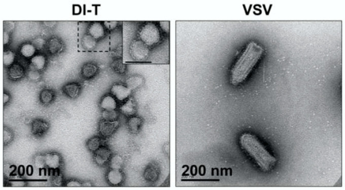
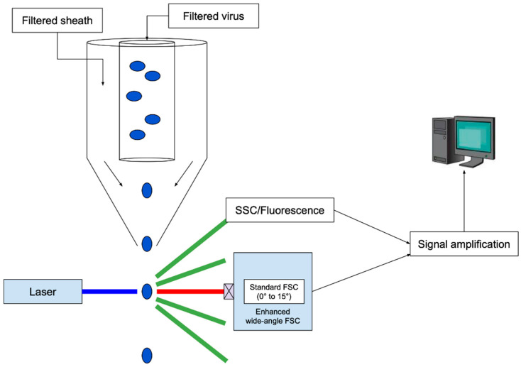
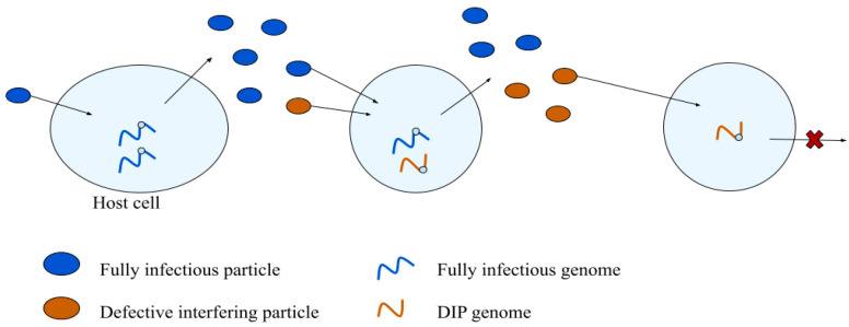
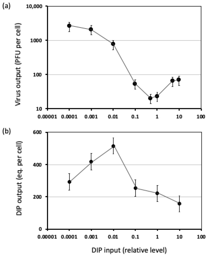
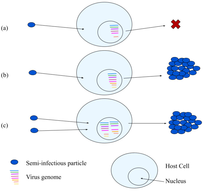
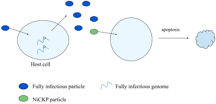
Virus-like particles resemble infectious virus particles in size, shape, and molecular composition; however, they fail to productively infect host cells. Historically, the presence of virus-like particles has been inferred from total particle counts by microscopy, and infectious particle counts or plaque-forming-units (PFUs) by plaque assay; the resulting ratio of particles-to-PFUs is often greater than one, easily 10 or 100, indicating that most particles are non-infectious. Despite their inability to hijack cells for their reproduction, virus-like particles and the defective genomes they carry can exhibit a broad range of behaviors: interference with normal virus growth during co-infections, cell killing, and activation or inhibition of innate immune signaling. In addition, some virus-like particles become productive as their multiplicities of infection increase, a sign of cooperation between particles. Here, we review established and emerging methods to count virus-like particles and characterize their biological functions. We take a critical look at evidence for defective interfering virus genomes in natural and clinical isolates, and we review their potential as antiviral therapeutics. In short, we highlight an urgent need to better understand how virus-like genomes and particles interact with intact functional viruses during co-infection of their hosts, and their impacts on the transmission, severity, and persistence of virus-associated diseases.
Keywords: cell killing particles; clonogenic assay; co-infection; coulter counting; defective interfering particles; defective viral genomes; flow virometry; multiplicity of infection; plaque forming unit; resistive pulse sensing; semi-infectious particles; transmission electron microscopy; virus-like particles.
Conflict of interest statement
The authors declare no conflict of interest. The funders had no role in the design of the study; in the collection, analyses, or interpretation of data; in the writing of the manuscript, or in the decision to publish the results.
Figures
References
-
- Stephenson B. Kepler’s Physical Astronomy. Springer; New York, NY, USA: 1987. Epitome of Copernican Astronomy; pp. 138–201.
-
- Darwin C. On the Origin of Species, 1859. Routledge; Oxford, UK: 2004.
-
- Von Borries B., Ruska E., Ruska H. Bakterien und Virus in übermikroskopischer Aufnahme. Klin. Wochenschr. 1938;17:921–925. doi: 10.1007/BF01775798. - DOI
Publication types
MeSH terms
LinkOut - more resources
Full Text Sources
Other Literature Sources
