. 2022 Feb 21;14(2):443.
doi: 10.3390/v14020443.

Collection of Monoclonal Antibodies Targeting SARS-CoV-2 Proteins

Affiliations

Collection of Monoclonal Antibodies Targeting SARS-CoV-2 Proteins

Marina Pribanić Matešić et al. Viruses. .

Abstract

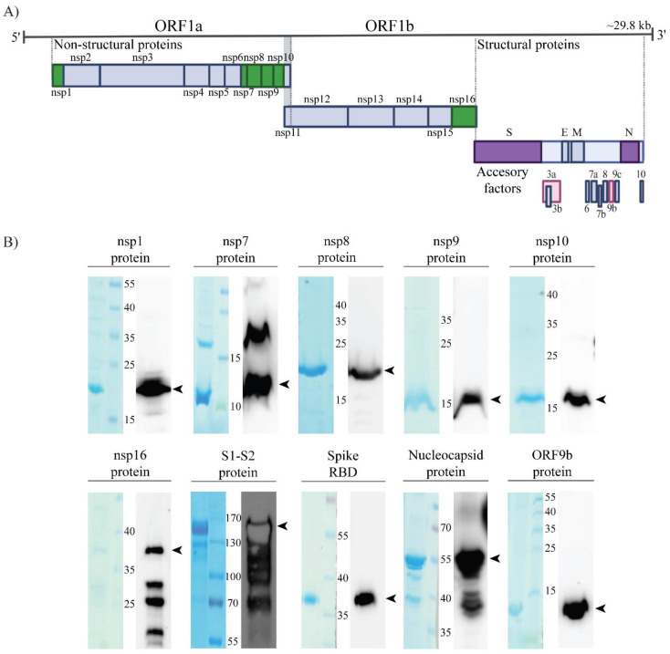
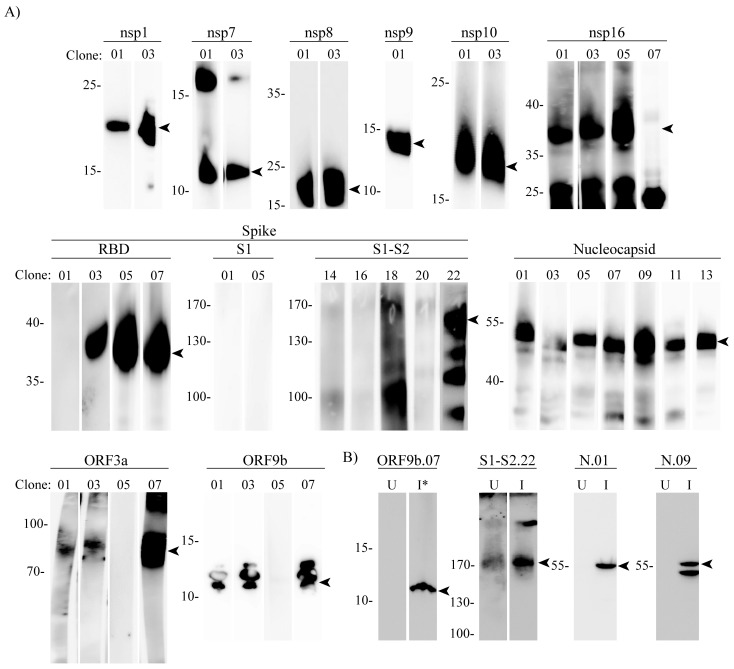
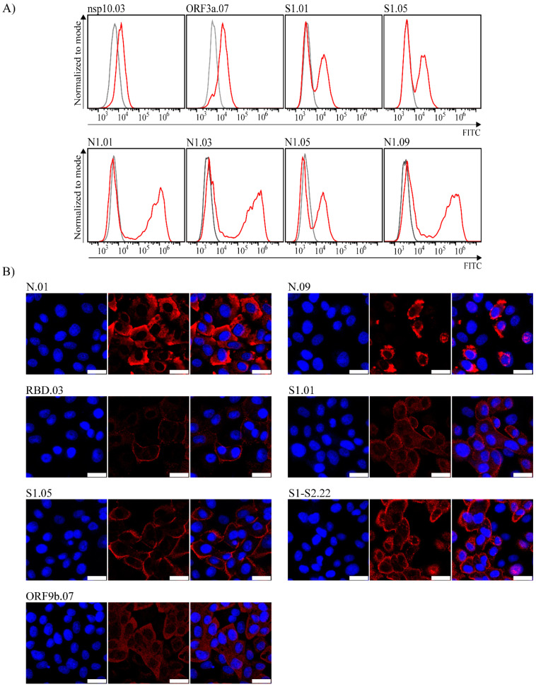
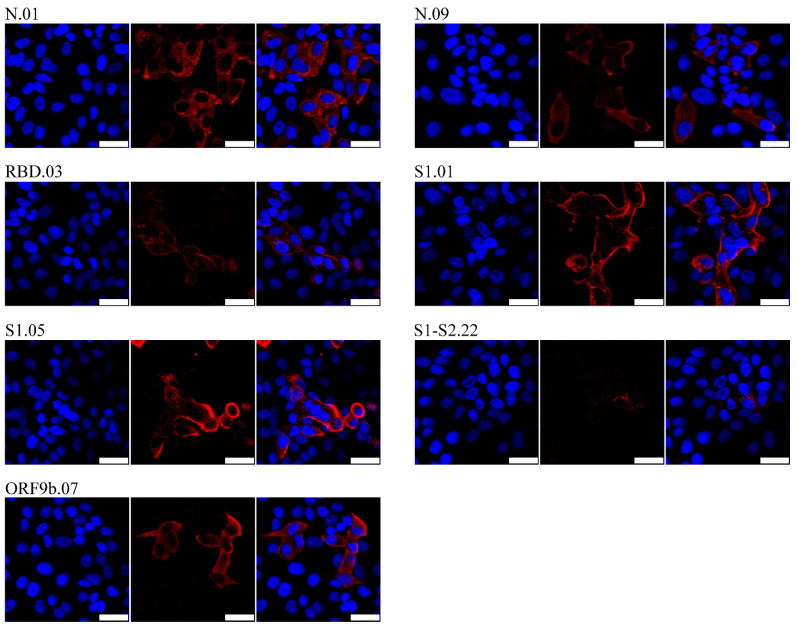
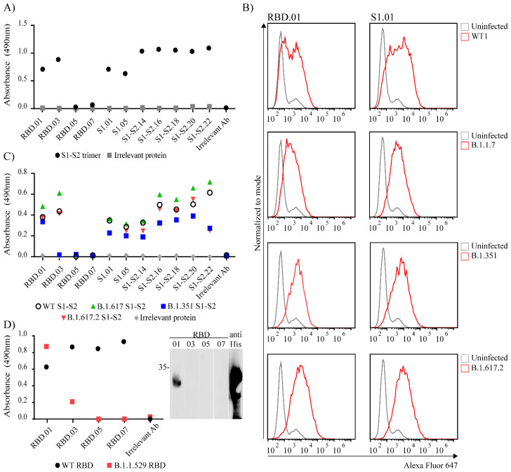
In early 2020, the COVID-19 pandemic sparked a global crisis that continues to pose a serious threat to human health and the economy. Further advancement in research is necessary and requires the availability of quality molecular tools, including monoclonal antibodies. Here, we present the development and characterization of a collection of over 40 new monoclonal antibodies directed against different SARS-CoV-2 proteins. Recombinant SARS-CoV-2 proteins were expressed, purified, and used as immunogens. Upon development of specific hybridomas, the obtained monoclonal antibody (mAb) clones were tested for binding to recombinant proteins and infected cells. We generated mAbs against structural proteins, the Spike and Nucleocapsid protein, several non-structural proteins (nsp1, nsp7, nsp8, nsp9, nsp10, nsp16) and accessory factors (ORF3a, ORF9b) applicable in flow cytometry, immunofluorescence, or Western blot. Our collection of mAbs provides a set of novel, highly specific tools that will allow a comprehensive analysis of the viral proteome, which will allow further understanding of SARS-CoV-2 pathogenesis and the design of therapeutic strategies.

Keywords: COVID-19; SARS-CoV-2; monoclonal antibodies; variants of concern.


Conflict of interest statement

The authors declare no conflict of interest.

Figures

Figure 1
Figure 1
Expression of SARS-CoV-2 recombinant proteins. (A) Graphical representation of SARS- CoV-2 viral genome. Immunogens generated in the study are shown as colored boxes: non-structural proteins (nsp1, nsp7, nsp8, nsp9, nsp10, nsp16) are shown as green boxes, Spike and Nucleocapsid protein as purple, and accessory factors ORF3a and ORF9b as pink boxes. (B) Analysis of expressed and purified His-tagged immunogens by SDS-PAGE (left) and Western blotting (right) with anti-His POD antibody. SDS-PAGE gel was stained with Coomassie blue. Lane shows purified protein, molecular mass markers (in kDa) are indicated in the middle. The arrowheads indicate bands near expected molecular weights.
Figure 2
Figure 2
Validation of SARS-CoV-2 specific antibodies by Western blot analysis. (A) Recombinant proteins or KLH-conjugated peptides (ORF3a) were subjected to gel electrophoresis and detected using SARS-CoV-2 mAbs raised against indicated immunogens. Molecular mass markers (in kDa) are indicated on the left. (B) Lysates of uninfected (U) or, WT2 infected (I) or Delta infected (I*) cells were prepared at 48 h post infection and analyzed by Western blotting using specific mAbs raised against the indicated proteins. Molecular mass markers (in kDa) are indicated on the right. Arrowheads indicate bands near expected molecular weights.
Figure 3
Figure 3
Detection of SARS-CoV-2 proteins in infected Vero E6 cells 24 h p.i. by flow cytometry or confocal microscopy. (A) Infected cells were analyzed by intracellular staining and flow cytometry with respective monoclonal antibodies following secondary FITC-coupled antibodies (red histogram). Mock infected cells were used as negative control (grey histogram). (B) Infected cells were stained by respective monoclonal antibodies following secondary TRITC-coupled antibodies (red) and nuclei staining by DAPI (blue). Images were obtained using Leica confocal microscope. The size bar (white line) corresponds to 25 µm. Representative mAb clones are shown and are indicated above the panel.
Figure 4
Figure 4
Detection of SARS-CoV-2 proteins in Delta SARS-CoV-2 infected cells by confocal microscopy. SARS-CoV-2 Delta infected Vero E6 cells were analyzed at 24 h p.i. Infected cells were stained by respective monoclonal antibodies following secondary TRITC-coupled antibodies (red) and nuclei staining by DAPI (blue). Images were obtained using a Leica confocal microscope. The size bar (white line) corresponds to 25 µm. Representative mAb clones are shown and indicated above the panel.
Figure 5
Figure 5
Testing specificity of Spike antibodies. (A) All anti-Spike clones were tested in ELISA for their recognition of trimeric Spike protein. (B) 293T-ACE2 cells were infected with the WT1 or different SARS-CoV-2 variants and after 24 h stained with indicated anti-Spike clones followed by Alexa Fluor 647-conjugated secondary antibodies (red line). Uninfected cells were used as negative control (grey line). (C) Anti-Spike clones generated using full Spike protein as immunogen were tested in ELISA for their specificity against mutated His-tagged Spike protein corresponding to different virus variants. (D) Anti-Spike clones generated using RBD protein as immunogen were tested in ELISA (left) and WB (right) for their specificity against mutated His-tagged RBD protein corresponding to B.1.1.529 variant. (A,C,D) For ELISA His-tagged Nucleocapsid protein (grey dot) was coated as negative control and anti-Nucleocapsid antibody (clone N.01) was used as irrelevant antibody. (D) For WB RBD B.1.1.529 protein was stained with anti-His POD.

Similar articles

Cited by

References

    1. Cui J., Li F., Shi Z.-L. Origin and evolution of pathogenic coronaviruses. Nat. Rev. Microbiol. 2019;17:181–192. doi: 10.1038/s41579-018-0118-9. - DOI - PMC - PubMed
    1. Lu R., Zhao X., Li J., Niu P., Yang B., Wu H., Wang W., Song H., Huang B., Zhu N., et al. Genomic characterisation and epidemiology of 2019 novel coronavirus: Implications for virus origins and receptor binding. Lancet. 2020;395:565–574. doi: 10.1016/S0140-6736(20)30251-8. - DOI - PMC - PubMed
    1. Wu Z., McGoogan J.M. Characteristics of and Important Lessons from the Coronavirus Disease 2019 (COVID-19) Outbreak in China: Summary of a Report of 72 314 Cases from the Chinese Center for Disease Control and Prevention. JAMA. 2020;323:1239–1242. doi: 10.1001/jama.2020.2648. - DOI - PubMed
    1. Hu B., Guo H., Zhou P., Shi Z.-L. Characteristics of SARS-CoV-2 and COVID-19. Nat. Rev. Microbiol. 2021;19:141–154. doi: 10.1038/s41579-020-00459-7. - DOI - PMC - PubMed
    1. Romano M., Ruggiero A., Squeglia F., Maga G., Berisio R. A Structural View of SARS-CoV-2 RNA Replication Machinery: RNA Synthesis, Proofreading and Final Capping. Cells. 2020;9:1267. doi: 10.3390/cells9051267. - DOI - PMC - PubMed

Publication types

MeSH terms

Supplementary concepts

LinkOut - more resources