. 2022 Feb 10;23(4):1955.
doi: 10.3390/ijms23041955.

Alpha 1 Antitrypsin Regulates Trophoblast Syncytialization and Inflammatory Factor Expression

Affiliations

Alpha 1 Antitrypsin Regulates Trophoblast Syncytialization and Inflammatory Factor Expression

Kanoko Yoshida et al. Int J Mol Sci. .

Abstract

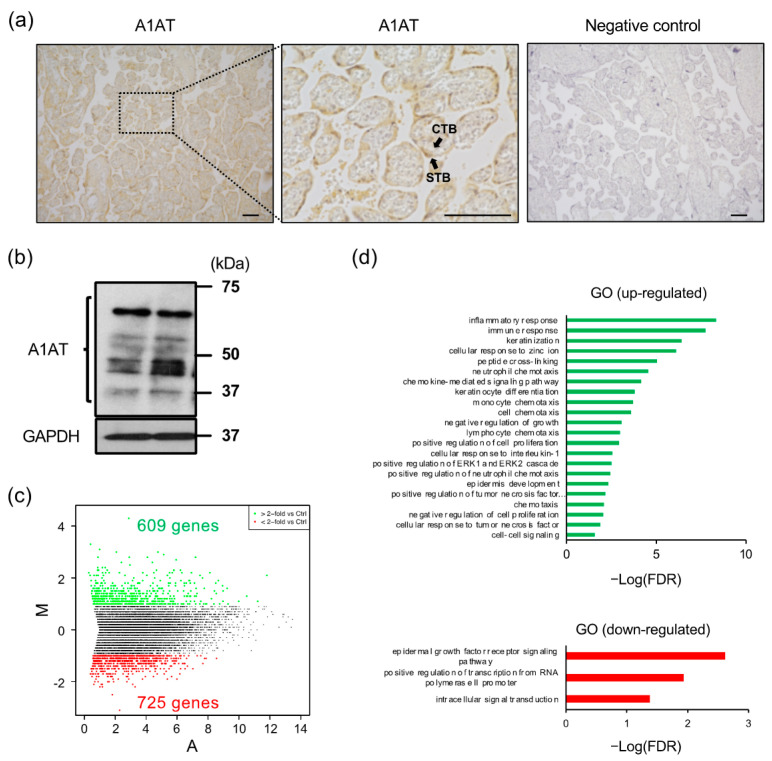
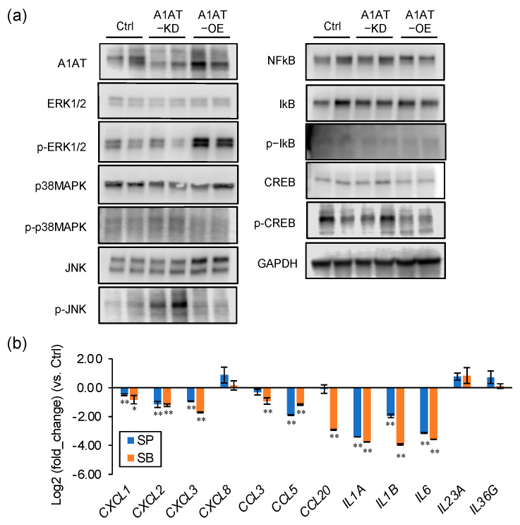
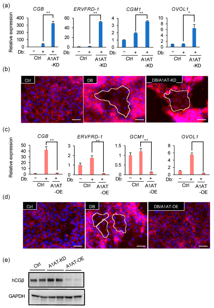
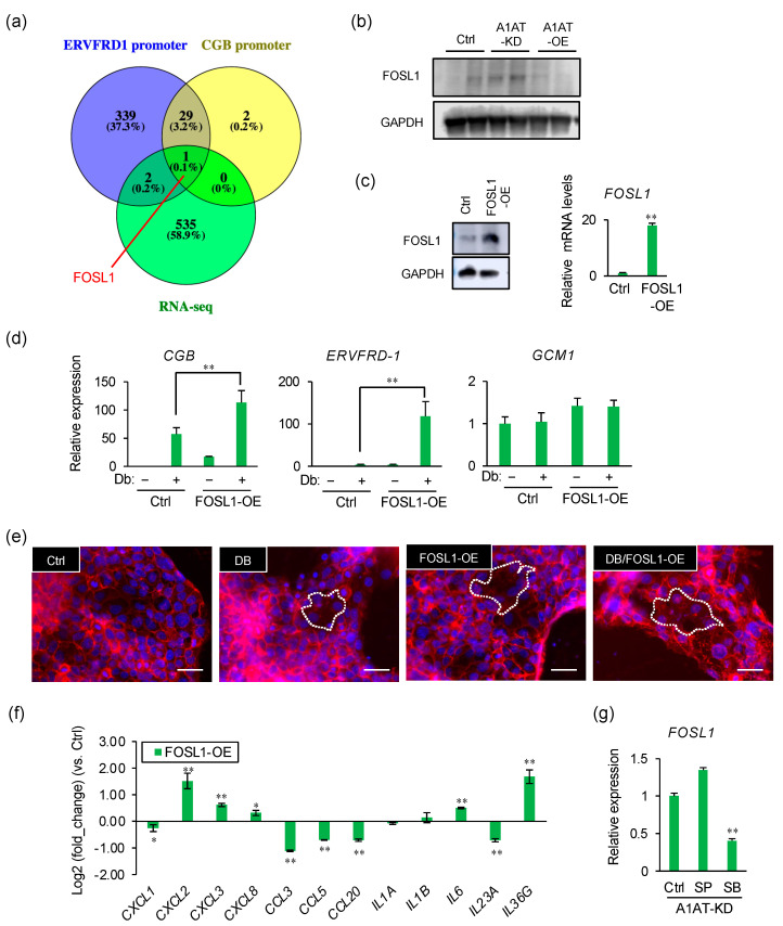
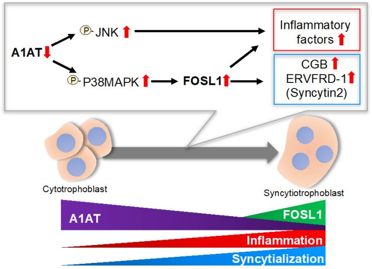
The serine protease inhibitor alpha1-antitrypsin (A1AT) may possess protective functions of impaired organs in a manner independent of its protease inhibitor activity. A1AT expression has been shown to fluctuate in patients with pregnancy-induced hypertension, which suggests that A1AT may play a role in the syncytialization of villous trophoblasts. A1AT expression was knocked down in primary trophoblasts. RNA was extracted from these cells and subjected to RNA-sequencing analysis to determine the levels of expression of markers of syncytialization and inflammation. In addition, A1AT protein was localized in trophoblastic cells in placental tissues. Knockdown of A1AT upregulated the expression of FOSL1 and markers of syncytialization, as well as cell fusion, whereas overexpression of A1AT had the opposite effects. FOSL1 overexpression stimulated syncytialization, similar to the effects of A1AT knock down. Inhibitors of p38MAPK and JNK reduce the expression of inflammatory factors, whereas a p38MAPK inhibitor suppressed FOSL1 expression. Collectively, these findings indicated A1AT may negatively regulate inflammatory responses by controlling the activation of p38MAPK and JNK, and that p38MAPK mediates trophoblast syncytialization by altering FOSL1 expression. Therefore, a dysfunction in A1AT could be responsible for abnormal placental formation and pregnancy-associated disorders.

Keywords: AP-1 transcription factor subunit (FOSL1); FOS like 1; alpha-1-antitrypsin (A1AT); inflammatory factor; syncytialization; trophoblasts.


Conflict of interest statement

The authors declare no conflict of interest.

Figures

Figure 1
Figure 1
Knockdown of A1AT upregulates the expression of inflammatory response-related genes in trophoblasts. (a) Expression of A1AT protein in human placentas. Immunostaining was performed using anti-A1AT antibody or normal rabbit IgG (negative control). The pictures display the chorionic villi. The magnified middle picture shows the rectangle in the left panel. Scale bar = 100 μm. CTB: cytotrophoblasts, STB: syncytiotrophoblast. (b) Lysates prepared from primary trophoblasts were subjected to immunoblotting, with GAPDH serving as the loading control. (c) MA plot showing the expression of transcripts identified by RNA-seq. The transcripts highlighted in red or green were more than 2-fold differentially expressed (p < 0.05). (d) Functional classification of differentially expressed genes (DEGs) by Gene Ontology analysis.
Figure 2
Figure 2
A1AT expression alters the levels of p38MAPK and JNK and the expression of inflammatory cytokines in trophoblasts. (a) Lysates prepared from BeWo cells with A1AT knockdown (A1AT-KD) or A1AT overexpression (A1AT-OE) were subjected to immunoblotting. GAPDH served as a loading control. (b) A1AT-KD BeWo cells were treated with the JNK inhibitor SP600125 (SP, 20 μM) or the p38 MAPK inhibitor SB203580 (SB, 20 μM) for 24 h, and the levels of mRNA encoding inflammatory cytokines were measured and normalized to that of GAPDH mRNA. The results shown are the means ± SEMs of three independent experiments. * p < 0.05, ** p < 0.01.
Figure 3
Figure 3
A1AT expression alters the syncytialization of trophoblasts. (a) A1AT-KD BeWo cells were treated for 72 h with Db (0.5 μM). Changes in syncytialization markers CGB, ERVFRD-1, GCM1, and OVOL1 mRNA expression were determined by qPCR. The results shown are the means ± SEMs of three independent experiments. * p < 0.05, ** p < 0.01. (b) A1AT-KD BeWo cells were treated for 72 h with Db (0.5 μM). Cells were immunostained with anti-E-cadherin antibody (red) and DAPI (blue) to visualize syncytialization. A representative picture from three independent experiments is shown and the syncytialized cells are marked with a stippled line. Scale bar = 50 μm. (c,d) A1AT-OE BeWo cells were treated for 72 h with Db (0.5 μM), and the expression of mRNAs encoding the syncytialization markers described in (a) was determined by qPCR (c). The results are the means ± SEMs of three independent experiments. * p < 0.05, ** p < 0.01. Cells were immunostained to visualize syncytialization (d) as in (b). A representative picture from three independent experiments is shown. Scale bar = 50 μm. (e) Immunoblottingshowing the protein levels of the syncytialization marker hCGβ in lysates from A1AT-KD and A1AT-OE BeWo cells. GAPDH served as the loading control.
Figure 4
Figure 4
FOSL1 mediates A1AT knockdown-induced syncytialization of trophoblasts. (a) Venn diagram showing the numbers of upregulated DEGs and of genes encoding proteins that may be capable of binding to the ERVFDR-1 or CGB promoter. (b) Immunoblotting showing the protein levels of FOSL1 in lysates from A1AT-KD and A1AT-OE BeWo cells. GAPDH served as the loading control. (c) Expression of FOSL1 in FOSL1-OE BeWo cells by Immunoblotting (left) and qPCR (right). Results shown are the means ± SEMs of three independent experiments. ** p < 0.01. (d) Expression of syncytialization markers in FOSL1-OE BeWo cells treated with Db (0.5 μM) by qPCR. Values are means ± SEMs of three independent experiments. ** p < 0.01. (e) Visualization of syncytialization by immunostaining cells with anti-E-cadherin antibody (red) and DAPI (blue). Representative pictures are shown, with syncytialized cells marked with a stippled line. Scale bar = 50 μm. (f) Expression of mRNAs encoding inflammatory cytokines in FOSL1-OE BeWo cells. GAPDH was used as the loading control. Results are reported as the means ± SEMs of three independent experiments. * p < 0.05, ** p < 0.01. (g) Expression of mRNAs encoding inflammatory cytokines in A1AT-KD BeWo cells treated with SP600125 (SP, 20 μM) or SB203580 (SB, 20 μM) for 24 h. GAPDH was used as the loading control. Results are reported as the means ± SEMs of three independent experiments. ** p < 0.01.
Figure 5
Figure 5
Schematic diagram illustrating the proposed A1AT/FOSL1 signaling in trophoblasts. A1AT controls the phosphorylation of p38MAPK and JNK, which may result in upregulation of several inflammatory cytokines, whereas p38MAPK regulates syncytialization by inducing FOSL1. P: phosphorylation. Red arrow: expression levels.

References

    1. Hunt J.M., Tuder R. Alpha 1 anti-trypsin: One protein, many functions. Curr. Mol. Med. 2012;12:827–835. doi: 10.2174/156652412801318755. - DOI - PubMed
    1. Tamura K., Takashima H., Fumoto K., Kajihara T., Uchino S., Ishihara O., Yoshie M., Kusama K., Tachikawa E. Possible Role of alpha1-Antitrypsin in Endometriosis-Like Grafts From a Mouse Model of Endometriosis. Reprod. Sci. 2015;22:1088–1097. doi: 10.1177/1933719115570901. - DOI - PubMed
    1. Ando Y., Kuroda A., Kusama K., Matsutani T., Matsuda A., Tamura K. Impact of serine protease inhibitor alpha1-antitrypsin on expression of endoplasmic reticulum stress-induced proinflammatory factors in adipocytes. Biochem. Biophys. Rep. 2021;26:100967. doi: 10.1016/j.bbrep.2021.100967. - DOI - PMC - PubMed
    1. Kaner Z., Ochayon D.E., Shahaf G., Baranovski B.M., Bahar N., Mizrahi M., Lewis E.C. Acute phase protein α1-antitrypsin reduces the bacterial burden in mice by selective modulation of innate cell responses. J. Infect. Dis. 2015;211:1489–1498. doi: 10.1093/infdis/jiu620. - DOI - PubMed
    1. Jonigk D., Al-Omari M., Maegel L., Müller M., Izykowski N., Hong J., Hong K., Kim S.H., Dorsch M., Mahadeva R., et al. Anti-inflammatory and immunomodulatory properties of α1-antitrypsin without inhibition of elastase. Proc. Natl. Acad. Sci. USA. 2013;110:15007–15012. doi: 10.1073/pnas.1309648110. - DOI - PMC - PubMed