. 2022 Feb 18;23(4):2274.
doi: 10.3390/ijms23042274.

Cytoskeleton Protein EB3 Contributes to Dendritic Spines Enlargement and Enhances Their Resilience to Toxic Effects of Beta-Amyloid

Affiliations

Cytoskeleton Protein EB3 Contributes to Dendritic Spines Enlargement and Enhances Their Resilience to Toxic Effects of Beta-Amyloid

Ekaterina Pchitskaya et al. Int J Mol Sci. .

Abstract

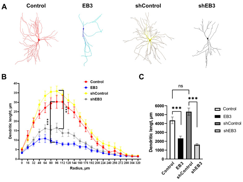
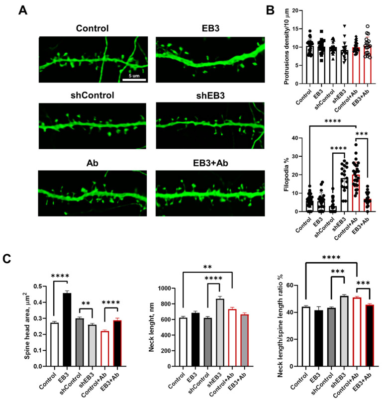
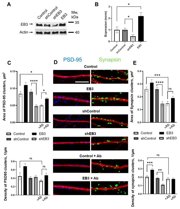
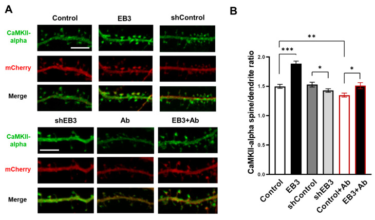
EB3 protein is expressed abundantly in the nervous system and transiently enters the dendritic spines at the tip of the growing microtubule, which leads to spine enlargement. Nevertheless, the role of dynamic microtubules, and particularly EB3 protein, in synapse function is still elusive. By manipulating the EB3 expression level, we have shown that this protein is required for a normal dendritogenesis. Nonetheless, EB3 overexpression also reduces hippocampal neurons dendritic branching and total dendritic length. This effect likely occurs due to the speeding neuronal development cycle from dendrite outgrowth to the step when dendritic spines are forming. Implementing direct morphometric characterization of dendritic spines, we showed that EB3 overexpression leads to a dramatic increase in the dendritic spine head area. EB3 knockout oppositely reduces spine head area and increases spine neck length and spine neck/spine length ratio. The same effect is observed in conditions of amyloid-beta toxicity, modeling Alzheimer`s disease. Neck elongation is supposed to be a common detrimental effect on the spine's shape, which makes them biochemically and electrically less connected to the dendrite. EB3 also potentiates the formation of presynaptic protein Synapsin clusters and CaMKII-alpha preferential localization in spines rather than in dendrites of hippocampal neurons, while its downregulation has an opposite effect and reduces the size of presynaptic protein clusters Synapsin and PSD95. EB3's role in spine development and maturation determines its neuroprotective effect. EB3 overexpression makes dendritic spines resilient to amyloid-beta toxicity, restores altered PSD95 clustering, and reduces CaMKII-alpha localization in spines observed in this pathological state.

Keywords: Alzheimer’s disease; EB3; PSD95; Synapsin CaMKII; beta-amyloid; dendritic spines; end-binding protein; neuronal morphology; neuroprotection.


Conflict of interest statement

The authors declare no conflict of interest. The funders had no role in the design of the study; in the collection, analyses, or interpretation of data; in the writing of the manuscript, or in the decision to publish the results.

Figures

Figure 1
Figure 1
EB3 expression level affects the dendritogenesis of hippocampal neurons. (A) Models of hippocampal neurons transfected with mCherry or co-transfected with mCherry and EB3, plasmid encoding control RNAi (shControl) or RNAi against EB3 (shEB3) at DIV 7−8 and fixed at DIV 15 build in Neuronstudio from confocal images. (B) Dendritic length between two Sholl rings separated by 1 μm radius (distance from the soma) for each group of neurons. (C) The total length of dendrites for each group of neurons, μm. ***: p < 0.001, ns-no statistically significant difference, n = 20 neurons from 4 batches of cultures. Mann–Whitney U test for Sholl analysis, Student`s t-test for dendritic length, normal distribution was checked by the Shapiro-Wilk test.
Figure 2
Figure 2
EB3 expression levels shape hippocampal dendritic spine morphology in WT neurons and in the condition of amyloid toxicity. (A) Confocal images of WT primary hippocampal dendrites and dendrites in the condition of amyloid toxicity (Ab) transfected with GFP or co-transfected with GFP and EB3, plasmid encoding control RNAi (shControl) or RNAi against EB3 (shEB3) at DIV 8-9, and fixed at DIV 16-17. Scale bar corresponds to 5μm. (B) The number of protrusions per 10 μm dendrite length and fraction of filopodia for each group of cells shown on panel A. (C) Direct morphometric measurements of dendritic spines morphology including spine head area, neck length, and neck length/spine length ratio. **: p < 0.01; ***: p < 0.001, ****: p < 0.0001. ANOVA test for protrusions density, Kruskal–Wallis test with post-hoc Dunn’s test for other data. n ≥ 18 neurons and n ≥ 230 spines from 3 batches of cultures.
Figure 3
Figure 3
EB3 affects PSD-95 and Synapsin protein clustering. (A) The efficiency of EB3 expression level regulation generated in HEK293T cells Lentiviral particles were analyzed by Western blotting the primary hippocampal cultures infected with lentiviruses encoding EB3, control RNAi (shControl), or RNAi against EB3 (shEB3) at DIV 7-8 and lysed at DIV 15. Actin was used as a loading control. (B) Quantification of EB3 expression level (normalized to actin levels). The mean density of each band was normalized to an actin signal in the same sample and averaged. n = 4 batches of culture, *: p < 0.05. (C) Area and density of PSD-95 protein clusters for each group of cells shown on panel D. (D) Binarized confocal images of WT hippocampal dendrites and dendrites treated with beta-amyloid, infected with lentiviruses encoding EB3, control RNAi (shControl) or RNAi against EB3 (shEB3) at DIV 7 and fixed at DIV 14 and stained anti-MAP2 (red), anti-PSD95 (blue), and anti-Synapsin (green). Scale bar corresponds to 5μm. (E) Area and density of Synapsin protein clusters for each group of cells shown on panel D. *: p < 0.05, **: p < 0.01, ***: p < 0.001, ****: p < 0.0001, ns-no statistically significant difference. Tamhane’s T3 test for PSD95 clusters area, Kruskal–Wallis test with post-hoc Dunn’s test for other data. n  ≥  25 neurons from 3 to 5 batches of cultures.
Figure 4
Figure 4
EB3 potentiates spine localization of CaMKII-alpha. (A) Confocal images of WT primary hippocampal dendrites and dendrites in condition of amyloid toxicity (Ab) co-transfected with Venus-CaMKII-alpha and mCherry infected with lentiviruses encoding EB3, control RNAi (shControl), or RNAi against EB3 (shEB3) at DIV 8-9 and fixed at DIV 16-17. Scale bar corresponds to 5μm. (B) The ratio of the CaMKII-alpha fluorescence intensity in spines to parent dendrite normalized to the mCherry. *: p < 0.05, **: p < 0.01, ***: p < 0.001. Student-t test. n ≥ 53 spines from 4 batches of cultures.

References

    1. Bailey C.H., Kandel E.R., Harris K. Structural Components of Synaptic Plasticity and Memory Consolidation. Cold Spring Harb. Perspect. Biol. 2015;7:a021758. doi: 10.1101/cshperspect.a021758. - DOI - PMC - PubMed
    1. Berry K.P., Nedivi E. Spine Dynamics: Are They All the Same? Neuron. 2017;96:43–55. doi: 10.1016/j.neuron.2017.08.008. - DOI - PMC - PubMed
    1. Kasai H., Ziv N.E., Okazaki H., Yagishita S., Toyoizumi T. Spine dynamics in the brain, mental disorders and artificial neural networks. Nat. Rev. Neurosci. 2021;22:407–422. doi: 10.1038/s41583-021-00467-3. - DOI - PubMed
    1. Alvarez V.A., Sabatini B.L. Anatomical and Physiological Plasticity of Dendritic Spines. Annu. Rev. Neurosci. 2007;30:79–97. doi: 10.1146/annurev.neuro.30.051606.094222. - DOI - PubMed
    1. Bourne J., Harris K.M. Do thin spines learn to be mushroom spines that remember? Curr. Opin. Neurobiol. 2007;17:381–386. doi: 10.1016/j.conb.2007.04.009. - DOI - PubMed

LinkOut - more resources