. 2022 Feb 20;23(4):2333.
doi: 10.3390/ijms23042333.

Employing CRISPR-Cas9 to Generate CD133 Synthetic Lethal Melanoma Stem Cells

Affiliations

Employing CRISPR-Cas9 to Generate CD133 Synthetic Lethal Melanoma Stem Cells

Cynthia M Simbulan-Rosenthal et al. Int J Mol Sci. .

Abstract

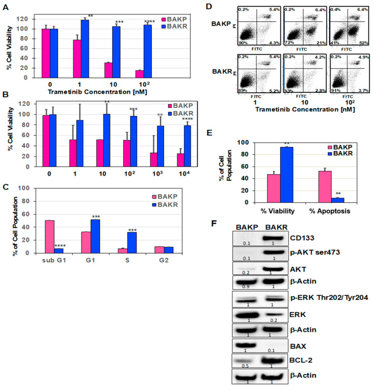
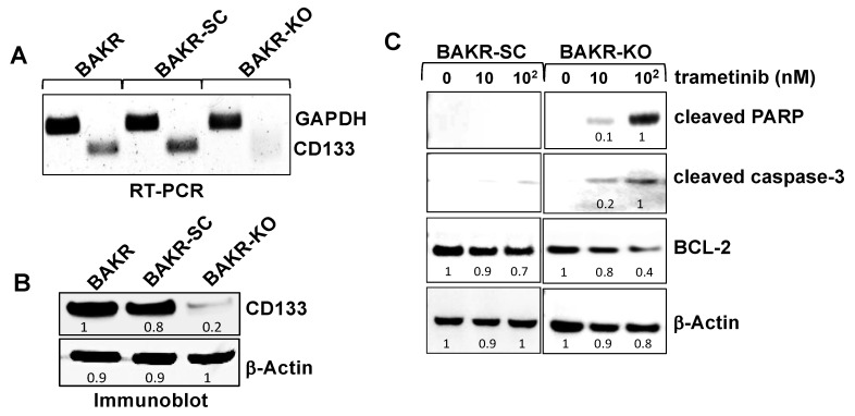
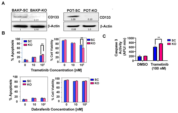
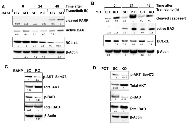
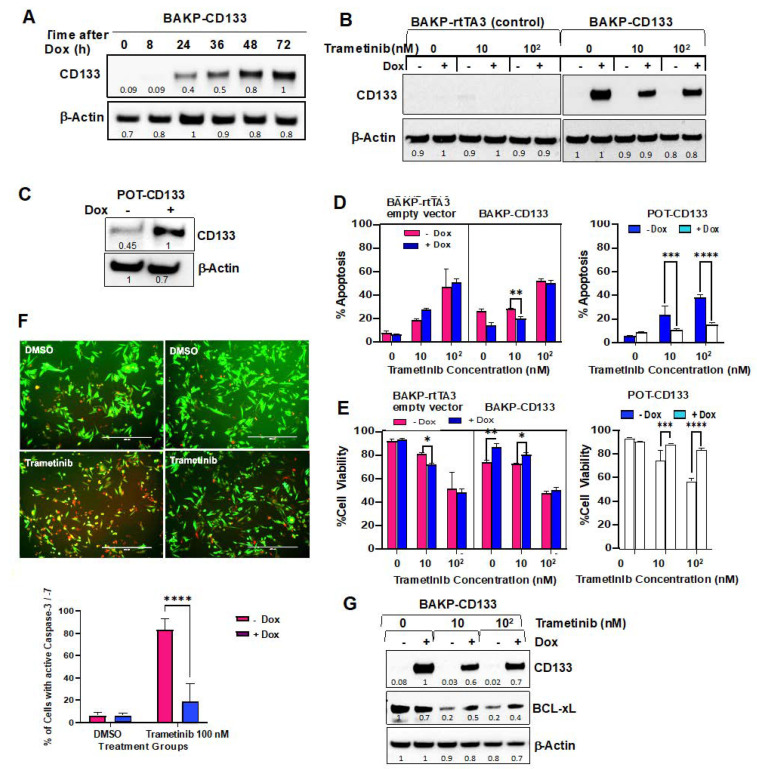
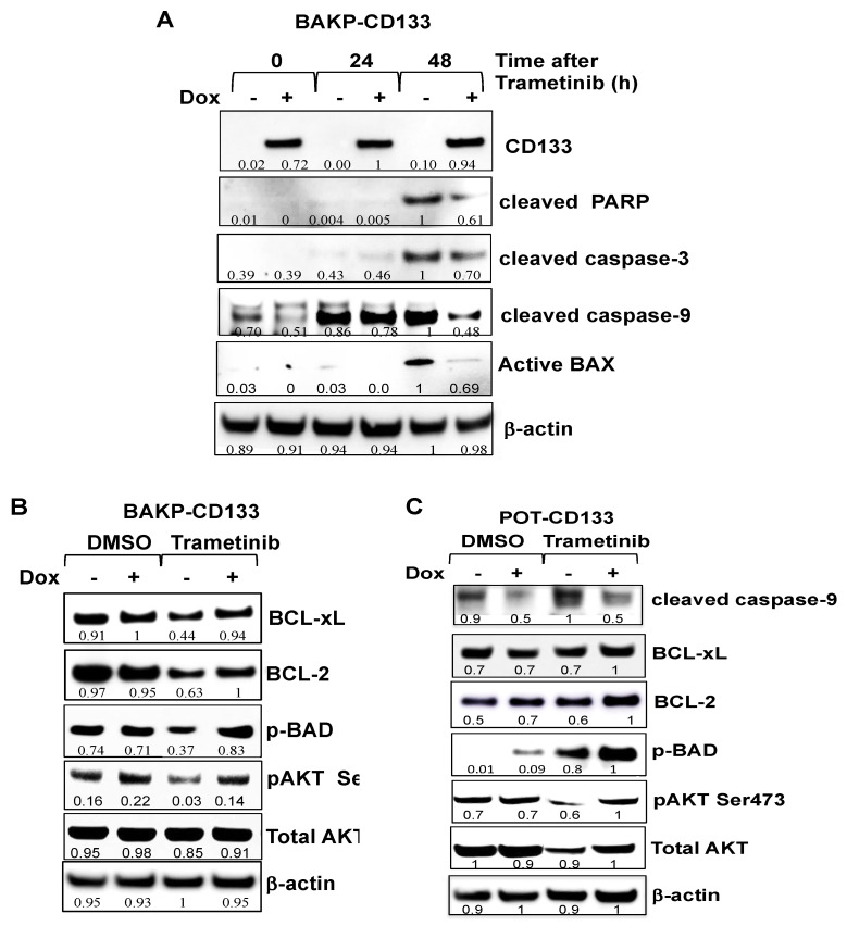
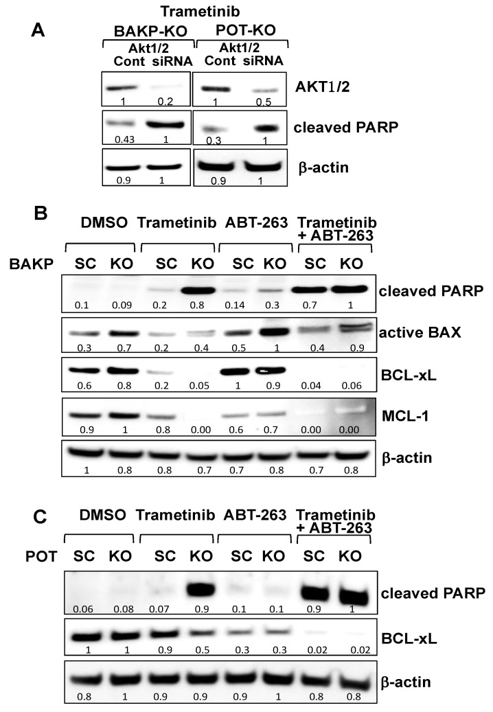
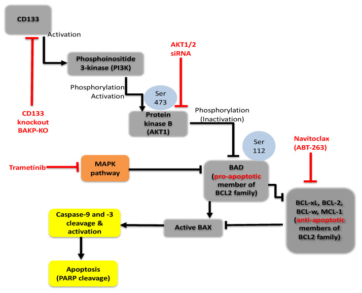
Malignant melanoma is a lethal skin cancer containing melanoma-initiating cells (MIC) implicated in tumorigenesis, invasion, and drug resistance, and is characterized by the elevated expression of stem cell markers, including CD133. The siRNA knockdown of CD133 enhances apoptosis induced by the MEK inhibitor trametinib in melanoma cells. This study investigates the underlying mechanisms of CD133's anti-apoptotic activity in patient-derived BAKP and POT cells, harboring difficult-to-treat NRASQ61K and NRASQ61R drivers, after CRISPR-Cas9 CD133 knockout or Dox-inducible expression of CD133. MACS-sorted CD133(+) BAKP cells were conditionally reprogrammed to derive BAKR cells with sustained CD133 expression and MIC features. Compared to BAKP, CD133(+) BAKR exhibit increased cell survival and reduced apoptosis in response to trametinib or the chemotherapeutic dacarbazine (DTIC). CRISPR-Cas9-mediated CD133 knockout in BAKR cells (BAKR-KO) re-sensitized cells to trametinib. CD133 knockout in BAKP and POT cells increased trametinib-induced apoptosis by reducing anti-apoptotic BCL-xL, p-AKT, and p-BAD and increasing pro-apoptotic BAX. Conversely, Dox-induced CD133 expression diminished apoptosis in both trametinib-treated cell lines, coincident with elevated p-AKT, p-BAD, BCL-2, and BCL-xL and decreased activation of BAX and caspases-3 and -9. AKT1/2 siRNA knockdown or inhibition of BCL-2 family members with navitoclax (ABT-263) in BAKP-KO cells further enhanced caspase-mediated apoptotic PARP cleavage. CD133 may therefore activate a survival pathway where (1) increased AKT phosphorylation and activation induces (2) BAD phosphorylation and inactivation, (3) decreases BAX activation, and (4) reduces caspases-3 and -9 activity and caspase-mediated PARP cleavage, leading to apoptosis suppression and drug resistance in melanoma. Targeting nodes of the CD133, AKT, or BCL-2 survival pathways with trametinib highlights the potential for combination therapies for NRAS-mutant melanoma stem cells for the development of more effective treatments for patients with high-risk melanoma.

Keywords: AKT; CD133; CRISPR/cas9 knockdown; melanoma stem cells; trametinib.


Conflict of interest statement

The authors declare no conflict of interest.

Figures

Figure 1
Figure 1
Sustained CD133 overexpression in BAKR cells increases cell viability and resistance to trametinib- and DTIC-induced apoptosis. CD133(+) BAKR melanoma cells and parental BAKP cells were exposed to increasing doses of trametinib (A) or DTIC (B) for 72 h, followed by XTT cell viability assays to assess drug sensitivity. (C) Cell cycle analysis of BAKR and BAKP cells after treatment with 100 nM trametinib for 72 h. (D) Representative dot plots of cells treated with indicated doses of trametinib and subjected to Annexin-FITC/PI staining and flow cytometry; and (E) % cell viability and apoptosis of cells in D. (F) Basal expression levels of pro-survival or pro-apoptotic proteins were compared in BAKP and BAKR cells by immunoblot analysis with antibodies to CD133, activated p-AKT (ser473), AKT, p-ERK (Thr202/Tyr204), ERK, BCL-2, BAX, and β-actin as loading control. Densitometric analysis comparing intensities of protein bands relative to bands with the highest intensity is shown in immunoblots. Scans of whole gel immunoblots for all Figures are shown in “Supplementary Materials” (Figure S1). Results shown are the means ± SD of three replicates of a representative experiment; essentially the same results were obtained in three independent experiments. **, ***, **** represent p < 0.01, p < 0.001, and p < 0.0001, respectively.
Figure 2
Figure 2
CRISPR-Cas9 CD133-knockout sensitizes BAKR-KO cells to trametinib-induced apoptosis via reduced BCL-2 levels. CD133 expression was compared between BAKR, CD133-knockout BAKR-KO, and sgRNA control BAKR-SC cells. (A) RT-PCR and (B) immunoblot analysis confirm depletion of CD133 RNA and protein, respectively, in BAKR-KO, compared to BAKR-SC and BAKR cells. (C) BAKR-SC and BAKR-KO cells were exposed to increasing trametinib concentrations for 72 h and subjected to immunoblot analysis with antibodies to the active cleaved form of caspase-3 (p17). Immunoblots were stripped of antibodies and re-probed with antibodies to cleaved PARP, BCL-2, and β-Actin for loading control. Densitometric analysis comparing intensities of protein bands relative to bands with the highest intensity is shown in immunoblots.
Figure 3
Figure 3
CRISPR-Cas9 knockout of CD133 expression in parental BAKP melanoma cells (A) increases caspase-3 mediated apoptosis (B,C) after exposure to trametinib. (A) Immunoblot analysis with anti-CD133 in BAKP-SC vs. BAKP-KO cells (left panel) and POT SC vs. POT-KO cells (right panel). (B) BAKP-SC and BAKP-KO cells were treated with indicated concentrations of trametinib (upper panels) or dabrafenib (lower panels) and subjected to Annexin FITC/PI apoptosis assays. Trametinib-treated CD133-depleted BAKP-KO cells display enhanced apoptosis relative to control BAKP-SC cells, an effect that was not seen with dabrafenib. (C) Fluorometric caspase-3 activity assays were performed 72 h after exposure of BAKP-SC and BAKP-KO cells to 100 nM trametinib. Results shown are the means ± SD of three biological replicates of a representative experiment; essentially the same results were obtained in three independent experiments. ** represents p < 0.01.
Figure 4
Figure 4
CRISPR-Cas9-mediated knockout of CD133 expression in BAKP (A) and POT (B) cells increases BAX activation, and PARP cleavage following trametinib treatment, sensitized by reduced basal levels of pro-survival BCL-xL, pAKT, and pBAD. (A) BAKP-KO, BAKP-SC cells, as well as (B) POT-SC and POT-KO cells were treated with 100 nM trametinib for 24 and 48 h, followed by immunoblot analysis with antibodies to cleaved PARP, cleaved active form of caspase-3, active BAX, BCL-xL, and β-actin as loading control. CD133 knockout in trametinib-exposed both BAKP-KO (A) and POT-KO (B) cells increases pro-apoptotic active BAX and decreases anti-apoptotic BCL-xL levels (A,B), sensitizing cells to apoptosis. Immunoblot analysis of pAKT, AKT, p-BAD, Bad, and β-actin as loading control in BAKP-KO (C) and POT-KO cells (D), compared to the SC controls. Densitometric analysis comparing intensities of protein bands relative to bands with the highest intensity is shown in immunoblots.
Figure 5
Figure 5
Dox-induced CD133 expression in BAKP (A,B) and POT (C) cells, as verified by immunoblot analysis, suppress caspase-3 mediated apoptotic cell death (D,F) and increases cell viability (E) after trametinib treatment (dose-response experiments). (A) Cells were incubated with Dox for the indicated times and subjected to immunoblot analysis with antibodies to CD133 and β-actin for normalization. (B) Immunoblot analyses with anti-CD133 of BAKP-CD133 and vector control BAKP-rtTA3 cells treated with increasing trametinib doses +/− Dox, as well as (C) POT-CD133 cells incubated +/− Dox for 72 h. (D) BAKP-CD133, BAKP-rtTA3 empty vector control, and POT-CD133 cells were exposed to increasing doses of trametinib for 72 h, and subjected to Annexin-FITC/PI apoptosis assays; apoptosis (%; D) and cell viability (%; E) of cells are shown. (F) Fluorescent FLICA Caspase 3/7 activity assays reveal a significant decline in caspase 3 activity in trametinib-treated CD133-expressing BAKP cells (+Dox), compared to uninduced cells (−Dox); representative merged images of red fluorescent FLICA- and GFP-positive cells (10×) (left panel); quantification of FLICA-positive cells (with active caspase-3, right panel). (G) Immunoblot analyses with antibodies to CD133, BCL-xL, and βActin of BAKP-CD133 treated with increasing trametinib doses +/− Dox. BAKP-CD133 cells were constitutively expressing GFP. Results shown are the means ± SD of three biological replicates of a representative experiment of three independent experiments. *, **, ***, **** represent p < 0.05, p < 0.01, p < 0.001, and p < 0.0001, respectively. Densitometric analysis comparing intensities of protein bands relative to bands with the highest intensity is shown in immunoblots.
Figure 6
Figure 6
Dox-inducible CD133 expression attenuates trametinib-induced apoptosis in BAKP-CD133 (A,B) and POT-CD133 cells (C), as evidenced by reduced PARP cleavage, proteolytic activation of caspases-3 and -9, and activation of BAX (A), and upregulation of anti-apoptotic proteins BCL-xL, BCL-2, p-BAD, and p-AKT (B), in response to trametinib treatment. (A) BAKP-CD133 cells were exposed to 100 nM trametinib for 24 or 48 h +/− Dox, followed by immunoblot analysis with antibodies to apoptotic markers cleaved PARP, cleaved caspase-3, cleaved caspase-9, and active Bax. BAKP-CD133 (B) and POT-CD133 (C) treated with 100 nM trametinib +/− Dox were subjected to immunoblot analysis with anti-apoptotic proteins BCL-xL, BCL-2, p-BAD, p-AKT, AKT, and β-actin for normalization. Densitometric analysis comparing intensities of protein bands relative to bands with the highest intensity is shown below each band in immunoblots.
Figure 7
Figure 7
siRNA knockdown of AKT1/2 in BAKP (A, left panel) and POT cells (A, right panel), or BCL-2 family inhibitor ABT-263 and/or CD133 knockdown reveals that AKT1/2 siRNA knockdown or BCL-2 family inhibition with ABT-263 plus CD133 knockdown both block the AKT pathway, inducing maximum apoptosis in the presence of trametinib in BAKP (B) and POT cells (C). (A) BAKP-KO and POT-KO cells were transfected with AKT1/2 siRNA pool or the scrambled control siRNA (“Cont”). Then, 48 h after transfection, cells were treated with 100 mM trametinib for 24 h, and subjected to immunoblot analysis with antibodies to AKT1, cleaved PARP, or β-actin. (B) BAKP-KO and (C) POT-KO cells or their respective controls BAKP-SC and POT-SC cells were treated for 48 h with trametinib (100 nM) or the BCL-2 family inhibitor ABT-263 (100 nM) alone or in combination, and then subjected to immunoblot analysis with antibodies to cleaved PARP, active BAX, BCL-xL, MCL-1, or β-actin. Densitometric analysis comparing intensities of protein bands relative to bands with the highest intensity is shown in immunoblots.
Figure 8
Figure 8
PI3K/Akt/ Bcl2 family pro-survival signaling pathway in CD133-positive melanoma initiating stem cells (MICs). CD133 may activate a survival pathway where (1) increased phosphorylation of AKT induces (2) phosphorylation and inactivation of BAD, (3) decreases the active form of BAX, and (4) reduces caspase activation and caspase-mediated PARP cleavage, indicating apoptosis suppression leading to drug resistance in melanomas.

References

    1. Cancer Stat Facts: Melanoma of the Skin. Bethesda; Rockville, MD, USA: 2021. [(accessed on 18 May 2021)]. Surveillance, Epidemiology, and End Results Program. Available online: https://seer.cancer.gov/statfacts/html/melan.html.
    1. Paluncic J., Kovacevic Z., Jansson P.J., Kalinowski D., Merlot A.M., Huang M.L.-H., Lok H.C., Sahni S., Lane D.J.R., Richardson D.R. Roads to melanoma: Key pathways and emerging players in melanoma progression and oncogenic signaling. Biochim. Biophys. Acta. 2016;1863:770–784. doi: 10.1016/j.bbamcr.2016.01.025. - DOI - PubMed
    1. Spagnolo F., Picasso V., Lambertini M., Ottaviano V., Dozin B., Queirolo P. Survival of patients with metastatic melanoma and brain metastases in the era of MAP-kinase inhibitors and immunologic checkpoint blockade antibodies: A systematic review. Cancer Treat. Rev. 2016;45:38–45. doi: 10.1016/j.ctrv.2016.03.003. - DOI - PubMed
    1. Ugurel S., Röhmel J., Ascierto P.A., Flaherty K.T., Grob J.J., Hauschild A., Larkin J., Long G.V., Lorigan P., McArthur G.A., et al. Survival of patients with advanced metastatic melanoma: The impact of novel therapies-update 2017. Eur. J. Cancer. 2017;83:247–257. doi: 10.1016/j.ejca.2017.06.028. - DOI - PubMed
    1. Sandri S., Faião-Flores F., Tiago M., Pennacchi P.C., Massaro R.R., Alves-Fernandes D.K., Berardinelli G.N., Evangelista A.F., Vazquez V.D., Reis R.M., et al. Vemurafenib resistance increases melanoma invasiveness and modulates the tumor microenvironment by MMP-2 upregulation. Pharmacol. Res. 2016;111:523–533. doi: 10.1016/j.phrs.2016.07.017. - DOI - PubMed

MeSH terms

Substances