. 2022 Feb 25;12(1):20.
doi: 10.1186/s13578-022-00764-z.

The dual role of p62 in ferroptosis of glioblastoma according to p53 status

Affiliations

The dual role of p62 in ferroptosis of glioblastoma according to p53 status

Fanen Yuan et al. Cell Biosci. .

Abstract

Background: Ferroptosis plays a key role in human cancer, but its function and mechanism in glioma is not clear. P62/SQSTM1 was reported to inhibit ferroptosis via the activation of NRF2 signaling pathway. In this study we reveal a dual role of p62 in ferroptosis of glioblastoma (GBM) according to p53 status.

Method: Lipid peroxidation analysis, transmission electron microscopy (TEM), GSH assay were performed to determine the level of ferroptosis. Western blot and qPCR were obtained to detect the expression of ferroptosis markers. Construction of mutant plasmids, immunoprecipitation, luciferase assay and rescue-experiments were performed to explore the regulatory mechanism.

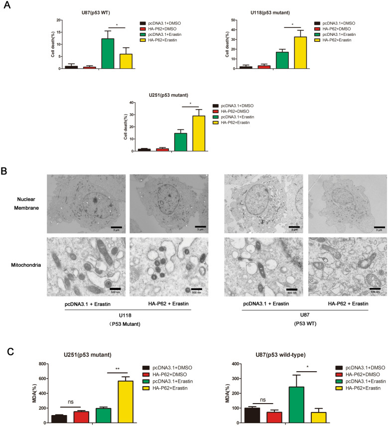
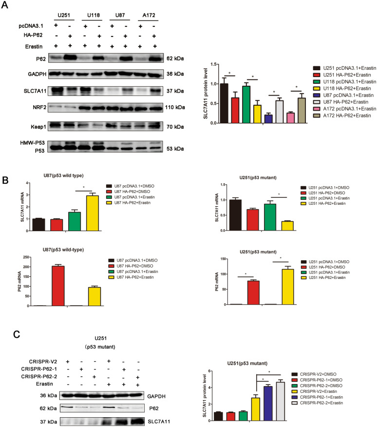
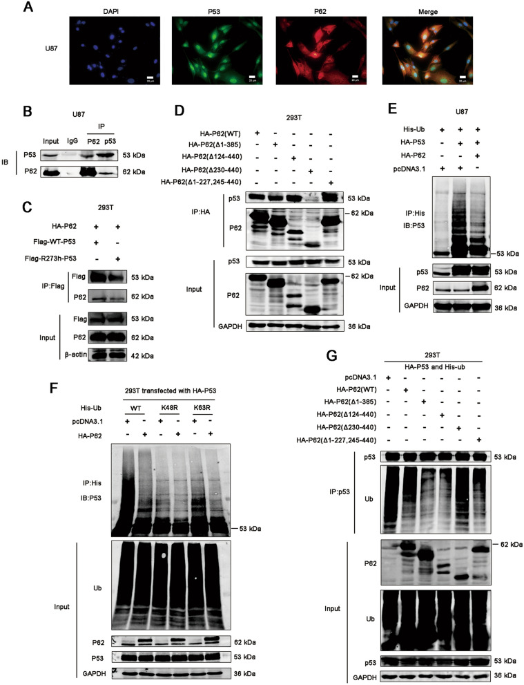
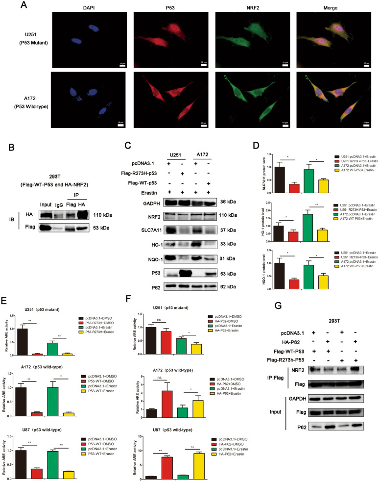
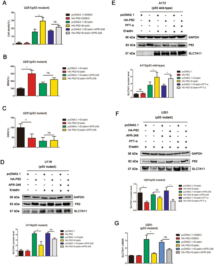
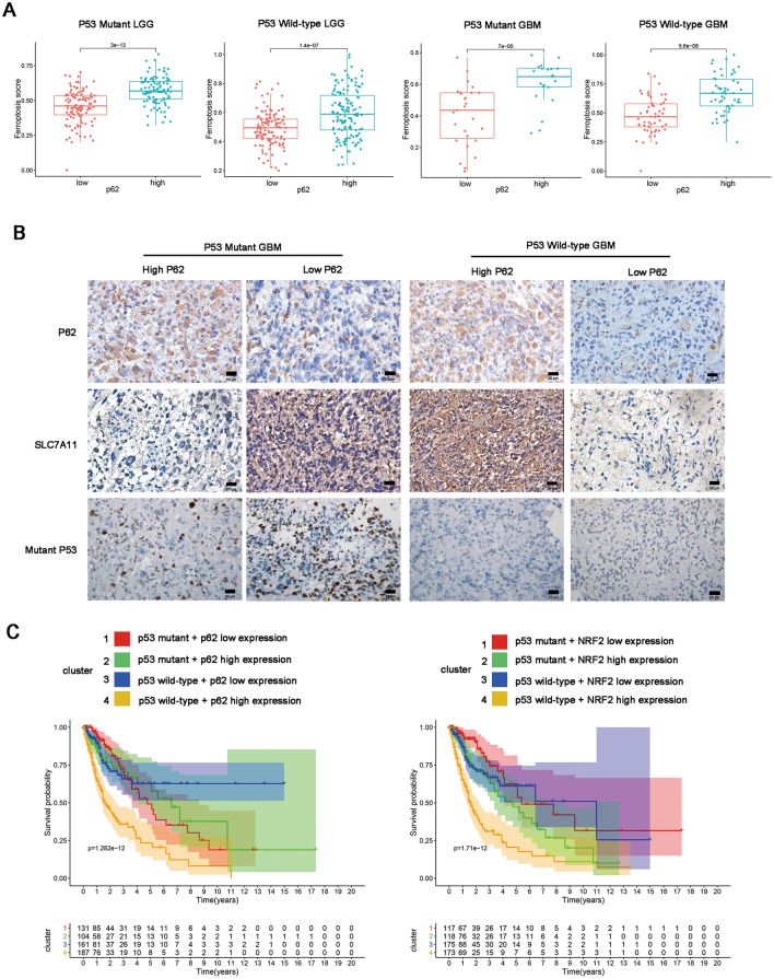
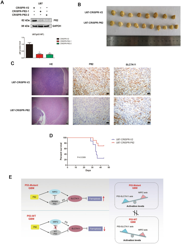
Results: P62 overexpression facilitates ferroptosis and inhibits SLC7A11 expression in p53 mutant GBM, while attenuates ferroptosis and promotes SLC7A11 expression in p53 wild-type GBM. P62 associates with p53 and inhibits its ubiquitination. The p53-NRF2 association and p53-mediated suppression of NRF2 antioxidant activity are diversely regulated by p62 according to p53 status. P53 mutation status is required for the dual regulation of p62 on ferroptosis. In wild-type p53 GBM, the classical p62-mediated NRF2 activation pathway plays a major regulatory role of ferroptosis, leading to increased SLC7A11 expression, resulting in a anti-ferroptosis role. In mutant p53 GBM, stronger interaction of mutant-p53/NRF2 by p62 enhance the inhibitory effect of mutant p53 on NRF2 signaling, which reversing the classical p62-mediated NRF2 activation pathway, together with increased p53's transcriptional suppression on SLC7A11 by p62, leading to a decrease of SLC7A11, resulting in a pro-ferroptosis role.

Conclusion: Together, this study shows novel molecular mechanisms of ferroptosis regulated by p62; the mutation status of p53 is an important factor that determines the therapeutic response to p62-mediated ferroptosis-targeted therapies in GBM.

Keywords: Ferroptosis; Glioblastoma; NRF2; p53; p62.


Conflict of interest statement

The authors declare that they have no conflict of interest.

Figures

Fig. 1
Fig. 1
P62 promotes ferroptosis in p53-mutant GBM cells but inhibits ferroptosis in p53-wild-type GBM cells. A U118, U251 or U87 cells were transfected with pcDNA3.1 or HA-P62 plasmids, followed by DMSO or erastin treatment. Cell death were determined by trypan blue staining. B U118 or U87 control cells and p62-overexpressed clones treated with erastin were subjected to transmission electron microscopy. C The levels of MDA were measured in U251 or U87 cells, which were transfected with pcDNA3.1 or HA-P62 plasmids, followed by DMSO or erastin treatment. pcDNA3.1: the control group. HA-P62: p62 overexpression group
Fig. 2
Fig. 2
P62 negatively regulates SLC7A11 expression in p53-mutant GBM cells but positively regulates SLC7A11 expression in p53-wild-type GBM cells. A U251, U118, U87 and A172 cells were transfected with pcDNA3.1 or HA-P62 plasmids, followed by erastin treatment. The protein expression of SLC7A11, NRF2, P53, Keap1 and P62 were detected by western blot analysis. The quantification of western blots were also presented. B U251 and U87 cells were transfected with pcDNA3.1 or HA-P62 plasmids, followed by DMSO or erastin treatment. The mRNA expression of P62 and SLC7A11 were measured by qPCR. mRNA expression was normalized to GAPDH using the 2−ΔΔCt method. C CRISPR/CAS9 system was performed to knockout p62 expression in U251 cells. SLC7A11 and p62 expression were detected by western blot analysis
Fig. 3
Fig. 3
P62 associates with p53 and inhibits its ubiquitination. A The localization of p62 and p53 was detected by immunofluorescence in U87 cells. B Co-immunoprecipitation assays were performed to assess the association between p62 and p53. Lysates of U87 cells were subjected to IP using anti-P62 or anti-P53 antibodies, followed by immunoblotting with anti-P62 and anti-P53 antibodies. Non-specific IgG was used as a control. Whole cell lysates were used as an input control. C 293 T cells were transfected with flag-WT-p53 or flag-R273h-p53 plasmids together with HA-P62 plasmids. The proteins were immunoprecipitated from cell extracts using anti-Flag antibody, followed by immunoblotting with anti-P62 antibodies. D 293 T cells were transfected with plasmids encoding wild-type p62 (HA-p62 wild type) or a p62 deletion mutant. The proteins were immunoprecipitated from cell extracts using anti-HA antibody, followed by immunoblotting with anti-P62 and anti-P53 antibodies. E Representative immunoblots of p53 and ubiquitinated p53 in U87 cells when p62 was upregulated or downregulated. The proteins were immunoprecipitated from cell extracts using anti-His antibody, followed by immunoblotting with anti-P53 antibodies. F 293 T cells were transfected with plasmids encoding HA-p53, pcDNA3.1 or HA-P62 together with His-ubiquitin or its indicated mutants (His-K48R-Ub, His-K63R-Ub). The proteins were immunoprecipitated from cell extracts using anti-His antibody, followed by immunoblotting with anti-P53 antibodies. G 293 T cells were transfected with plasmids encoding HA-p53 and His-ubiquitin together with HA-P62 or its indicated mutants. The proteins were immunoprecipitated from cell extracts using anti-P53 antibody, followed by immunoblotting with anti-P62 and anti-Ubiquitin antibodies
Fig. 4
Fig. 4
P53-NRF2 interaction and p53-mediated suppression of NRF2 activity are diversely regulated by p62 according to p53 status. A The localization of p53 and NRF2 was detected by immunofluorescence in U251 and A172 cells. Scar bars = 10 um. B Co-immunoprecipitation assays were performed to assess the association between NRF2 and p53. 293 T cells were co-transfected with HA-NRF2 and Flag-WT-p53 plasmids. Lysates of 293 T cells were subjected to IP using anti-Flag or anti-HA antibodies, followed by immunoblotting with anti-Flag and anti-HA antibodies. Non-specific IgG was used as a control. Whole cell lysates were used as an input control. C U251 cells were transfected with pcDNA3.1 or Flag-R273h-p53 plasmids, followed by erastin treatment or not. A172 cells were transfected with pcDNA3.1 or Flag-WT-p53 plasmids, followed by erastin treatment or not. The protein expression of SLC7A11, NRF2, p53, p62, HO-1 and NOQ-1 were detected by western blot analysis. D The quantification in Fig. 4C. E The overexpression of R273h mutant p53 inhibited the NRF2-driven luciferase activity in U251 cells by dual luciferase reporter assays. The overexpression of wild-type p53 inhibited the NRF2-driven luciferase activity in A172 and U87 cells by dual luciferase reporter assays. Cells were treated with DMSO or Erastin. The luciferase activity was calculated as Firefly luciferase/Renilla luciferase. F The effect of p62 overexpression on NRF2-driven luciferase activity in U251, U87 and A172 cells were determined by dual luciferase reporter assays. Cells were treated with DMSO or Erastin. G The effect of p62 overexpression on p53-NRF2 association in different p53 status was determined by IP analysis
Fig. 5
Fig. 5
P53 mutation status is responsible for the dual regulation of p62 on GBM ferroptosis. A Cells were transfected with pcDNA3.1 or HA-P62 plasmids, followed by DMSO, erastin or erastin + APR-246 treatment. Cell death were determined by trypan blue staining. B The levels of MDA were measured in U251 cells, which were transfected with pcDNA3.1 or HA-P62 plasmids, followed by erastin or erastin + APR-246 treatment. C The levels of GSH were measured in U251 cells, which were transfected with pcDNA3.1 or HA-P62 plasmids, followed by erastin or erastin + APR-246 treatment. D U118 cells were transfected with pcDNA3.1 or HA-P62 plasmids, followed by DMSO or erastin or erastin + APR-246 treatment. The protein expression of SLC7A11 and P62 were detected by western blot analysis. E A172 cells were transfected with pcDNA3.1 or HA-P62 plasmids, followed by DMSO or erastin or erastin + PFT-a treatment. The protein expression of SLC7A11 and P62 were detected by western blot analysis. F U251 cells were transfected with pcDNA3.1 or HA-P62 plasmids, followed by DMSO or erastin or erastin + APR-246 treatment. The protein expression of SLC7A11 and P62 were detected by western blot analysis. G U251 cells were transfected with pcDNA3.1 or HA-P62 plasmids, followed by DMSO or erastin or erastin + APR-246 treatment. The mRNA expression of SLC7A11 were detected by qPCR
Fig. 6
Fig. 6
The clinical expression pattern and survival analysis of p62 in p53-wild-type and p53-mutant gliomas. A Difference analysis of ferroptosis enrichment score (ES) among different subtypes. According to the median value of p62, the level of ES were compared among low and high expression group in p53 mutant LGG, p53 wild-type LGG, p53 mutant GBM and p53 wild-type GBM. B Representatives of IHC staining of p62 and SLC7A11 expression in p53 mutant GBM and p53 wild-type GBM tissues. C Survival analysis of OS in four clusters in p62 (left) and NRF2 (right) respectively. Cluster 1: p53 mutant + p62 or NRF2 low expression, cluster 2: p53 mutant + p62 or NRF2 high expression, cluster 3: p53 wild-type + p62 or NRF2 low expression, cluster 4: p53 wild-type + p62 or NRF2 high expression
Fig. 7
Fig. 7
Depletion of p62 promotes ferroptosis in p53 wild-type GBM with an intracranial xenograft model. A The CRISPR/CAS9 system was applied to construct stable p62 knockout U87 cells (CRISPR-p62-1 and CRISPR-p62-2). Knockout efficacy was verified by western blot analysis. B Representative images of pLenti-CRISPR-V2 (CTRL) (n = 8) and pLenti-CRISPR-p62-1 (CRISPR-p62) (n = 10) cells from mouse brains. C H&E and IHC analyses of p62 and SLC7A11 in orthotopic tumour sections. D Mouse survival is shown by Kaplan–Meier curves. U87-CRISPR-V2 groups, n = 8. U87-CRISPR-P62 groups, n = 10. P values were calculated using the log-rank test. E Mechanistic model for dual role of p62 in ferroptosis according to p53 status in GBM

Similar articles

Cited by

References

    1. Ostrom QT, Bauchet L, Davis FG, Deltour I, Fisher JL, Langer CE, Pekmezci M, Schwartzbaum JA, Turner MC, Walsh KM, Wrensch MR, Barnholtz-Sloan JS. The epidemiology of glioma in adults: a "state of the science" review. Neuro Oncol. 2014;16:896–913. - PMC - PubMed
    1. Stupp R, Mason WP, van den Bent MJ, Weller M, Fisher B, Taphoorn MJ, Belanger K, Brandes AA, Marosi C, Bogdahn U, Curschmann J, Janzer RC, Ludwin SK, Gorlia T, Allgeier A, Lacombe D, Cairncross JG, Eisenhauer E, Mirimanoff RO. Radiotherapy plus concomitant and adjuvant temozolomide for glioblastoma. N Engl J Med. 2005;352:987–996. - PubMed
    1. Dixon SJ, Lemberg KM, Lamprecht MR, Skouta R, Zaitsev EM, Gleason CE, Patel DN, Bauer AJ, Cantley AM, Yang WS, Morrison BR, Stockwell BR. Ferroptosis: an iron-dependent form of nonapoptotic cell death. Cell. 2012;149:1060–1072. - PMC - PubMed
    1. Stockwell BR, Friedmann AJ, Bayir H, Bush AI, Conrad M, Dixon SJ, Fulda S, Gascon S, Hatzios SK, Kagan VE, Noel K, Jiang X, Linkermann A, Murphy ME, Overholtzer M, Oyagi A, Pagnussat GC, Park J, Ran Q, Rosenfeld CS, Salnikow K, Tang D, Torti FM, Torti SV, Toyokuni S, Woerpel KA, Zhang DD. Ferroptosis: a regulated cell death nexus linking metabolism. Redox Biol Dis Cell. 2017;171:273–285. - PMC - PubMed
    1. Su Y, Zhao B, Zhou L, Zhang Z, Shen Y, Lv H, AlQudsy L, Shang P. Ferroptosis, a novel pharmacological mechanism of anti-cancer drugs. Cancer Lett. 2020;483:127–136. - PubMed