Review
. 2022 May;81(5):476-488.
doi: 10.1016/j.eururo.2022.01.040. Epub 2022 Feb 22.

Novel Imaging Methods for Renal Mass Characterization: A Collaborative Review

Affiliations
Review

Novel Imaging Methods for Renal Mass Characterization: A Collaborative Review

Eduard Roussel et al. Eur Urol. 2022 May.

Abstract

Context: The incidental detection of localized renal masses has been rising steadily, but a significant proportion of these tumors are benign or indolent and, in most cases, do not require treatment. At the present time, a majority of patients with an incidentally detected renal tumor undergo treatment for the presumption of cancer, leading to a significant number of unnecessary surgical interventions that can result in complications including loss of renal function. Thus, there exists a clinical need for improved tools to aid in the pretreatment characterization of renal tumors to inform patient management.

Objective: To systematically review the evidence on noninvasive, imaging-based tools for solid renal mass characterization.

Evidence acquisition: The MEDLINE database was systematically searched for relevant studies on novel imaging techniques and interpretative tools for the characterization of solid renal masses, published in the past 10 yr.

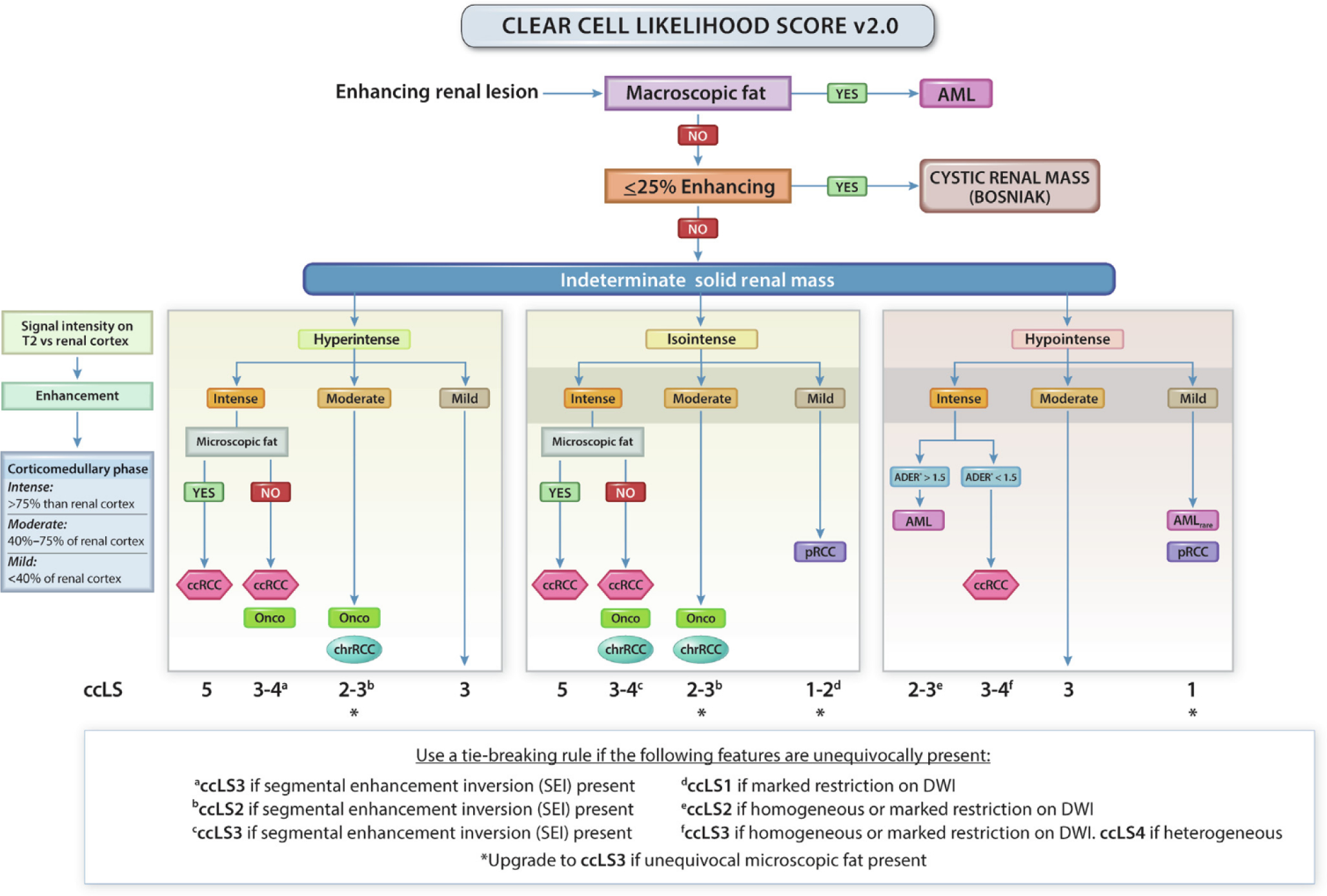
Evidence synthesis: Over the past decade, several novel imaging tools have offered promise for the improved characterization of indeterminate renal masses. Technologies of particular note include multiparametric magnetic resonance imaging of the kidney, molecular imaging with targeted radiopharmaceutical agents, and use of radiomics as well as artificial intelligence to enhance the interpretation of imaging studies. Among these, 99mTc-sestamibi single photon emission computed tomography/computed tomography (CT) for the identification of benign renal oncocytomas and hybrid oncocytic chromophobe tumors, and positron emission tomography/CT imaging with radiolabeled girentuximab for the identification of clear cell renal cell carcinoma, are likely to be closest to implementation in clinical practice.

Conclusions: A number of novel imaging tools stand poised to aid in the noninvasive characterization of indeterminate renal masses. In the future, these tools may aid in patient management by providing a comprehensive virtual biopsy, complete with information on tumor histology, underlying molecular abnormalities, and ultimately disease prognosis.

Patient summary: Not all renal tumors require treatment, as a significant proportion are either benign or have limited metastatic potential. Several innovative imaging tools have shown promise for their ability to improve the characterization of renal tumors and provide guidance in terms of patient management.

Keywords: (99m)Tc-sestamibi; Artificial intelligence; Girentuximab; Kidney cancer; Machine learning; Multiparametric magnetic resonance imaging; PET; Radiomics; Renal cell carcinoma; SPECT; Virtual biopsy.


Conflict of interest statement

Financial disclosures: Eduard Roussel certifies that all conflicts of interest, including specific financial interests and relationships and affiliations relevant to the subject matter or materials discussed in the manuscript (eg, employment/affiliation, grants or funding, consultancies, honoraria, stock ownership or options, expert testimony, royalties, or patents filed, received, or pending), are the following: Eduard Roussel has received an unconditional research grant from Ipsen and Pfizer. Ivan Pedrosa served as a scientific advisor for both Bayer Healthcare and Merck, for which he received honoraria; served as a scientific advisor for Health Tech International, for which he received stock options; and is a coinventor of patents with Philips Healthcare, for which he receives no royalties. Steven Rowe receives salary support and serves as a consultant to Lantheus Medical Imaging, Inc.; owns stock in, has licensed patents to, and serves as a consultant to Precision Molecular, Inc.; and owns stock in and has licensed patents to Plenary AI, Inc. Michael Gorin serves as a paid consultant to Ambu A/S; Blue Earth Diagnostics, Inc; Corbin Clinical Resources, Inc. (Perineologic); Galvanize Therapeutics; KOELIS, Inc.; Lanthius Medical Imaging, Inc; and Simulated Inanimate Models, LLC. Dr. Gorin also has licensed a patent to Precision Molecular, Inc. for which he receives royalties and owns stock in Simulated Inanimate Models, Inc. All other authors have nothing to disclose.

Figures

Fig. 1 –
Fig. 1 –
PRISMA flowchart showing study selection. PRISMA = Preferred Reporting Items for Systematic Reviews and Meta-analyses.
Fig. 2 –
Fig. 2 –
Clear cell likelihood score (ccLS) algorithm version 2.0. Presence of enhancement in ≥25% of the mass volume and absence of macroscopic fat represent eligibility criteria for the use of the ccLS algorithm. Signal intensity on T2-weighted single-shot fast/turbo spin echo imaging (T2) relative to renal cortex, enhancement during the corticomedullary phase relative to renal cortex, and presence of microscopic fat are the major criteria evaluated in every mass. Restriction in DWI, segmental enhancement inversion, and arterial-to-delayed enhancement ratio (ADER) are ancillary findings, which are assessed when indicated in the algorithm. AML = angiomyolipoma; ccRCC = clear cell renal cell carcinoma; chrRCC = chromophobe renal cell carcinoma; DWI = diffusion weighted imaging; Onco = oncocytoma; pRCC = papillary renal cell carcinoma. (Figure was reproduced with permission from Pedrosa and Cadeddu [120].)
Fig. 3 –
Fig. 3 –
Differentiation of renal tumors on the basis of 99mTc-sestamibi uptake. (A) Axial contrast-enhanced CT, (B) axial 99mTc-sestamibi SPECT, and (C) axial 99mTc-sestamibi SPECT/CT images of a patient with a right renal mass (red arrow heads). Uptake in the mass is heterogeneous but overall similar to kidney background. There is relative photopenia in the less enhancing central portion of the tumor. The patient underwent percutaneous biopsy that demonstrated an oncocytic neoplasm and was then enrolled in active surveillance. (D) Axial contrast-enhanced CT, (E) axial 99mTc-sestamibi SPECT, and (F) axial 99mTc-sestamibi SPECT/CT images of a patient with a right renal mass (red arrow heads). There is no appreciable radiotracer uptake in the tumor. The patient underwent a partial nephrectomy with surgical pathology revealing a clear cell renal cell carcinoma. CT = computed tomography; SPECT = single photon emission computed tomography.
Fig. 4 –
Fig. 4 –
Differentiation of a renal tumor on the basis of 89Zr-girentuximab uptake. (A) Axial contrast-enhanced CT, (B) axial 89Zr-girentuximab PET, and (C) axial fused 89Zr-girentuximab PET/CT images in a patient with a left renal mass (red arrow heads). There is clear uptake in the mass, with very little background uptake. The patient underwent a partial nephrectomy with surgical pathology revealing a clear cell renal cell carcinoma. CT = computed tomography; PET = positron emission tomography.
Fig. 5 –
Fig. 5 –
Schematic representation of a radiomic workflow.

Comment in

References

    1. Capitanio U, Bensalah K, Bex A, et al. Epidemiology of renal cell carcinoma. Eur Urol 2019;75:74–84. - PMC - PubMed
    1. Znaor A, Lortet-Tieulent J, Laversanne M, Jemal A, Bray F. International variations and trends in renal cell carcinoma incidence and mortality. Eur Urol 2015;67:519–30. - PubMed
    1. Smaldone MC, Egleston B, Hollingsworth JM, et al. Understanding treatment disconnect and mortality trends in renal cell carcinoma using tumor registry data. Med Care 2017;55:398–404. - PMC - PubMed
    1. Kang SK, Huang WC, Pandharipande PV, Chandarana H. Solid renal masses: what the numbers tell us. Am J Roentgenol 2014;202:1196–206. - PMC - PubMed
    1. Sevcenco S, Heinz-Peer G, Ponhold L, et al. Utility and limitations of 3-Tesla diffusion-weighted magnetic resonance imaging for differentiation of renal tumors. Eur J Radiol 2014;83:909–13. - PubMed

Substances