

Case Reports
doi: 10.1136/bcr-2021-248542.
Andersson lesion in ankylosing spondylitis
Affiliations
- PMID: 35217558
- PMCID: PMC8883225
- DOI: 10.1136/bcr-2021-248542
Item in Clipboard
Case Reports
Andersson lesion in ankylosing spondylitis
BMJ Case Rep.
.
No abstract available
Keywords: andersson lesion; ankylosing spondylitis; neurosurgery; orthopaedics; rheumatology.
Conflict of interest statement
Competing interests: None declared.
Figures

(A) Antero-posterior and (B) lateral radiograph of lumbosacral spine, (C) dorsal spine radiograph showing bilateral decreased SI joint space, and sclerosis around SI joints, suggestive of sacroiliitis (red arrows). Syndesmophytes are seen (blue arrows) as paravertebral ossifications, causing the spine to have diffuse syndesmophytic ankylosis and giving the bamboo spine appearance (green arrow). Irregularities, erosions and sclerosis of vertebral end plates of D11–D12 are noted (yellow arrow), which are suggestive of Andersson lesion. SI, sacroiliac.
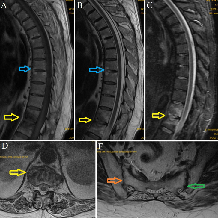
MRI showing classical features of Andersson lesion (yellow arrows) in D11–D12 of hemispherical shaped lesion, which is hypointense on (A) T1 weighted sequences, hyperintense on (B) T2 weighted sequences and (C) STIR sequences. Modic II changes (blue arrows) being hyperintense on T1 and T2 sequences are noted in D4, D5, D7 and D8 vertebra. (D) Axial section showing vertebral end plate erosions, irregularity and reactive sclerosis. (E) SI joints showing decreased joint space (orange arrow) and subchondral erosions (green arrow). STIR, short tau inversion recovery.

(A, B) Antero-posterior and lateral postoperative radiographs following posterior spinal stabilisation and instrumented fusion of D9–L2 vertebra, later followed by (C and D) minimally invasive anterior discectomy, anterolateral interbody fusion of D11–D12 with cage and bone grafting. (E and F) Follow-up radiographs 1 year following surgery, showing no recurrence of Andersson lesion.
References
-
- Kurugoglu S, Mihmanli I, Kanberoglu K, et al. . Destructive diskovertebral lesions in ankylosing spondylitis: appearance on magnetic resonance imaging. South Med J 2001;94:837–41. - PubMed
Publication types
MeSH terms
LinkOut - more resources
Full Text Sources
Medical
Research Materials