. 2022 Mar 1;119(9):e2114401119.
doi: 10.1073/pnas.2114401119.

PRDM9 losses in vertebrates are coupled to those of paralogs ZCWPW1 and ZCWPW2

Affiliations

PRDM9 losses in vertebrates are coupled to those of paralogs ZCWPW1 and ZCWPW2

Maria Izabel A Cavassim et al. Proc Natl Acad Sci U S A. .

Abstract

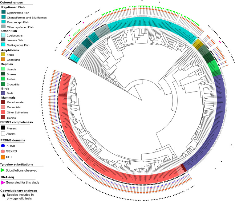
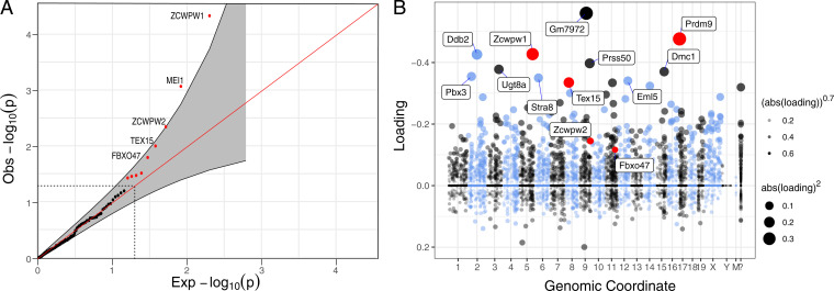
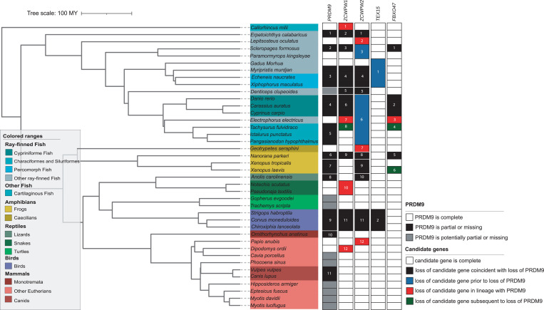
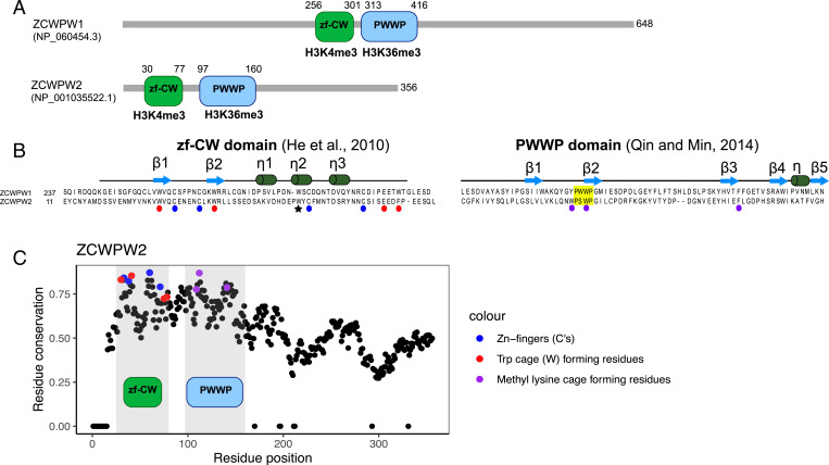
In most mammals and likely throughout vertebrates, the gene PRDM9 specifies the locations of meiotic double strand breaks; in mice and humans at least, it also aids in their repair. For both roles, many of the molecular partners remain unknown. Here, we take a phylogenetic approach to identify genes that may be interacting with PRDM9 by leveraging the fact that PRDM9 arose before the origin of vertebrates but was lost many times, either partially or entirely-and with it, its role in recombination. As a first step, we characterize PRDM9 domain composition across 446 vertebrate species, inferring at least 13 independent losses. We then use the interdigitation of PRDM9 orthologs across vertebrates to test whether it coevolved with any of 241 candidate genes coexpressed with PRDM9 in mice or associated with recombination phenotypes in mammals. Accounting for the phylogenetic relationship among a subsample of 189 species, we find two genes whose presence and absence is unexpectedly coincident with that of PRDM9: ZCWPW1, which was recently shown to facilitate double strand break repair, and its paralog ZCWPW2, as well as, more tentatively, TEX15 and FBXO47ZCWPW2 is expected to be recruited to sites of PRDM9 binding; its tight coevolution with PRDM9 across vertebrates suggests that it is a key interactor within mammals and beyond, with a role either in recruiting the recombination machinery or in double strand break repair.

Keywords: PRDM9 evolution; comparative genomics; genetics; phylogenetics; recombination.


Conflict of interest statement

The authors declare no competing interest.

Figures

Fig. 1.
Fig. 1.
The phylogenetic distribution of PRDM9 and its domain architecture across vertebrates. The inferred PRDM9 status of 432 vertebrate species is shown. Branch lengths were computed based on the TimeTree database. For 28 species not present in the database, we used branch length information from a close evolutionary relative; for 14 species in which we made PRDM9 calls, we were unable to find such a substitute, so they are not represented. Different vertebrate clades are indicated by colored segments, with salmon for mammals, cyan for fish, mustard for amphibians, green for reptiles, and purple for birds. In the inner circle, squares indicate whether PRDM9 is complete (solid black) or incomplete/absent (open black); for species with an uncertain PRDM9 status, no box is shown. The PRDM9 domain architecture of each species is shown with a cartoon, in which the presence of a KRAB domain is indicated in blue, SSXRD in pink, and the SET domain in orange. Green triangles indicate species that only carry PRDM9 orthologs with substitutions at putatively important catalytic residues in the SET domain (see SI Appendix, Table S4). The tree was drawn using itool (https://itol.embl.de/); an interactive version is available at https://itol.embl.de/shared/izabelcavassim.
Fig. 2.
Fig. 2.
Phylogenetic tests and genes coexpressed with PRDM9 in single-cell mouse testes data. (A) Quantile-quantile plot of the P values obtained from the phylogenetic tests run on 139 genes that appeared to have been lost at least once in the 189 vertebrate species considered. Genes that are significant at the 5% level are shown in red (outside the dashed lines), and a pointwise 95% confidence interval is shown in gray. Genes with an FDR ≤ 50% are annotated. (B) Loadings for one of 46 components (component 5) inferred from single-cell–expression data in mouse testes (31), in which PRDM9 is most highly expressed. The dot sizes are proportional to the square of the absolute value of the loading. PRDM9 and the three genes identified in our phylogenetic tests with P < 0.05 are shown in red. Mouse genomic coordinates are displayed. Panel B was made from summary statistics provided by ref. , using SDAtools (https://github.com/marchinilab/SDAtools/).
Fig. 3.
Fig. 3.
A summary of the phylogenetic distribution of PRDM9 and the four candidate genes across 189 species. Ortholog calls for candidate genes were based on a search of gene models within whole-genome sequences (Methods), and the phylogenetic test for coevolution with PRDM9 was rerun on these updated calls; updated P values for the phylogenetic test are shown in Table 1. Solid white and black rectangles indicate whether PRDM9 is present or absent, respectively, and gray rectangles lineages for which the status of PRDM9 is uncertain (Methods). For candidate genes, white rectangles are instances in which the gene is present and complete and solid black rectangles indicate when loss of candidate gene is coincident with that of PRDM9. Solid blue rectangles point to instances in which loss of candidate gene occurred prior to that of PRDM9 and green rectangles when it occurred subsequent to that of PRDM9. Red rectangles denote cases in which loss of the candidate gene occurred in lineages with a complete PRDM9. The full phylogenetic distribution of PRDM9 and candidate genes is in SI Appendix, Fig. S10.
Fig. 4.
Fig. 4.
Domain architecture ZCWPW1 and ZCWPW2. (A) Amino acid sequence and domain structure composition of genes ZCWPW1 and ZCWPW2 in humans. (B) The ZF-CW domain structure includes the fingers (residues indicated by blue circles) and an aromatic cage (red) expected to bind to H3K4me3 (61), and the star indicates the third Trp residue that is thought to stabilize the fold by hydrophobic interactions (61). The PWWP domain (yellow) is expected to bind to histone H3K36me3 through a hydrophobic cavity composed of three aromatic residues (purple) (62). The secondary structures of zf-CW and PWWP domains are represented above sequences. (C) Conservation of residues in ZCWPW2 across vertebrates, with those residues recognizing modifications on the histone tail colored in blue, red and purple. Positions in the ZCWPW2 alignment with >30% of gaps were ignored, and the conservation score was set to 0. A similar plot for the conservation of residues in ZCWPW1 was previously reported (figure 1B in Ref. 27).

References

    1. Keeney S., Neale M. J., Initiation of meiotic recombination by formation of DNA double-strand breaks: Mechanism and regulation. Biochem. Soc. Trans. 34, 523–525 (2006). - PubMed
    1. Baudat F., et al. , PRDM9 is a major determinant of meiotic recombination hotspots in humans and mice. Science 327, 836–840 (2010). - PMC - PubMed
    1. Myers S., et al. , Drive against hotspot motifs in primates implicates the PRDM9 gene in meiotic recombination. Science 327, 876–879 (2010). - PMC - PubMed
    1. Parvanov E. D., Petkov P. M., Paigen K., Prdm9 controls activation of mammalian recombination hotspots. Science 327, 835 (2010). - PMC - PubMed
    1. Altemose N., et al. , A map of human PRDM9 binding provides evidence for novel behaviors of PRDM9 and other zinc-finger proteins in meiosis. eLife 6, e28383 (2017). - PMC - PubMed

LinkOut - more resources