. 2022 Mar 1;119(9):e2118286119.
doi: 10.1073/pnas.2118286119.

Casein kinase 1 and disordered clock proteins form functionally equivalent, phospho-based circadian modules in fungi and mammals

Affiliations

Casein kinase 1 and disordered clock proteins form functionally equivalent, phospho-based circadian modules in fungi and mammals

Daniela Marzoll et al. Proc Natl Acad Sci U S A. .

Abstract

Circadian clocks are timing systems that rhythmically adjust physiology and metabolism to the 24-h day-night cycle. Eukaryotic circadian clocks are based on transcriptional-translational feedback loops (TTFLs). Yet TTFL-core components such as Frequency (FRQ) in Neurospora and Periods (PERs) in animals are not conserved, leaving unclear how a 24-h period is measured on the molecular level. Here, we show that CK1 is sufficient to promote FRQ and mouse PER2 (mPER2) hyperphosphorylation on a circadian timescale by targeting a large number of low-affinity phosphorylation sites. Slow phosphorylation kinetics rely on site-specific recruitment of Casein Kinase 1 (CK1) and access of intrinsically disordered segments of FRQ or mPER2 to bound CK1 and on CK1 autoinhibition. Compromising CK1 activity and substrate binding affects the circadian clock in Neurospora and mammalian cells, respectively. We propose that CK1 and the clock proteins FRQ and PERs form functionally equivalent, phospho-based timing modules in the core of the circadian clocks of fungi and animals.

Keywords: CK1; FRQ; PER; circadian clock; intrinsically disordered.


Conflict of interest statement

The authors declare no competing interest.

Figures
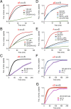
Fig. 1.
Fig. 1.
Activity of CK1a and CK1δ variants. (A) Priming-dependent activity of CK1a variants. ChEF assay with 135 nM of the indicated recombinant CK1a version and 10 µM primed SOX-labeled peptide (pS-x-x-S). The curve represents the average of six measurements. (B) CK1a displays low affinity for unprimed peptide. A ChEF assay was carried out with 10 µM unprimed SOX-labeled peptide (A-x-x-S) and 2.7 µM (20-fold higher than in A) of the indicated recombinant CK1a version. (C) Temperature-dependent activity of CK1a. ChEF assay was carried out at 20 °C as described in A but with 42 nM recombinant CK1a. Solid curve represents the average of six measurements (gray curves). Phosphorylation of pS-x-x-S at 4 °C was measured at t = 0, 30, 60, 90, 120, 180, and 240 min as described in experimental procedures (further time points are shown in Dataset S6). Solid purple curve represents the average of four replicates (light purple curves). (D) Priming-dependent activity of CK1δ variants. ChEF assay was carried as in A. CK1δ and CK1δ-R178Q were unphosphorylated, and CK1δ (-λPPase) was autophosphorylated. (E) Priming-independent activity of CK1δ variants. ChEF assay was carried out as in B. (F and G) Temperature-dependent activity of CK1δ (F) and CK1δΔC (G). ChEF assay was carried out at 20 °C as described in A. The solid red curve represents the average of six measurements (light red curves). Phosphorylation of pS-x-x-S at 4 °C was measured at t = 0, 10, 40, 70, 100, 130, 160, 190, and 250 min as described in experimental procedures (further time points are shown in Dataset S6). The number of replicates is indicated.
Fig. 2.
Fig. 2.
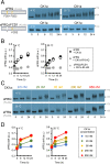
CK1a supports progressive hyperphosphorylation of sFRQ in vitro. (A) Hyperphosphorylation kinetics of sFRQ are independent of priming but requires CK1a recruitment. (Upper) sFRQ (8.3 nM) was incubated with CK1a (95 nM) and an ATP regenerating system at 4 and 20 °C for up to 24 h. (Lower) CK1a does not hyperphosphorylate sFRQΔFCD1. Phosphorylation kinetics were analyzed by Western blot with FLAG antibodies. Molecular mass standards are indicated. (B) Quantification of phosphorylation kinetics such as shown in A. The electrophoretic position of the center of mass of sFRQ was determined by densitometry, and the electrophoretic shift (sFRQ shift) was blotted relative to molecular mass standards. The electrophoretic positions of the 95- and 170-kDa molecular mass markers were set arbitrarily to 0 and 1, respectively. Error bars indicate ± SEM, n = 3 for CK1a and range, and n = 2 for CK1a-R181Q, except n = 1 for 3 h time points and n = 1 for sFRQΔFCD1. (C) Phosphorylation kinetics of sFRQ are dependent on CK1a concentration and slightly more efficient at 4 than at 20 °C. FRQ (8.3 nM) was incubated with the indicated concentrations of CK1a for the indicated time periods (n = 1). (D) Quantification of phosphorylation kinetics shown in C.
Fig. 3.
Fig. 3.
Priming-dependent phosphorylation of sFRQ by CK1a. (A) Kinases present in yWCL and CK1a-R181Q, deficient in priming-dependent phosphorylation, do not support efficient phosphorylation of sFRQΔFCD1. sFRQΔFCD1 (8.3 nM) was incubated with 200 µg yWCL with and without CK1a-R181Q (95 nM) for the indicated time periods. The asterisk indicates the position of phosphospecies generated with low efficiency. (B) CK1a supports priming-dependent phosphorylation in an FCD1-independent manner. YWCL (200 µg) was incubated with CK1a (95 nM) and FRQΔFCD1 (8.3 nM) at 4 and 20 °C. The generated phosphospecies is indicated by the asterisk. (C) Priming-dependent phosphorylation delays priming-independent phosphorylation of sFRQ. Phosphorylation kinetics of sFRQ (8.3 nM) by CK1a (95 nM) in the presence of yWCL at 4 °C are similar to the priming-independent phosphorylation in absence of WCL (Fig. 3A). At 20 °C, phosphospecies dependent on yWCL and CK1 are rapidly generated (asterisk) and priming-independent progressive phosphorylation is delayed (arrow). (D) Densitometric traces show the electrophoretic mobility shift of sFRQ (shown in C) upon phosphorylation by CK1a at 4 °C (black traces) and 20 °C (gray traces). The asterisk indicates the position of the priming-dependent phosphospecies, and the arrow indicates the delayed generation of highly phosphorylated sFRQ at 20 °C.
Fig. 4.
Fig. 4.
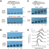
Hyperphosphorylation of mPER2 by CK1δ in vitro is facilitated by substrate-binding and does not require priming. (A) Hyperphosphorylation of mPER2 in vitro is strongly inhibited by autophosphorylation of CK1δ. In total, 250 nM unphosphorylated CK1δ (+λPP) and autophosphorylated CK1δ (−λPP), respectively, were incubated with V5-tagged mPER2 (mPER2) transiently expressed in HEK293 cells (SI Appendix, SI Methods). The phosphorylation kinetics of mPER2 by CK1δ were analyzed by Western blot. (B) CK1δ and the Tau-like version CK1δ-R178Q hyperphosphorylate mPER2 with similar kinetics and in a temperature-independent fashion. Quantification of mPER2 phosphorylation kinetics by CK1δ and CK1δ-R178Q at 4 (Left) and 20 °C (Right) is shown. mPER2 was incubated with 42 nM CK1δ (open circles) or CK1δ-R178Q (black circles) for the indicated time periods and then analyzed by Western blot as shown in A. The electrophoretic position of the center of mass of mPER2 was determined by densitometry and blotted relative to the molecular mass standards. The electrophoretic positions of the 130- and 170-kDa molecular mass markers were set arbitrarily to 0 and 0.5, respectively (gray circle: 24-h mock incubation without added kinase). Error bars are ± SEM, n = 3 for CK1δ, and n = 2 for CK1δ-R178Q and mock. (C) Phosphorylation of mPER2 is dependent on CK1δ concentration at 4 and at 20 °C. Quantification was performed as in B (n = 1). Ctrl = no kinase added, 0 min incubation. (D) Hyperphosphorylation of mPER2 by CK1δ is facilitated by CKBD. (Top) Schematic of PER2ΔCKBD, lacking the CK1-binding domain, aa 554 through 763 (32). (Bottom) mPER2ΔCKBD is not hyperphosphorylated by 42 nM CK1δ but efficiently hyperphosphorylated by 840 nM CK1δ. (E) CK1BD-A and CK1BD-B facilitate hyperphosphorylation of mPER2 by CK1δ. (Left) PER2–CK1 interaction domain A (CK1BD-A) and CK1BD-B(37, 49) have the potential to form amphipathic helices (https://heliquest.ipmc.cnrs.fr) (67). (Right) Phosphorylation kinetics of mPER2, mPER2ΔCK1BD-A, and mPER2ΔCK1BD-B by 25 nM CK1δ.
Fig. 5.
Fig. 5.
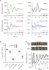
CK1δ variants affect the circadian period in T-REx-U2OS cells. (A) Luciferase reporter assay. T-REx-U2OS control cells (Upper Left) and T-REx-U2OS cells expressing the indicated CK1δ versions in a DOX-inducible manner were transiently transfected with a pBmal1-luc reporter plasmid (51). Bioluminescence data were detrended. Dotted lines represent SD (± SD) of four technical replicates. Circadian period length in presence and absence of DOX is indicated (note that the period length of CK1δ and CK1δ-R178Q cells in absence of DOX is already shortened compared to control cells). (B) Expression of CK1δ versions affects circadian period length. Each symbol represents an independent biological replicate with four technical replicates as shown in A. Data are presented as mean ± SD. (C) Overexpression of CK1a in Neurospora shortens circadian period length. (Upper) CK1a was overexpressed in Neurospora, and the circadian conidiation rhythm was analyzed by race-tube assay. (Lower) CK1a was overexpressed in a Neurospora strain harboring a frq-lucPEST reporter gene (68). A 96-well plate with luciferin medium was inoculated with conidia, and bioluminescence was recorded at 25 °C in darkness as described (69). Traces are averages of at least eight replicates.
Fig. 6.
Fig. 6.
CK1 forms equivalent complexes with FRQ and mPER2. (A) FRQ and mPER2 share a similar architecture and IDRs of similar size. Schematic and disorder plot of FRQ (Upper) and mPER2 (Lower). Dimerization domains are indicated by red boxes: coiled-coil domain (CC) in FRQ (A147 through A209) and PAS-PAS domains in mPER2 (I179 through P436). Binding of CK1a and CK1δ/ε to the central portions and of FRH and CRYPTOCHROMEs (CRY1/2) to the C-terminal portions of FRQ and mPER2, respectively, is shown. Plots were generated by IUPred2A-long (red) and ANCHOR2 (blue). The total lengths of the regions N- and C-terminal of the dimerization domains and their seryl and threonyl content are indicated. They are largely predicted to be disordered and hence referred to as N + C disorder. (B) Model of functionally equivalent circadian phospho-timers in fungi and mammals. (Left) FRQ and PER contain regions of similar length that display low folding propensity. These putative IDRs may adopt a rather compact conformation, potentially around FRH and CRYs, respectively. CK1, recruited through specific interaction domains, progressively phosphorylates, on a circadian time scale, low-affinity sites in the flexible IDRs. (Right) The increasing phosphorylation status becomes incompatible with the compact conformation of IDRs and favors open conformations, which may render the clock proteins inactive and prone to degradation (the dimerization domains of FRQ and PER2 are indicated by a red box, but only a monomeric clock protein is shown).

Comment in

Similar articles

Cited by

References

    1. Rosbash M., The implications of multiple circadian clock origins. PLoS Biol. 7, e62 (2009). - PMC - PubMed
    1. Cox K. H., Takahashi J. S., Circadian clock genes and the transcriptional architecture of the clock mechanism. J. Mol. Endocrinol. 63, R93–R102 (2019). - PMC - PubMed
    1. Patke A., Young M. W., Axelrod S., Molecular mechanisms and physiological importance of circadian rhythms. Nat. Rev. Mol. Cell Biol. 21, 67–84 (2020). - PubMed
    1. Dunlap J. C., Loros J. J., Making time: Conservation of biological clocks from fungi to animals. Microbiol. Spectr. 5 (2017). - PMC - PubMed
    1. Diernfellner A. C. R., Brunner M., Phosphorylation timers in the Neurospora crassa circadian clock. J. Mol. Biol. 432, 3449–3465 (2020). - PubMed