. 2022 Mar 1;119(9):e2118818119.
doi: 10.1073/pnas.2118818119.

Kurarinone alleviated Parkinson's disease via stabilization of epoxyeicosatrienoic acids in animal model

Affiliations

Kurarinone alleviated Parkinson's disease via stabilization of epoxyeicosatrienoic acids in animal model

Cheng-Peng Sun et al. Proc Natl Acad Sci U S A. .

Abstract

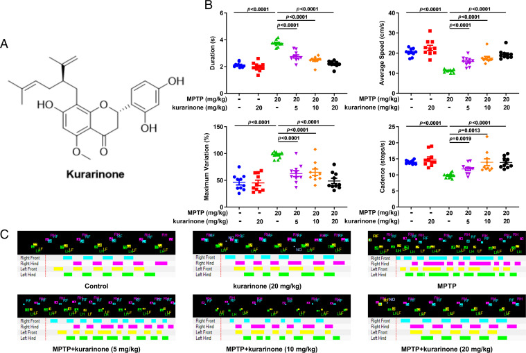
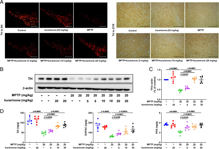
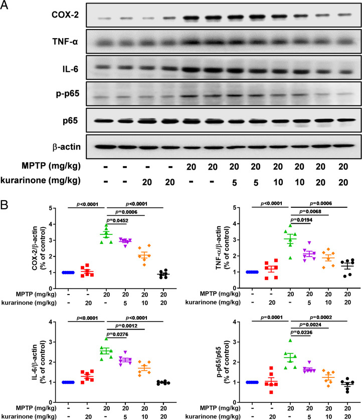
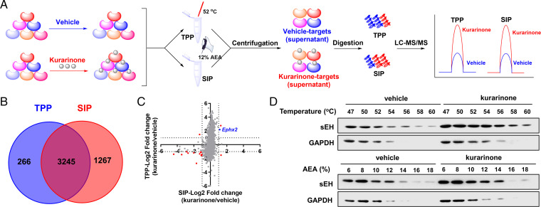
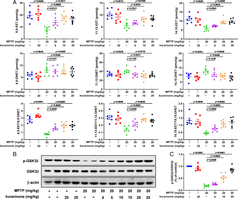
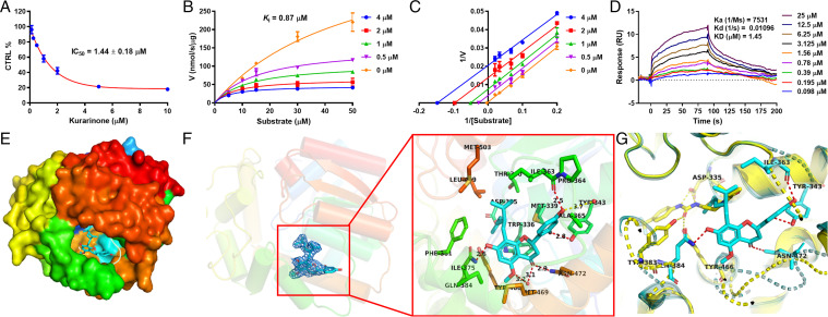
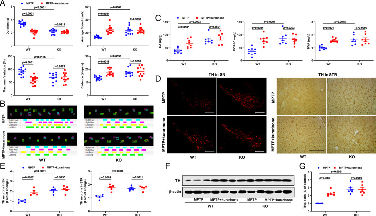
Parkinson's disease (PD) is one of the most common neurodegenerative disorders and is characterized by loss of dopaminergic neurons in the substantia nigra (SN), causing bradykinesia and rest tremors. Although the molecular mechanism of PD is still not fully understood, neuroinflammation has a key role in the damage of dopaminergic neurons. Herein, we found that kurarinone, a unique natural product from Sophora flavescens, alleviated the 1-methyl-4-phenyl-1,2,3,6-tetrahydropyridine (MPTP)-induced behavioral deficits and dopaminergic neurotoxicity, including the losses of neurotransmitters and tyrosine hydroxylase (TH)-positive cells (SN and striatum [STR]). Furthermore, kurarinone attenuated the MPTP-mediated neuroinflammation via suppressing the activation of microglia involved in the nuclear factor kappa B signaling pathway. The proteomics result of the solvent-induced protein precipitation and thermal proteome profiling suggest that the soluble epoxide hydrolase (sEH) enzyme, which is associated with the neuroinflammation of PD, is a promising target of kurarinone. This is supported by the increase of plasma epoxyeicosatrienoic acids (sEH substrates) and the decrease of dihydroxyeicosatrienoic acids (sEH products), and the results of in vitro inhibition kinetics, surface plasmon resonance, and cocrystallization of kurarinone with sEH revealed that this natural compound is an uncompetitive inhibitor. In addition, sEH knockout (KO) attenuated the progression of PD, and sEH KO plus kurarinone did not further reduce the protection of PD in MPTP-induced PD mice. These findings suggest that kurarinone could be a potential natural candidate for the treatment of PD, possibly through sEH inhibition.

Keywords: Parkinson’s disease; Sophora flavescens; kurarinone; soluble epoxide hydrolase.


Conflict of interest statement

The authors declare no competing interest.

Figures

Fig. 1.
Fig. 1.
Kurarinone from S. flavescens alleviated the behavior of MPTP-induced PD mice. (A) The structure of kurarinone. (B) The behavioristic parameters. Data represent mean ± SEM, n = 10. The significance was determined by one-way ANOVA followed by Tukey’s test. (C) The representative footprints. LF, left front; LH, left hind; RF, right front; RH, right hind; NO, noise.
Fig. 2.
Fig. 2.
Kurarinone enhanced TH expression and neurotransmitter levels in MPTP-induced PD mice. (A) The representative staining of TH in the SN (scale bar: 300 μm) and STR (scale bar: 100 μm). (B) Effect of kurarinone on the TH expression level in the SN. (C) Quantitative data of TH. Data represent mean ± SEM, n = 6. The significance of difference was determined by one-way ANOVA followed by Tukey’s test. (D) Effects of kurarinone on neurotransmitters DA, DOPAC, and HVA. Data represent mean ± SEM, n = 6. The significance of difference was determined by one-way ANOVA followed by Tukey’s test.
Fig. 3.
Fig. 3.
Kurarinone form S. flavescens inhibited neuroinflammation in MPTP-induced PD mice. (A) Effects of kurarinone on expression levels of COX-2, TNF-α, IL-6, p-p65, and p65. (B) Quantitative data on COX-2, TNF-α, IL-6, and p-p65/p65 levels. Data represent mean ± SEM, n = 6. The significance of difference was determined by one-way ANOVA followed by Tukey’s test.
Fig. 4.
Fig. 4.
sEH was identified as the potential target of kurarinone by TPP and SIP assays. (A) Schematic representation of the TPP and SIP methods for screening of target proteins of kurarinone. (B) The Venn diagram of potential target proteins identified by the TPP and SIP methods. (C) Fourteen candidate targets were screened by filtering the log2 (fold change) of each protein identified in the TPP and SIP assays. Red and blue dots are differential proteins affected by kurarinone in the TPP and SIP assays, respectively. (D) Western blotting demonstrated that sEH was stabilized after the incubation with kurarinone by using the TPP and SIP methods. AEA, an organic solvent mixture of acetone: ethanol: acetic acid; GAPDH, glyceraldehyde 3-phosphate dehydrogenase.
Fig. 5.
Fig. 5.
Kurarinone alleviated the increase of sEH activity and suppressed the GSK3β signaling pathway via stabilizing the level of EETs in MPTP-induced PD mice. (A) Effect of kurarinone on 8,9-EET; 11,12-EET; 14,15-EET; 8,9-DHET; 11,12-DHET; 14,15-DHET; 8,9-EET/8,9-DHET; 11,12-EET/11,12-DHET; and 14,15-EET/14,15-DHET in MPTP-induced PD mice. Data represent mean ± SEM, n = 6. The significance of difference was determined by one-way ANOVA followed by Tukey’s test. (B) Effects of kurarinone on p-GSK3β and GSK3β expression levels in MPTP-induced PD mice. (C) Quantitative data of phosphorylated GSK3β (p-GSK3β)/GSK3β. Data represent mean ± SEM, n = 6. The significance of difference was determined by one-way ANOVA followed by Tukey’s test.
Fig. 6.
Fig. 6.
Kurarinone inhibited the sEH activity in vitro and interacted with sEH. (A) IC50 plot of kurarinone against sEH. (B) Michaelis-Menten plot of kurarinone against sEH. (C) Lineweaver-Burk plot of kurarinone against sEH. (D) SPR plot of kurarinone with sEH. (E) Kurarinone bound to the cavity of sEH (PDB code 7EBA). (F) Electronic cloud plot of kurarinone and the interaction of kurarinone with sEH. (G) Interaction differences of kurarinone (cyan) and TPPU (yellow, PDB code 4OCZ) with sEH. CTRL, control.
Fig. 7.
Fig. 7.
Ephx2 genetic deletion abolished the an-PD effect of kurarinone. (A) Ephx2 genetic deletion abolished the effect of kurarinone on gait disorders. Data represent mean ± SEM, n = 10 to 12. (B) The representative footprints. (C) Ephx2 genetic deletion abolished the effect of kurarinone on neurotransmitters DA, DOPAC, and HVA. Data represent mean ± SEM, n = 7 or 8. (D) Ephx2 genetic deletion abolished the effect of kurarinone on TH-positive neurons in the SN (scale bar: 300 μm) and STR (scale bar: 100 μm). The representative staining of TH in the SN and STR. (E) The data of TH-positive neurons in the SN and STR. Data represent mean ± SEM, n = 5. The significance of difference was determined by two-way ANOVA followed by Tukey’s test. (F) Ephx2 genetic deletion abolished the effect of kurarinone on the TH expression level in the SN. (G) Quantitative data of TH. Data represent mean ± SEM, n = 6. The significant difference was determined by two-way ANOVA followed by Tukey’s test. CTRL, control.

Similar articles

Cited by

References

    1. Dickson D. W., Parkinson’s disease and parkinsonism: Neuropathology. Cold Spring Harb. Perspect. Med. 2, a009258 (2012). - PMC - PubMed
    1. Selikhova M., et al. , A clinico-pathological study of subtypes in Parkinson’s disease. Brain 132, 2947–2957 (2009). - PubMed
    1. Dickson D. W., et al. , Neuropathological assessment of Parkinson’s disease: Refining the diagnostic criteria. Lancet Neurol. 8, 1150–1157 (2009). - PubMed
    1. Grayson M., Parkinson’s disease. Nature 538, S1 (2016). - PubMed
    1. Stoker T. B., Barker R. A., Recent developments in the treatment of Parkinson’s disease. F1000 Res. 9, 862 (2020). - PMC - PubMed