. 2022 Mar 1;119(9):e2121478119.
doi: 10.1073/pnas.2121478119.

Role of ubiquitin-protein ligase UBR5 in the disassembly of mitotic checkpoint complexes

Affiliations

Role of ubiquitin-protein ligase UBR5 in the disassembly of mitotic checkpoint complexes

Sharon Kaisari et al. Proc Natl Acad Sci U S A. .

Abstract

The mitotic (or spindle assembly) checkpoint system ensures accurate chromosome segregation in mitosis by preventing the onset of anaphase until correct bipolar attachment of sister chromosomes to the mitotic spindle is attained. It acts by promoting the assembly of a mitotic checkpoint complex (MCC), composed of mitotic checkpoint proteins BubR1, Bub3, Mad2, and Cdc20. MCC binds to and inhibits the action of ubiquitin ligase APC/C (anaphase-promoting complex/cyclosome), which targets for degradation regulators of anaphase initiation. When the checkpoint system is satisfied, MCCs are disassembled, allowing the recovery of APC/C activity and initiation of anaphase. Many of the pathways of the disassembly of the different MCCs have been elucidated, but the mode of their regulation remained unknown. We find that UBR5 (ubiquitin-protein ligase N-recognin 5) is associated with the APC/C*MCC complex immunopurified from extracts of nocodazole-arrested HeLa cells. UBR5 binds to mitotic checkpoint proteins BubR1, Bub3, and Cdc20 and promotes their polyubiquitylation in vitro. The dissociation of a Bub3*BubR1 subcomplex of MCC is stimulated by UBR5-dependent ubiquitylation, as suggested by observations that this process in mitotic extracts requires UBR5 and α-β bond hydrolysis of adenosine triphosphate. Furthermore, a system reconstituted from purified recombinant components carries out UBR5- and ubiquitylation-dependent dissociation of Bub3*BubR1. Immunodepletion of UBR5 from mitotic extracts slows down the release of MCC components from APC/C and prolongs the lag period in the recovery of APC/C activity in the exit from mitotic checkpoint arrest. We suggest that UBR5 may be involved in the regulation of the inactivation of the mitotic checkpoint.

Keywords: cell cycle; mitosis; ubiquitin.


Conflict of interest statement

The authors declare no competing interest.

Figures

Fig. 1.
Fig. 1.
UBR5 binds to mitotic checkpoint proteins and promotes their ubiquitylation. (A) Binding of mitotic checkpoint proteins to UBR5 in extracts from mitotic and asynchronous HeLa cells. Extracts from nocodazole-arrested (“mitotic”) or logarithmically growing (“asynchronous”) HeLa cells were prepared as described previously (7). Immunoprecipitation (IP) of extracts with an anti-UBR5 antibody or with nonimmune rabbit IgG was carried out as described in SI Appendix, SI Materials and Methods. Results are expressed as the percentage of immunoprecipitated proteins relative to the corresponding inputs. Inputs show 10% of the amounts of the indicated proteins in mitotic or asynchronous extracts used for immunoprecipitation. Numbers on the Right indicate the electrophoretic migration of marker proteins (in kDa). (B) Binding of UBR5 to individual recombinant mitotic checkpoint proteins. The indicated recombinant mitotic checkpoint proteins (100 nM) were incubated with recombinant his6-UBR5 (at a quantity similar to that in 40 μg protein of mitotic HeLa cell extract) in a buffer containing: 50 mM Tris⋅HCl (pH 7.2), 20% (vol/vol) glycerol, 1 mg/mL bovine serum albumin (BSA), and 1 mM dithiothreitol (DTT). Following incubation for 1 h at 23 °C, the samples were immunoprecipitated with anti-UBR5 polyclonal antibody or with nonimmune rabbit IgG. Immunoprecipitated material was resolved by sodium dodecyl sulphate–polyacrylamide gel electrophoresis (SDS-PAGE) and immunoblotted for the indicated proteins. Results are expressed as the percentage of immunoprecipitated material relative to input. Numbers on the Right (kDa) indicate the electrophoretic migration position of marker proteins. (C) Ubiquitylation of individual mitotic checkpoint proteins by recombinant UBR5. The indicated purified recombinant checkpoint proteins (100 nM, each) were incubated in a volume of 30 μL with a ubiquitylation mixture consisting of 40 mM Tris⋅HCl (pH 7.6), 5 mM MgCl2, 1 mg/mL BSA, 1 mM DTT, 150 nM E1 (Enzo BML-UW9410), 600 nM UbcH5ɑ/UBE2D1 (Boston Biochem E2-616), 100 μM ubiquitin, and 2 mM adenosine 5′-(β,γ-imido)triphosphate (AMP-PNP), in the presence or absence of recombinant his6-UBR5 (supplemented at a quantity similar to its amount in 40 μg protein of HeLa cell extract). Following incubation at 37 °C for 1 h, samples were subjected to SDS-PAGE and immunoblotting for the indicated proteins. Numbers on the Right indicate the electrophoretic migration position of marker proteins.
Fig. 2.
Fig. 2.
UBR5 stimulates the release of Bub3 from BubR1. (A) Effect of immunodepletion of UBR5 from extracts on the release of Bub3 from BubR1. Samples of UBR5-depleted or sham-treated mitotic extracts (1.6 mg of protein) were incubated in a reaction volume of 100 μL containing 40 nM his6-Bub3*BubR1 subcomplex, 40 mM Tris HCl (pH 7.6), 5 mM MgCl2, 1 mM DTT, 2 mM ATP, 10 mM phosphocreatine, and 100 μg/mL creatine phosphokinase. Samples were incubated at 23 °C for the indicated time periods. Subsequently, samples were immunoprecipitated with anti-BubR1- beads (10 μL, packed), with rotation for 2 h at 4 °C. Beads were then washed and treated with lambda phosphatase as described in SI Appendix, SI Materials and Methods. The release of His6-Bub3 from anti-BubR1 beads was determined by immunoblotting of immunoprecipitated material with anti-Bub3. Numbers on the Right indicate the position of molecular size marker proteins (kDa). (B) Quantitation of data from A. Ratios of his6-Bub3/BubR1 were determined for each lane and were expressed as percentage of this ratio at time 0. (C) The release of Bub3 from BubR1 in HeLa cell extracts requires ATP but not proteasome activity. The His6-Bub3*BubR1 subcomplex was incubated with sham-treated extracts as described in A. Where indicated, the following additions were made: 2 mM ATP together with 10 mM phosphocreatine and 100 μg/mL creatine phosphokinase; 2 mM adenosine 5′-(β,γ-imido)triphosphate (AMP-PNP); 10 μM MG-132; a mixture of hexokinase (Roche 11426362001, 0.4 mg/mL) and 2-deoxyglucose (10 mM) (“HK+DG”). Reaction mixtures were incubated at 23 °C for the indicated time periods. Samples were subjected to immunoprecipitation with anti-BubR1, and the release of His6-Bub3 from anti-BubR1 beads was analyzed as in A. Results (shown at Bottom) were expressed as the percentage of His6-Bub3 bound to BubR1 relative to the initial value. (D) The dissociation of BubR1*Bub3 subcomplex in a purified system is stimulated by UBR5-dependent ubiquitylation. Recombinant BubR1*Bub3 subcomplex (100 nM) and his6-UbR5 (at a quantity similar to that in 40-μg HeLa cell extract) were supplemented as indicated to a reaction mixture containing the following: 40 mM Tris HCl (pH 7.6), 5 mM MgCl2, 1 mM DTT, and 1 mg/mL BSA. Where indicated, a ubiquitylation system consisting of 2 mM AMP-PNP, 150 nM E1, 600 nM UbcH5ɑ, and 100 μM ubiquitin was added. Following incubation at 37 °C for 1 h, Nonidet P-40 (0.2%) and NaCl (150 mM) were added and samples were subjected to immunoprecipitation with anti-BubR1 beads. Supernatants were collected and beads were washed as described under SI Appendix, SI Materials and Methods. Samples of immunoprecipitated material (Left) and of supernatants (Right) were immunoblotted for the indicated proteins. Numbers below blots for Bub3 express the percentage of Bub3/BubR1 ratio relative to the initial value (Lower Left) and the percentage of Bub3 released to the supernatant (Right). Electrophoretic migration positions of molecular size marker proteins are indicated on the Right (kDa).
Fig. 3.
Fig. 3.
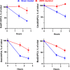
Effect of UBR5 depletion on the release of MCC components from APC/C. Quantitation of immunoblots shown in SI Appendix, Fig. S3. The ratios of the different mitotic checkpoint proteins to APC4 were determined for each lane and were expressed as the percentage of corresponding ratios at time 0. All values of quantitation of APC4 immunoblots were in the linear range of the assay, and thus, APC4 blots could serve as valid loading controls for the estimation of the levels of the other proteins shown in the figure.
Fig. 4.
Fig. 4.
UBR5 shortens the lag period in the activation of APC/C in exit from mitotic checkpoint. (A) Influence of UBR5 on the degradation of myc-securin in checkpoint extracts incubated with ATP. Recombinant Myc-securin (2.5 ng) was added to samples of UBR5-depleted or sham-treated HeLa cell extracts (150 μg of protein) and was incubated in a reaction volume of 10 μL containing 40 mM Tris⋅HCl (pH 7.6), 5 mM MgCl2, 1 mM DTT, 2 mM ATP, 10 mM phosphocreatine, and 100 μg/mL creatine phosphokinase. Following incubation at 23 °C for the time periods indicated, samples were resolved by SDS-PAGE and were blotted with Myc-tag antibody. (B) Quantitation of data from A. Results were expressed as the percentage of Myc-securin at time 0. (C) Influence of immunodepletion of UBR5 on activation of APC/C in checkpoint extract incubated with ATP. Checkpoint extracts subjected to UBR5-depletion or to sham depletion were incubated with ATP as described under A, for the time periods indicated. Samples were mixed with anti-Cdc27 beads for 2 h at 4 °C, and then beads were washed three times with a buffer consisting of 50 mM Tris⋅HCl (pH 7.2), 20% (vol/vol) glycerol, 1 mg/mL BSA, and 1 mM DTT. APC/C activity was determined in samples of 1-μL packed beads contained by the ubiquitylation of an 125I-labeled N-terminal fragment of cyclin B, as described previously (41). (D) Quantitation of data from C. The data were expressed as the percentage of I125-cyclin B-(Ub)n conjugates formed relative to the total amount of I125-cyclin B in each lane.

Similar articles

Cited by

References

    1. Musacchio A., Salmon E. D., The spindle-assembly checkpoint in space and time. Nat. Rev. Mol. Cell Biol. 8, 379–393 (2007). - PubMed
    1. Lara-Gonzalez P., Westhorpe F. G., Taylor S. S., The spindle assembly checkpoint. Curr. Biol. 22, R966–R980 (2012). - PubMed
    1. Musacchio A., The molecular biology of spindle assembly checkpoint signaling dynamics. Curr. Biol. 25, R1002–R1018 (2015). - PubMed
    1. Watson E. R., Brown N. G., Peters J. M., Stark H., Schulman B. A., Posing the APC/C E3 ubiquitin ligase to orchestrate cell division. Trends Cell Biol. 29, 117–134 (2019). - PMC - PubMed
    1. London N., Biggins S., Signalling dynamics in the spindle checkpoint response. Nat. Rev. Mol. Cell Biol. 15, 736–747 (2014). - PMC - PubMed

LinkOut - more resources