. 2022 Mar 1;119(9):e2122090119.
doi: 10.1073/pnas.2122090119.

Lack of association between pandemic chilblains and SARS-CoV-2 infection

Collaborators, Affiliations

Lack of association between pandemic chilblains and SARS-CoV-2 infection

Jeff R Gehlhausen et al. Proc Natl Acad Sci U S A. .

Abstract

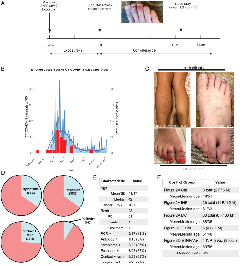
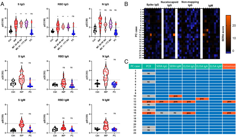
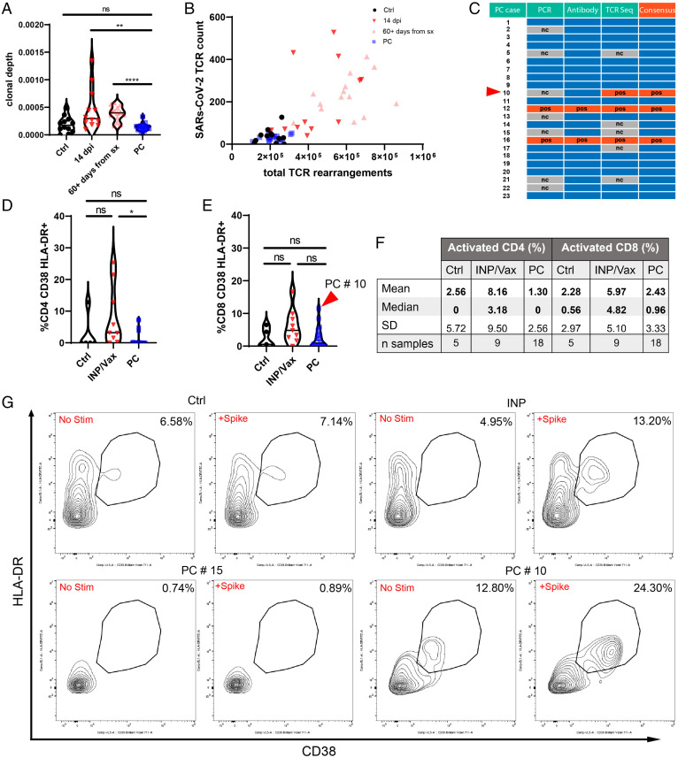
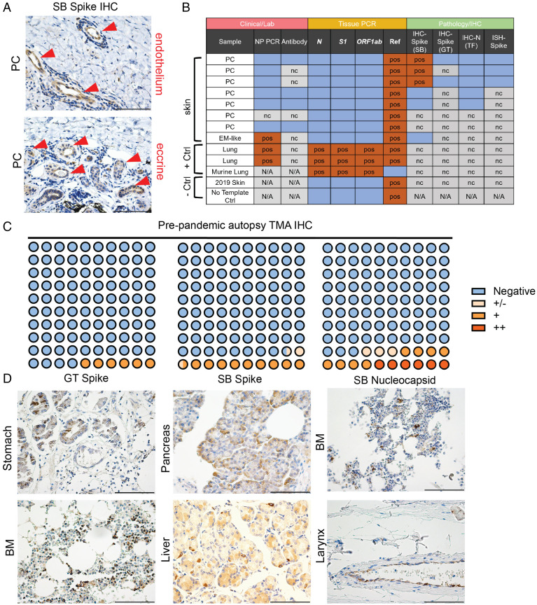
An increased incidence of chilblains has been observed during the severe acute respiratory syndrome coronavirus 2 (SARS-CoV-2) pandemic and attributed to viral infection. Direct evidence of this relationship has been limited, however, as most cases do not have molecular evidence of prior SARS-CoV-2 infection with PCR or antibodies. We enrolled a cohort of 23 patients who were diagnosed and managed as having SARS-CoV-2-associated skin eruptions (including 21 pandemic chilblains [PC]) during the first wave of the pandemic in Connecticut. Antibody responses were determined through endpoint titration enzyme-linked immunosorbent assay and serum epitope repertoire analysis. T cell responses to SARS-CoV-2 were assessed by T cell receptor sequencing and in vitro SARS-CoV-2 antigen-specific peptide stimulation assays. Immunohistochemical and PCR studies of PC biopsies and tissue microarrays for evidence of SARS-CoV-2 were performed. Among patients diagnosed and managed as "covid toes" during the pandemic, we find a percentage of prior SARS-CoV-2 infection (9.5%) that approximates background seroprevalence (8.5%) at the time. Immunohistochemistry studies suggest that SARS-CoV-2 staining in PC biopsies may not be from SARS-CoV-2. Our results do not support SARS-CoV-2 as the causative agent of pandemic chilblains; however, our study does not exclude the possibility of SARS-CoV-2 seronegative abortive infections.

Keywords: SARS-CoV-2; chilblain; covid toe; interferon; pernio.


Conflict of interest statement

The authors declare no competing interest.

Figures

Fig. 1.
Fig. 1.
Clinical characteristics of the PC cohort. (A) Schema of our clinical study of the PC cohort (created in BioRender). Patients had a preceding potential exposure period of up to 2 mo; mean time to blood draw after onset of rash was 3.3 mo. All cases were resolved at the time of blood draw. (B) Histogram comparing case counts in the first wave of COVID-19 in 2020 in Connecticut (CT) to the timing of rashes in our cohort. (C) Clinical photos of two different households (four patients total) with SARS-CoV-2–associated rashes that developed in a concurrent time line. (D) Pie charts of clinical characteristics of the PC cohort, including symptoms, suspected or confirmed SARS-CoV-2 exposure, contacts with a rash attributed to SARS-CoV-2 infection (chilblains or livedo), and diagnostic testing results. (E) More detailed breakdown of demographics and clinical characteristics of the PC cohort. (F) Demographic and numeric data of control groups used in Figs. 2 and 3. Ab, antibody; Ctrl, control; F, female; M, male.
Fig. 2.
Fig. 2.
Convalescent antibody studies of PC cases compared with controls. (A) ELISA IgG, IgA, and IgM assays targeting S, RBD, and N antigens revealing antibody responses in indicated cohorts. The dotted lines at p(EC50) of two indicate the threshold for positivity. For details on p(EC50) please see Methods. Red dots in the PC group indicate previous confirmed positive cases (two total). (B) Heat map of SERA responses in the PC cohort for individual cases. Values at or above 25 are considered positive. (C) Consensus table summarizing positivity from various assays for each PC case. Blue boxes are negative. For all experiments with significance testing, Kruskal–Wallis test with post hoc Dunn’s test for multiple comparisons was employed. Ctrl, no exposure negative control; nc, not completed; ns, not significant; pos, positive. *P < 0.05; **P < 0.01; ***P < 0.001.
Fig. 3.
Fig. 3.
T cell studies of PC cases compared with controls. (A) Clonal depth of PC cases compared with indicated cohorts as assessed by TCR sequencing of the CD8 CDR3 region. Clonal depth is a ratio of the sum of antigen-specific (SARS-CoV-2) TCRs compared with the total productive TCR rearrangements. (B) Scatterplot relating the number of SARS-CoV-2–mapped TCRs to the number of rearranged TCRs per sample. For A and B, outliers that were more than three SDs from the median of each cohort were removed for visualization purposes: Ctrl (one), 14 dpi (two), 60+ d from symptoms (sx) (three), and PC (one). (C) Consensus table summarizing the results for antibody and T cell studies for PC cases. Blue boxes are negative. The red arrowhead denotes PC #10, which was not confirmed positive by antibody testing but was by TCR sequencing. (D and E) Violin plots of T cell responses for CD4 (D) and CD8 (E) responses to S-peptide stimulation. Percentages reflect the stimulated responses subtracting the baseline activation. PC #10 had the most robust CD8+ T cell activation in response to S in the PC cohort. (F) Descriptive statistics of S peptide stimulation studies. (G) Flow plots for representative CD8 T cell responses for Ctrl, INP/Vax, and PC cases in addition to PC #10, which was identified by TCR sequencing and peptide stimulation studies as having prior SARS-CoV-2 exposure. For all experiments with significance testing, Kruskal–Wallis test with post hoc Dunn’s test for multiple comparisons was employed. There were five INP and four Vax. Ctrl, no exposure negative control; nc, not completed; ns, not significant; pos, positive. *P < 0.05; **P < 0.01; ****P < 0.0001.
Fig. 4.
Fig. 4.
Immunohistochemical and PCR analyses of PC cases and TMAs. (A) Representative staining of the three positive PC cases of S IHC previously published by our group with focal staining in endothelium and eccrine glands (red arrowheads). (B) Summary of laboratory, PCR, and immunohistochemical analyses of SARS-CoV-2–associated skin rashes (chilblains and erythema multiforme [EM]–like lesions) compared with controls. Blue boxes are negative. (C and D) Dot plot summarizing TMA IHC results for respective antibodies. Representative staining for indicated antibodies is seen in D. Methods has a detailed discussion of antibodies and staining as well as PCR protocol. All images are 400× original magnification. BM, bone marrow; GT, GeneTex; N/A, not available; nc, not completed; NP, nasopharyngeal; pos, positive; SB, Sino Biologics; TF, ThermoFisher. (Scale bars: 100 μM.)

Comment in

References

    1. Freeman E. E., et al. ; American Academy of Dermatology Ad Hoc Task Force on COVID-19, Pernio-like skin lesions associated with COVID-19: A case series of 318 patients from 8 countries. J. Am. Acad. Dermatol. 83, 486–492 (2020). - PMC - PubMed
    1. Hébert V., et al. , Lack of association between chilblains outbreak and severe acute respiratory syndrome coronavirus 2: Histologic and serologic findings from a new immunoassay. J. Am. Acad. Dermatol. 83, 1434–1436 (2020). - PMC - PubMed
    1. Herman A., et al. , Evaluation of chilblains as a manifestation of the COVID-19 pandemic. JAMA Dermatol. 156, 998–1003 (2020). - PMC - PubMed
    1. Fernandez-Nieto D., et al. , Characterization of acute acral skin lesions in nonhospitalized patients: A case series of 132 patients during the COVID-19 outbreak. J. Am. Acad. Dermatol. 83, e61–e63 (2020). - PMC - PubMed
    1. Freeman E. E., et al. , The spectrum of COVID-19-associated dermatologic manifestations: An international registry of 716 patients from 31 countries. J. Am. Acad. Dermatol. 83, 1118–1129 (2020). - PMC - PubMed

Publication types