. 2022 Mar 1;119(9):e2123163119.
doi: 10.1073/pnas.2123163119.

CSB-independent, XPC-dependent transcription-coupled repair in Drosophila

Affiliations

CSB-independent, XPC-dependent transcription-coupled repair in Drosophila

Nazli Deger et al. Proc Natl Acad Sci U S A. .

Abstract

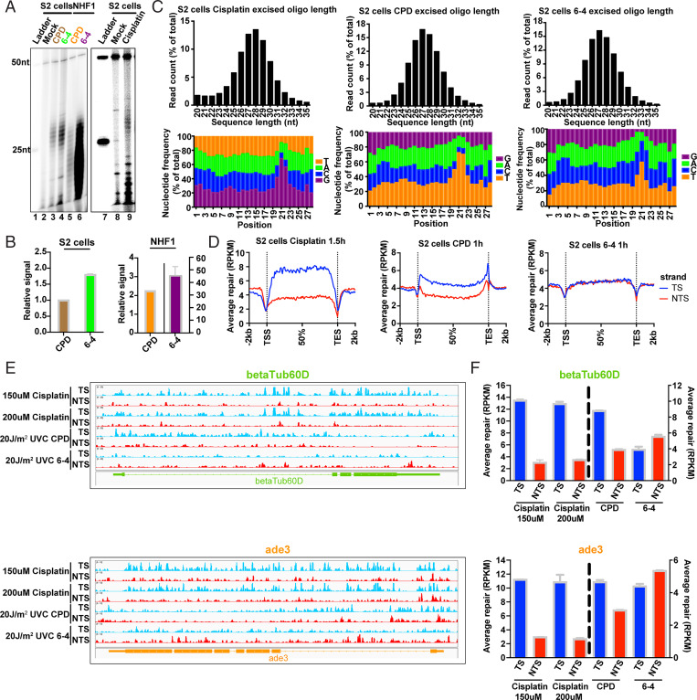
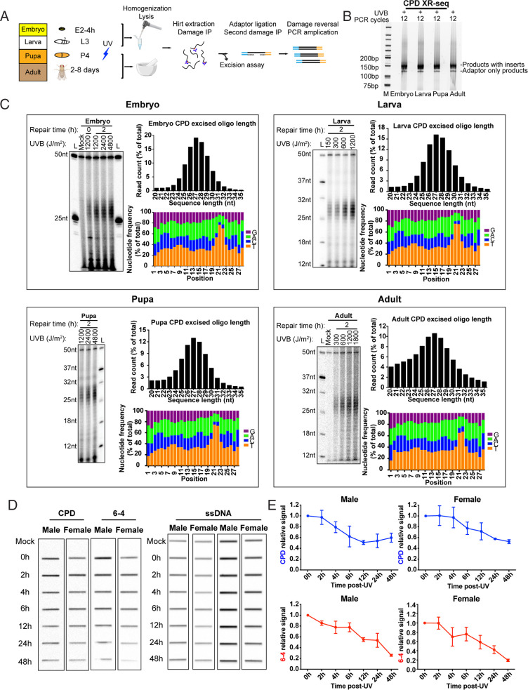
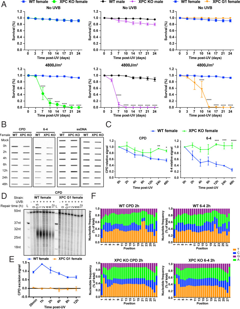
Drosophila melanogaster has been extensively used as a model system to study ionizing radiation and chemical-induced mutagenesis, double-strand break repair, and recombination. However, there are only limited studies on nucleotide excision repair in this important model organism. An early study reported that Drosophila lacks the transcription-coupled repair (TCR) form of nucleotide excision repair. This conclusion was seemingly supported by the Drosophila genome sequencing project, which revealed that Drosophila lacks a homolog to CSB, which is known to be required for TCR in mammals and yeasts. However, by using excision repair sequencing (XR-seq) genome-wide repair mapping technology, we recently found that the Drosophila S2 cell line performs TCR comparable to human cells. Here, we have extended this work to Drosophila at all its developmental stages. We find TCR takes place throughout the life cycle of the organism. Moreover, we find that in contrast to humans and other multicellular organisms previously studied, the XPC repair factor is required for both global and transcription-coupled repair in Drosophila.

Keywords: XPC; XR-seq; transcription-coupled repair.


Conflict of interest statement

The authors declare no competing interest.

Figures

Fig. 1.
Fig. 1.
Repair of cisplatin-d(GpG) adducts and UV photoproducts in Drosophila S2 cells. (A) Excision assay of cisplatin adducts, CPDs, and (6-4)PPs in S2 cells and human NHF1 cells. All three adducts are excised in S2 cells predominantly as 26 to 29 mers, with less degradation in S2 cells than in NHF1 cells. (B) Quantitation of excision in A. Excision repair of (6-4)PPs predominates over CPDs in both Drosophila and human cells at the 1-h repair time point tested. Data points reflect means and SEs obtained from two experiments. (C) Characterization of XR-seq excision repair reads. Results are shown for cisplatin adducts (Left), CPDs (Middle), and (6-4)PPs (Right). Top shows frequency distribution profiles of excision products as percent of total reads (y axis) versus excision product length (x axis). In agreement with the excision assay results in A, reads of length 26 to 29 nt predominate. The peak read length is 28 nt for cisplatin and 27 nt for CPDs and (6-4)PPs. Bottom shows the 28-nt excision products, the relative frequency (y axis) of each nucleotide at each position (x axis). The 5′ end of the excision products is located at position 1. Enrichment of G residues on the Left indicates the likely site of platination at G-G dinucleotides, and enrichment of pyrimidine residues indicates the likely site of formation of CPDs (Middle) and (6-4)PPs (Right). With respect to these damage sites, sites of dual incision are located 6 nt 3′ from each adduct, and 20 nt 5′ from the cisplatin adducts and 19 nt 5′ from the CPDs and (6-4)PPs. (D) Repair of cisplatin and UV photoproducts in the TS (blue) and NTS (red). Excision product reads were mapped to the Drosophila genome and reads across all genes were scaled to a unit gene, which represents average repair in a Drosophila gene. Results are shown for cisplatin adducts (Left), CPDs (Middle), and (6-4)PPs (Right). Transcription-coupled repair is indicated by enhanced TS repair and is evident in cisplatin and CPD but not (6-4)PP repair. (E). Screenshots showing repair reads (y axis) across two representative housekeeping genes betaTub60D (Top) and ade3 (Bottom) and (F) quantitation of repair in each strand of the two genes. TCR is evident with cisplatin and CPDs but not (6-4)PPs. Small antisense transcripts are present in ade3 but not shown in E; apparently this antisense transcription is too weak to affect the overall TCR shown. Data points reflect means and SEs obtained from two experiments.
Fig. 2.
Fig. 2.
Excision repair of CPDs in Drosophila in vivo. (A) Method developed for excision and XR-seq assay of embryo, larva, pupa, and adult. Drosophila at each developmental stage was irradiated directly with UVB. Following repair, samples were lysed by various means to release excision products (see Materials and Methods and Results). Excision products were processed for excision assay and XR-seq as was done with cultured cells. (B) XR-seq libraries generated from different developmental stages. The libraries are of good quality (5). (C) Analysis of excision repair in vivo. For each developmental stage, excision assay results are shown alongside plots characterizing repair reads generated by XR-seq. The results show that excision product lengths peak at 26 to 28 nt among all stages in a dose-independent manner. Also, relatively little degradation of excision products is seen with Drosophila both in vivo and in vitro. (D and E) Slot blot analysis of CPD and (6-4)PP repair following 1,200 J/m2 UVB given to adults shows more rapid removal of (6-4)PPs from the genome overall. Data points reflect means and SEs obtained from two experiments.
Fig. 3.
Fig. 3.
Genome-wide analysis of TCR in Drosophila in vivo. (A) Drosophila adult XR-seq data for the indicated time points are plotted. For each analysis, CPD repair reads were mapped to the genome, and reads from the two strands of each gene were scaled to a unit gene, which represents the average repair in each strand of genes included. Each plot includes results obtained with nonoverlapping genes longer than 1 kb and shown to be transcribed by RNA-seq (RPKM > 10). Expression levels were obtained from FlyBase RNA-seq reports (37). Reads 2 kb upstream and downstream of each gene were averaged and plotted. (B) Change in TCR with time. TCR is plotted on the y axis as average Log2(TS/NTS). TCR peaks at ∼2 h post-UVB and remains high until 8 to 12 h post-UV. (C) XR-seq results from Drosophila embryo, larva, pupa, and adults of each sex are plotted. TCR is evident in all samples, which were tested following 2-h repair.
Fig. 4.
Fig. 4.
XPC ortholog mus210 is required for repair in Drosophila. (A) Survival of wild type (WT, W1118), mus210 mutant (XPCG1), and mus210 knockout (XPCKO) adult flies without and with 4,800 J/m2 UVB exposure. Group data were analyzed by two-way ANOVA (Tukey’s multiple comparison test for more than two groups by using GraphPad Prism 8 software) and expressed as means ± SEM, n = 3. *P < 0.05, **P < 0.01, ***P < 0.001, and ****P < 0.0001 were considered to be statistically significant. XPCG1 males exhibited poor survival in the absence of UV and were not characterized due to this complication. (B) Representative slot blot showing CPD and (6-4)PP formation and repair in wild-type and XPCKO female flies. The Left shows anti-CPD and anti-(6-4)PP immunoreactivity, and anti-ssDNA reactivity is shown to the Right. (C) Plot of slot blot results. The CPD signal was normalized to the amount of ssDNA detected in each slot. Group data were analyzed by two-way ANOVA (Šidák’s multiple comparisons test for two groups by using GraphPad Prism 8 software) and expressed as means ± SEM, n = 3. *P < 0.05, **P < 0.01, ***P < 0.001, and ****P < 0.0001 were considered to be statistically significant. Plotted are means and SEs from two or more experiments. (D) Excision assay of wild-type (WT, W1118) and XPCG1 (mus210) mutant flies. Assay was done with anti-CPD immunoprecipitation. (E) Plot of quantitative values from experiments represented in D. For plotting, results were normalized to the 50-mer labeling control oligo (see D), and averages and SDs from two experiments are shown. (F) Characterization of XR-seq excision repair reads of wild-type and XPCKO adults. Wild-type excision products 28 nt in length plotted as in Fig. 2C show enrichment of pyrimidine residues with the same pattern as in Fig. 2C, indicating the likely site of formation of CPDs and (6-4)PPs. XPCKO 28-nt ligation products do not show dipyrimidine enrichment, confirming our excision assay results in showing an absence of both TCR and global repair in the absence of XPC.

Similar articles

Cited by

References

    1. Sancar A., DNA excision repair. Annu. Rev. Biochem. 65, 43–81 (1996). - PubMed
    1. Huang J. C., Svoboda D. L., Reardon J. T., Sancar A., Human nucleotide excision nuclease removes thymine dimers from DNA by incising the 22nd phosphodiester bond 5′ and the 6th phosphodiester bond 3′ to the photodimer. Proc. Natl. Acad. Sci. U.S.A. 89, 3664–3668 (1992). - PMC - PubMed
    1. Wood R. D., Nucleotide excision repair in mammalian cells. J. Biol. Chem. 272, 23465–23468 (1997). - PubMed
    1. Hu J., Adar S., Selby C. P., Lieb J. D., Sancar A., Genome-wide analysis of human global and transcription-coupled excision repair of UV damage at single-nucleotide resolution. Genes Dev. 29, 948–960 (2015). - PMC - PubMed
    1. Hu J., et al. , Genome-wide mapping of nucleotide excision repair with XR-seq. Nat. Protoc. 14, 248–282 (2019). - PMC - PubMed