. 2022 Feb 25;5(1):169.
doi: 10.1038/s42003-022-03090-9.

Identification of SARS-CoV-2 inhibitors targeting Mpro and PLpro using in-cell-protease assay

Affiliations

Identification of SARS-CoV-2 inhibitors targeting Mpro and PLpro using in-cell-protease assay

Anoop Narayanan et al. Commun Biol. .

Abstract

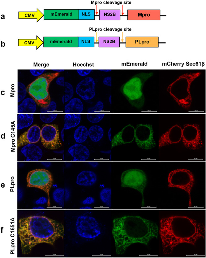
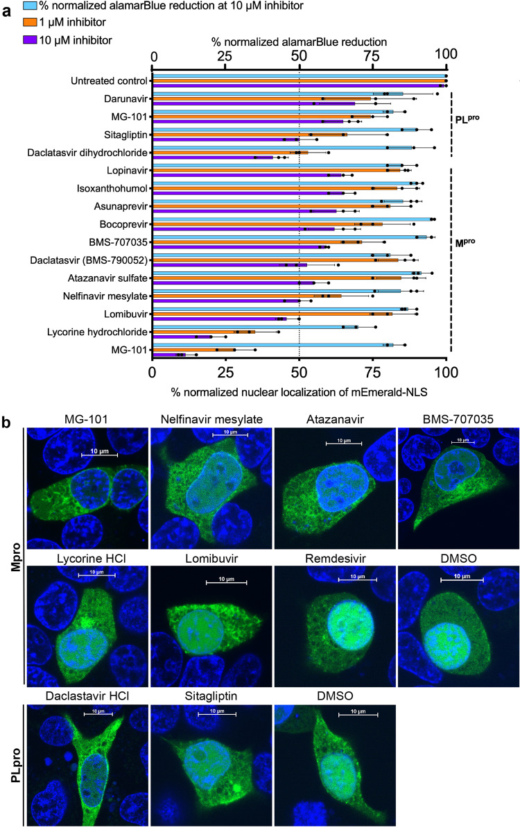
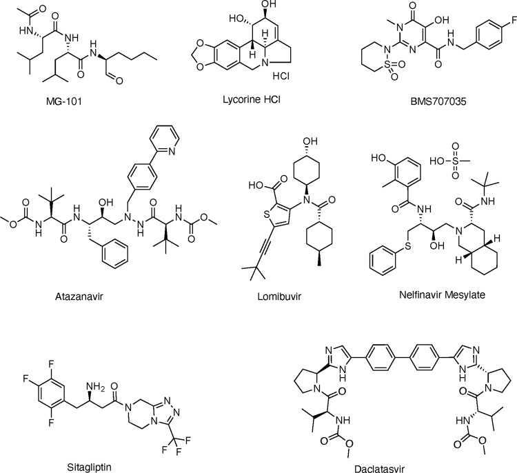
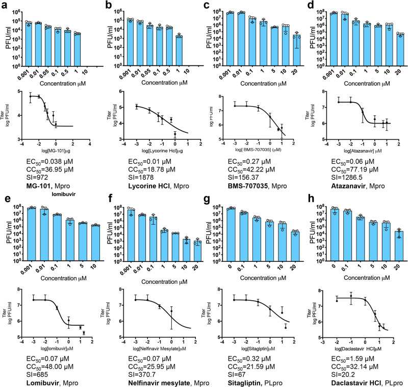
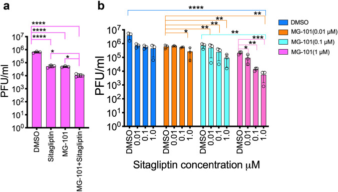
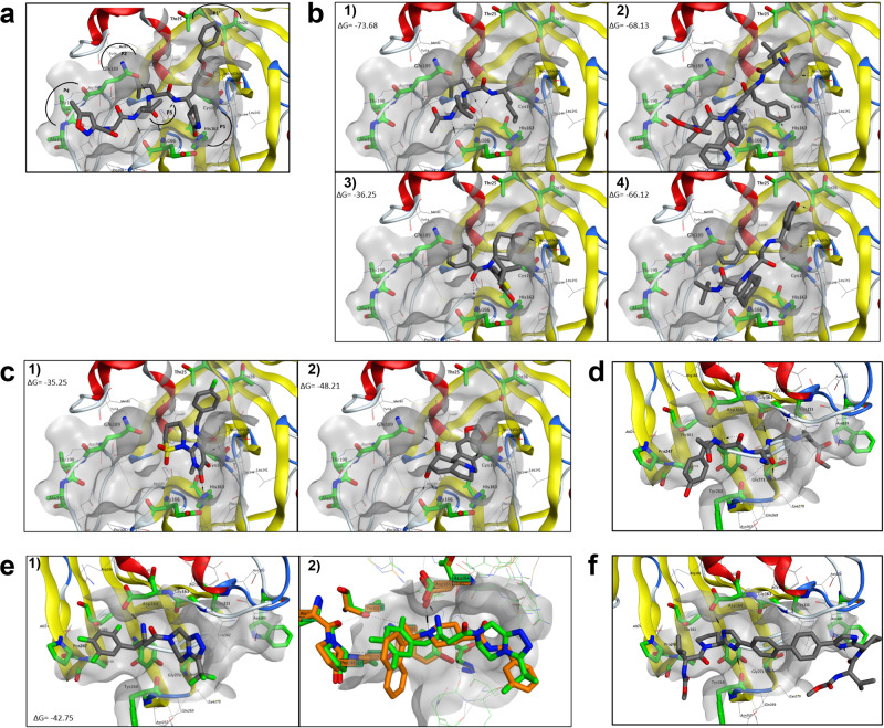
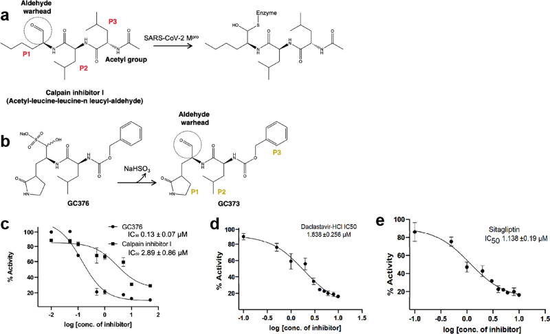
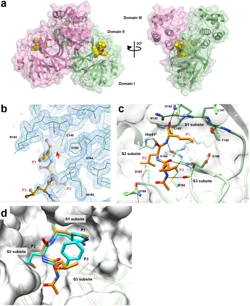
SARS-CoV-2 proteases Mpro and PLpro are promising targets for antiviral drug development. In this study, we present an antiviral screening strategy involving a novel in-cell protease assay, antiviral and biochemical activity assessments, as well as structural determinations for rapid identification of protease inhibitors with low cytotoxicity. We identified eight compounds with anti-SARS-CoV-2 activity from a library of 64 repurposed drugs and modeled at protease active sites by in silico docking. We demonstrate that Sitagliptin and Daclatasvir inhibit PLpro, and MG-101, Lycorine HCl, and Nelfinavir mesylate inhibit Mpro of SARS-CoV-2. The X-ray crystal structure of Mpro in complex with MG-101 shows a covalent bond formation between the inhibitor and the active site Cys145 residue indicating its mechanism of inhibition is by blocking the substrate binding at the active site. Thus, we provide methods for rapid and effective screening and development of inhibitors for blocking virus polyprotein processing as SARS-CoV-2 antivirals. Additionally, we show that the combined inhibition of Mpro and PLpro is more effective in inhibiting SARS-CoV-2 and the delta variant.


Conflict of interest statement

The authors declare no competing interests.

Figures

Fig. 1
Fig. 1. In-cell protease (ICP) assay for screening inhibitors of SARS-CoV-2 proteases.
a, b Constructs designed for the ICP assay containing mEmerald with nuclear localization signal (NLS), cleavage site for Mpro or PLpro, Zika virus NS2B followed by Mpro (a) or PLpro (amino acids 1541–1855 of nsp3) (b). cf Localization of mEmerald-NLS (green) and ER marker mCherry-Sec61 β (red) at 6 h post-transfection and stained with nuclear stain Hoechst 33342 dye (blue), in ICP assay. c Cells transfected with ICP construct A, d cells transfected with ICP construct A with inactive Mpro mutant (C145A). e Cells transfected with ICP construct B, f cells transfected with ICP construct B with inactive PLpro mutant (C1651A).
Fig. 2
Fig. 2. SARS-CoV-2 protease inhibitors selected after ICP assay and quantification.
a ImageJ quantification of mEmerald-NLS localization to the nucleus identifies six Mpro inhibitors and two PLpro inhibitors with approximately 50% reduction in protease activity. The selection criteria were 50% reduction at 10 µM and 25% at 1 µM concentrations without cytotoxicity. The distribution of mEmerald-NLS was quantified from cells in each image (n = 3) using ImageJ, the ratio between fluorescence in the nucleus, and total fluorescence calculated and normalized to that of untreated cells. b Live confocal images of HEK293T cells expressing PLpro or Mpro at 6 h post-transfection. Cells were treated with 10 µM inhibitors as indicated and nuclei were stained using Hoechst stain. Cells treated with DMSO and Remdesivir are negative controls.
Fig. 3
Fig. 3. Chemical structures of selected inhibitors with antiviral activity against SARS-CoV-2.
MG-101, Lycorine HCl, BMS 707035, Atazanavir, Lomibuvir, and Nelfinavir mesylate are inhibitors of Mpro. Sitagliptin and Daclastavir HCl are inhibitors of PLpro.
Fig. 4
Fig. 4. Inhibition of SARS-CoV-2 replication in Huh-7.5 cells by compounds selected from ICP assay.
Reduction in virus titers for different concentrations of compounds against Mpro (af) and PLpro (g, h) was determined by plaque assays (n = 3). Dose-response curves were plotted, EC50 (50% Effective concentration) was determined from plaque assay and CC50 (50% cytotoxic concentration) was determined by alamarBlue reduction data. Selectivity index SI = CC50/EC50.
Fig. 5
Fig. 5. Effect of compounds selected from ICP assay on SARS-CoV-2 replication.
a Huh-7.5 cells positive for SARS-CoV-2 determined by immunofluorescence assay. Huh-7.5 cells pre-treated for 24 h with inhibitors were infected with SARS-CoV-2 at an MOI of 0.1. At 24 h.p.i., cells were fixed with paraformaldehyde and probed with anti-N (SARS CoV-2 nucleocapsid) primary antibody and FITC-conjugated goat anti-rabbit secondary antibody (green, infected cells). Nuclei were stained with Hoechst stain and confocal images were acquired. b The ratio of infected to uninfected cells for each treatment was calculated using Nikon Elements software and normalized to untreated controls (n = 5). c Quantification of SARS-CoV-2 RNA molecules from infected cells treated with inhibitors as indicated (n = 3).
Fig. 6
Fig. 6. Antiviral activity of combinations of Mpro and PLpro inhibitors.
Effect of combination of Mpro inhibitor MG-101 and PLpro inhibitor Sitagliptin on the growth of SARS-CoV-2 (a) and the delta variant (b) (n = 3). Huh7.5 cells were treated with the inhibitors as shown, and reduction in virus titer at 24 h.p.i. was determined by plaque assays. Data are shown as mean ± SEM. p Values were considered significant when p < 0.05 (*), p < 0.01 (**), p < 0.001(***), and p < 0.0001(****).
Fig. 7
Fig. 7. Docking of SARS-CoV-2 inhibitors.
The key residues forming the binding pocket are highlighted in green. H-bonds are depicted as dashed black lines. a Co-crystallographic pose of peptidomimetic inhibitor N3 (PDB 7BQY) in the active site of SARS-CoV-2 Mpro. b Predicted binding mode for (1) MG-101, (2) Atazanavir, (3) Lomibuvir, and (4) Nelfinavir in the Mpro- active site. c Predicted binding mode for (1) BMS-707035, and (2) Lycorine HCl in the Mpro- active site. d Co-crystallographic poses of VIR251 (PDB 6WX4) in the active site of SARS‐CoV-2 PLpro. e (1) Predicted binding mode for Sitagliptin in the PLpro-active site, (2) Superposition of SARS-CoV crystal structure (4OW0, orange) and SARS-CoV-2 crystal structure (PDB 6WX4, green) the docked Sitagliptin. f Predicted binding mode for Daclatasvir in the PLpro- active site.
Fig. 8
Fig. 8. Inhibitions of the Mpro and PLpro activities in vitro by screened compounds.
a Schematic representation of the inhibition of Mpro by MG-101. Chemical groups and substrate positions (P1–P3) are indicated. b Chemical structures of GC376 (prodrug) and GC373 (active drug). c IC50 values of MG-101 and GC376 for the cleavage of (Dabcyl)-KTSAVLQ*SGFRKME(Edans) substrate by Mpro. A cleavage site is indicated by an asterisk. The inhibitory activities of d Daclastavir-HCl and e Sitagliptin against PLpro were tested using fluorogenic peptide Z-RLRGG-AMC as the substrate. N = 3, values are represented as mean ± SE.
Fig. 9
Fig. 9. Structural basis for the Mpro inhibition.
a Crystal structure of the Mpro and MG-101 complex. Mpro dimer is depicted as a cartoon model with a transparent surface and each protomer is colored green and pink. MG-101 is shown as CPK representation. b Electron density map (2Fo–Fc, blue mesh) of MG-101 bound at the active site of Mpro. The inhibitor and Mpro are depicted as ball-and-stick and wire models, respectively. A thiohemiacetal covalent between the inhibitor and C145 residue of Mpro is indicated by a red arrow. c Interactions between the inhibitor and active site of Mpro. The binding of MG-101 is stabilized by H-bonds (black dot lines) with Gly143, Cys145, His164, and Glu166. d Comparison of the binding of MG-101 (orange) and GC373 (cyan) at the active site of Mpro. S1–S3 subsites of Mpro and P1–P3 of inhibitors are indicated.

References

    1. Zhou P, et al. A pneumonia outbreak associated with a new coronavirus of probable bat origin. Nature. 2020;579:270–273. - PMC - PubMed
    1. Guan WJ, et al. Clinical Characteristics of Coronavirus Disease 2019 in China. N. Engl. J. Med. 2020;382:1708–1720. - PMC - PubMed
    1. Wu F, et al. A new coronavirus associated with human respiratory disease in China. Nature. 2020;579:265–269. - PMC - PubMed
    1. Hassan AO, et al. A SARS-CoV-2 infection model in mice demonstrates protection by neutralizing antibodies. Cell. 2020;182:744–753 e744. - PMC - PubMed
    1. Wang Z, et al. mRNA vaccine-elicited antibodies to SARS-CoV-2 and circulating variants. Nature. 2021;592:616–622. - PMC - PubMed

Publication types

MeSH terms

Substances