. 2022 Oct;43(10):2624-2635.
doi: 10.1038/s41401-022-00885-8. Epub 2022 Feb 25.

Direct cardio-protection of Dapagliflozin against obesity-related cardiomyopathy via NHE1/MAPK signaling

Affiliations

Direct cardio-protection of Dapagliflozin against obesity-related cardiomyopathy via NHE1/MAPK signaling

Ke Lin et al. Acta Pharmacol Sin. 2022 Oct.

Abstract

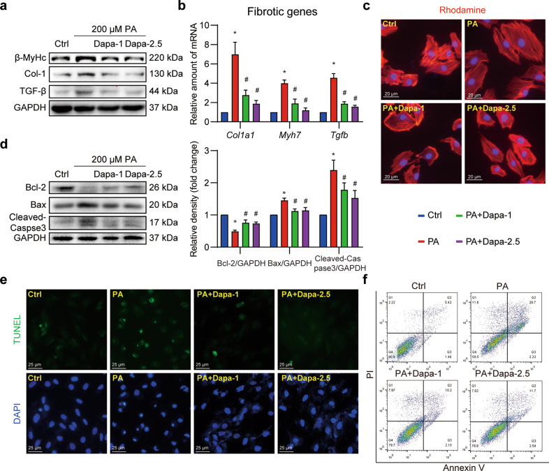
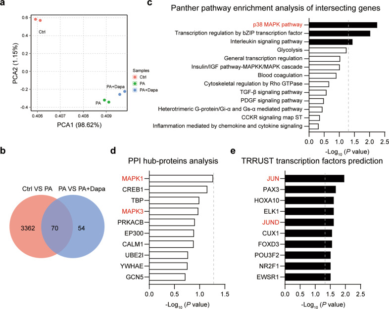
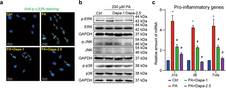
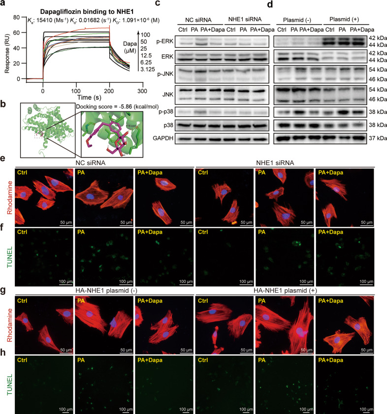
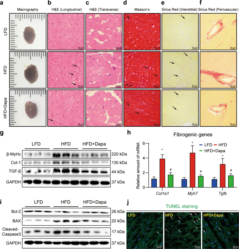
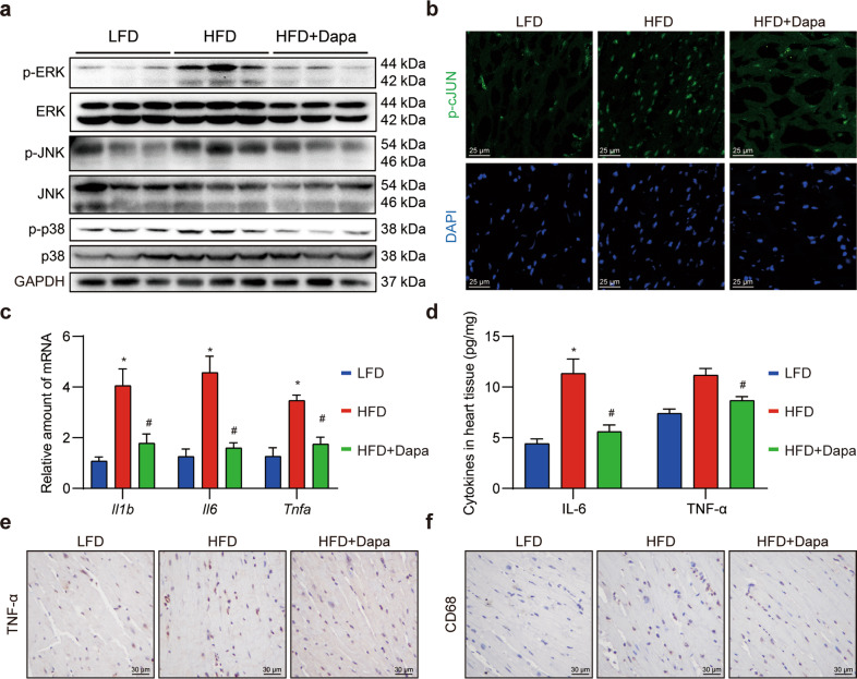
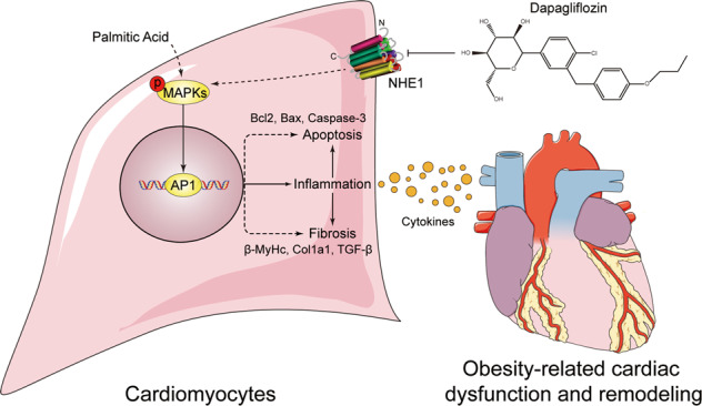
Obesity is an important independent risk factor for cardiovascular diseases, remaining an important health concern worldwide. Evidence shows that saturated fatty acid-induced inflammation in cardiomyocytes contributes to obesity-related cardiomyopathy. Dapagliflozin (Dapa), a selective SGLT2 inhibitor, exerts a favorable preventive activity in heart failure. In this study, we investigated the protective effect of Dapa against cardiomyopathy caused by high fat diet-induced obesity in vitro and in vivo. Cultured rat cardiomyocyte H9c2 cells were pretreated with Dapa (1, 2.5 μM) for 1.5 h, followed by treatment with palmitic acid (PA, 200 μM) for 24 h. We showed that Dapa pretreatment concentration-dependently attenuated PA-induced cell hypertrophy, fibrosis and apoptosis. Transcriptome analysis revealed that inhibition of PA-activated MAPK/AP-1 pathway contributed to the protective effect of Dapa in H9c2 cells, and this was confirmed by anti-p-cJUN fluorescence staining assay. Using surface plasmon resonance analysis we found the direct binding of Dapa with NHE1. Gain and loss of function experiments further demonstrated the role of NHE1 in the protection of Dapa. In vivo experiments were conducted in mice fed a high fat diet for 5 months. The mice were administered Dapa (1 mg·kg-1·d-1, i.g.) in the last 2 months. Dapa administration significantly reduced the body weight and improved the serum lipid profiles. Dapa administration also alleviated HFD-induced cardiac dysfunction and cardiac aberrant remodeling via inhibiting MAPK/AP-1 pathway and ameliorating cardiac inflammation. In conclusion, Dapa exerts a direct protective effect against saturated fatty acid-induced cardiomyocyte injury in addition to the lowering effect on serum lipids. The protective effect results from negative regulating MAPK/AP-1 pathway in a NHE1-dependent way. The current study highlights the potential of clinical use of Dapa in the prevention of obesity-related cardiac dysfunction.

Keywords: Dapagliflozin; MAPK pathway; NHE1; cardiomyopathy; inflammation; obesity; palmitic acid; rat cardiomyocyte H9c2 cell line.


Conflict of interest statement

The authors declare no competing interests.

Figures

Fig. 1
Fig. 1. Dapa directly protects cardiomyocytes against palmitic acid-induced cellular fibrosis and apoptosis.
ac Dapa attenuated PA-induced cardiomyocyte fibrosis and hypertrophy. H9c2 cells were pretreated with Dapa, and were then stimulated with PA for 24 h. a Cell lysates were collected and proteins of β-MyHc, Col-1, and TGF-β were detected with GAPDH as loading control. b Total mRNA of H9c2 cells was isolated, and mRNA levels of fibrotic genes were detected with β-actin as the normalization control. c H9c2 cells were stained with rhodamine-phalloidin, and counterstained with DAPI. Representative images were shown (scale bar = 20 μm). d–f Dapa attenuated PA-induced cardiomyocyte apoptosis. H9c2 cells were pretreated with Dapa, and were then stimulated with PA for 24 h. d Cell lysates were collected and proteins of Bcl-2, Bax and Cleaved-Caspase-3 were detected with GAPDH as loading control. Quantifications were shown in the right panel. e H9c2 cells were stained with TUNEL kit, and counterstained with DAPI. Representative images were shown (scale bar = 25 μm). f H9c2 cells were digested and stained with Annexin V-PI apoptosis kit, cells were then detected using flow cytometry. Representative images were shown. n = 3; Means ± SEM; One-way ANOVA followed by Turkey post-hoc tests; *P < 0.05, compared with Ctrl group, #P < 0.05, compared with PA group.
Fig. 2
Fig. 2. MAPK signaling contributes to the direct cardiomyocyte protection of Dapa.
H9c2 cells were pretreated with Dapa, and were then stimulated with PA for 24 h. Samples were isolated using TRIzol, and transcriptome sequencing was conducted. a PCA analysis of the sequencing results. b Venn diagram of differential genes in Ctrl VS PA, and PA VS PA + Dapa. c Panther pathway enrichment analysis of 70 intersecting genes. d PPI hub-proteins analysis of 70 intersecting genes. e TRRUST transcription analysis of 70 intersecting genes. Dotted lines represented P = 0.05. Black column showed significant changed items.
Fig. 3
Fig. 3. Dapa inhibits MAPK signaling and inflammation in cardiomyocytes.
H9c2 cells were pretreated with Dapa, and were then stimulated with PA for 1 h. a H9c2 cells were fixed and stained with anti-p-cJUN antibody, and counterstained with DAPI. Representative images were shown (scale bar = 25 μm). b Cell lysates were collected and proteins of p-ERK, p-JNK and p-p38 were detected with total proteins as loading control. c Cells were pretreated with Dapa, and were then stimulated with PA for 8 h. Total mRNA was isolated using TRIzol, and mRNA levels of pro-inflammatory genes were detected with β-actin as the normalization control. n = 3; Means ± SEM; One-way ANOVA followed by Turkey post-hoc tests; *P < 0.05, compared with Ctrl group, #P < 0.05, compared with PA group.
Fig. 4
Fig. 4. Dapa targets NHE1 hence regulating MAPK/AP-1 pathway to protect cardiomyocytes.
a Direct binding of Dapa and NHE1 protein was shown by SPR assay. Ka binding constant, Kd dissociation constant, KD equilibrium dissociation constant. b Molecular docking of Dapa with NHE1. c–h H9c2 cells were transfected with NHE1 siRNA or HA-NHE1 plasmid, and were then treated with Dapa and PA. Cell lysates from knockdown (c) or overexpression (d) assay were collected and proteins of p-ERK, p-JNK, and p-p38 were detected with total proteins as loading control. H9c2 cells after knockdown of NHE1 were stained with rhodamine-phalloidin (e) or TUNEL kit (f), and counterstained with DAPI. Representative images were shown. g, h H9c2 cells after overexpression of NHE1 were stained with TUNEL kit, and counterstained with DAPI (e, g, scale bar = 50 μm; f, h, scale bar = 100 μm). Representative images were shown. n = 3; Means ± SEM; One-way ANOVA followed by Turkey post-hoc tests.
Fig. 5
Fig. 5. Dapa attenuates HFD-induced cardiac fibrosis, remodeling and cardiomyocytes apoptosis.
Mice were randomly divided into 3 groups, including LFD, HFD and HFD + Dapa. After 3 months of high fat diet, mice were treated with 1 mg·kg−1·d−1 Dapa for another 2 months in HFD + Dapa group. a Representative macrographs of hearts from different groups of mice. Hematoxylin-eosin staining of heart sections. Representative images of longitudinal views (b, scale bar = 30 μm) and transverse views (c, scale bar = 10 μm). d Representative images of Masson’s staining of heart sections (scale bar = 50 μm). Sirius Red staining of heart sections. Representative images of interstitial views (e, scale bar = 50 μm) and perivascular views (f, scale bar = 30 μm). g Western blot analysis of fibrotic and hypertrophy protein markers in heart tissue. GAPDH was used as loading control. h Quantitative PCR analysis of fibro-genic genes in heart tissue, and β-actin was used as normalization control. i Western blot analysis of apoptotic protein markers in heart tissue. GAPDH was used as loading control. j Representative images of TUNEL staining of heart sections. White arrows showed positive TUNEL staining cells in heart tissue (scale bar = 25 μm). n = 8 (a-f, h, j), n = 6 (g, i); Means ± SEM; One-way ANOVA followed by Turkey post-hoc tests; *P < 0.05, compared with LFD group, #P < 0.05, compared with HFD group.
Fig. 6
Fig. 6. Dapa attenuates MAPK/AP1 cascades and cardiac inflammation in HFD mice.
Mice were randomly divided into 3 groups, including LFD, HFD and HFD + Dapa. After 3 months of high fat diet, mice were treated with 1 mg·kg−1·d−1 Dapa for another 2 months. a Western blot analysis of proteins in MAPK pathway in heart tissue. GAPDH was used as loading control. b Representative images of anti-p-cJUN immunofluorescence staining in heart sections (scale bar = 25 μm). c Quantitative analysis of inflammatory genes in heart tissue, and β-actin was used as normalization control. d ELISA assay of inflammatory cytokines in heart tissues. e, f Representative images of anti-TNF-α and anti-CD68 immunochemistry staining in heart sections (scale bar = 30 μm). n = 8; Means ± SEM; One-way ANOVA followed by Turkey post-hoc tests; *P < 0.05, compared with LFD group, #P < 0.05, compared with HFD group.
Fig. 7
Fig. 7. Schematic illustration of the main findings.
Dapa directly inhibits saturated fatty acid (PA)-induced MAPK signaling and inflammation, thus attenuating cell apoptosis and fibrosis in cardiomyocytes, which might be associated with the function of NHE1 receptor.

Similar articles

Cited by

References

    1. Horwich TB, Fonarow GC, Clark AL. Obesity and the obesity paradox in heart failure. Prog Cardiovasc Dis. 2018;61:151–6. doi: 10.1016/j.pcad.2018.05.005. - DOI - PubMed
    1. Abel ED, Litwin SE, Sweeney G. Cardiac remodeling in obesity. Physiol Rev. 2008;88:389–419. doi: 10.1152/physrev.00017.2007. - DOI - PMC - PubMed
    1. Tiwari S, Ndisang JF. The role of obesity in cardiomyopathy and nephropathy. Curr Pharmacol Des. 2014;20:1409–17. doi: 10.2174/13816128113199990562. - DOI - PubMed
    1. Kennedy A, Martinez K, Chuang CC, LaPoint K, McIntosh M. Saturated fatty acid-mediated inflammation and insulin resistance in adipose tissue: mechanisms of action and implications. J Nutr. 2009;139:1–4. doi: 10.3945/jn.108.098269. - DOI - PubMed
    1. Schaeffler A, Gross P, Buettner R, Bollheimer C, Buechler C, Neumeier M, et al. Fatty acid-induced induction of Toll-like receptor-4/nuclear factor-kappaB pathway in adipocytes links nutritional signalling with innate immunity. Immunology. 2009;126:233–45. doi: 10.1111/j.1365-2567.2008.02892.x. - DOI - PMC - PubMed

MeSH terms