Review
. 2022 May;43(5):1103-1119.
doi: 10.1038/s41401-022-00880-z. Epub 2022 Feb 25.

Bile acid and receptors: biology and drug discovery for nonalcoholic fatty liver disease

Affiliations
Review

Bile acid and receptors: biology and drug discovery for nonalcoholic fatty liver disease

Ting-Ying Jiao et al. Acta Pharmacol Sin. 2022 May.

Abstract

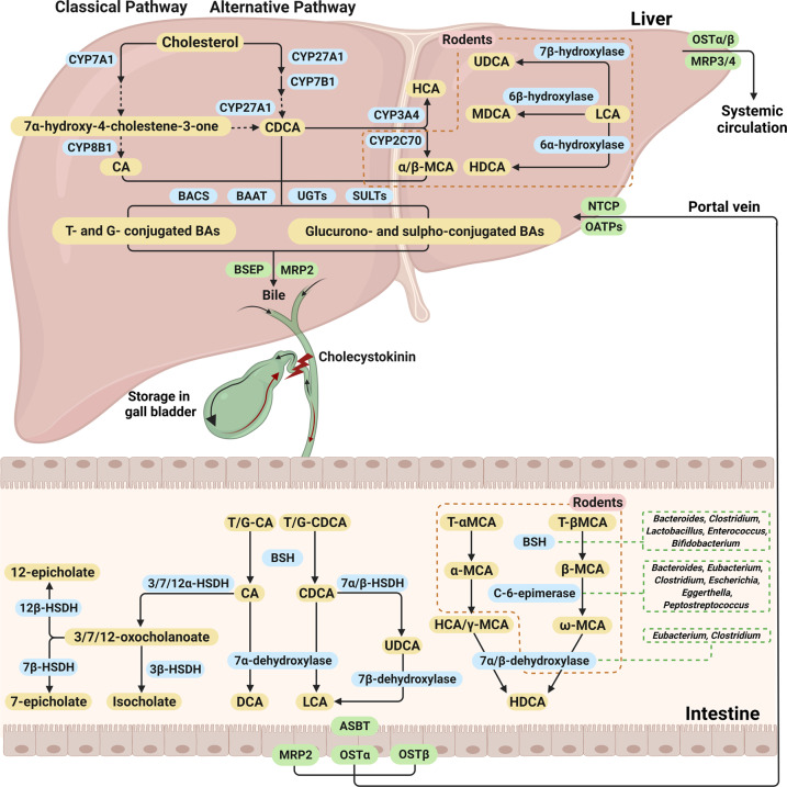
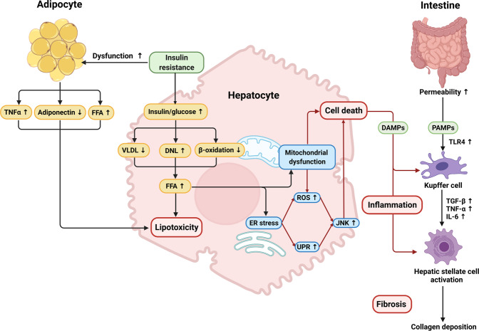
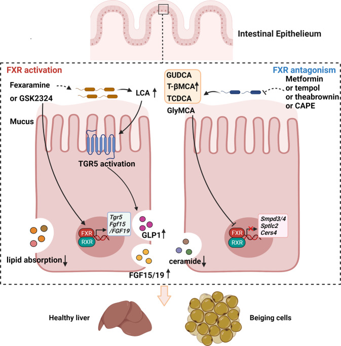
Nonalcoholic fatty liver disease (NAFLD), a series of liver metabolic disorders manifested by lipid accumulation within hepatocytes, has become the primary cause of chronic liver diseases worldwide. About 20%-30% of NAFLD patients advance to nonalcoholic steatohepatitis (NASH), along with cell death, inflammation response and fibrogenesis. The pathogenesis of NASH is complex and its development is strongly related to multiple metabolic disorders (e.g. obesity, type 2 diabetes and cardiovascular diseases). The clinical outcomes include liver failure and hepatocellular cancer. There is no FDA-approved NASH drug so far, and thus effective therapeutics are urgently needed. Bile acids are synthesized in hepatocytes, transported into the intestine, metabolized by gut bacteria and recirculated back to the liver by the enterohepatic system. They exert pleiotropic roles in the absorption of fats and regulation of metabolism. Studies on the relevance of bile acid disturbance with NASH render it as an etiological factor in NASH pathogenesis. Recent findings on the functional identification of bile acid receptors have led to a further understanding of the pathophysiology of NASH such as metabolic dysregulation and inflammation, and bile acid receptors are recognized as attractive targets for NASH treatment. In this review, we summarize the current knowledge on the role of bile acids and the receptors in the development of NAFLD and NASH, especially the functions of farnesoid X receptor (FXR) in different tissues including liver and intestine. The progress in the development of bile acid and its receptors-based drugs for the treatment of NASH including bile acid analogs and non-bile acid modulators on bile acid metabolism is also discussed.

Keywords: Farnesoid X receptor; G protein-coupled bile acid receptor; bile acids; drug target; nonalcoholic fatty liver disease; nonalcoholic steatohepatitis.


Conflict of interest statement

The authors declare no competing interests.

Figures

Fig. 1
Fig. 1. Bile acid synthesis and enterohepatic circulation.
Hepatocytes produce primary bile acids via the classical and alternative pathways. The classical pathway starts with cholesterol 7α-hydroxylase (CYP7A1) and produces cholic acid (CA) and chenodeoxycholic acid (CDCA) through the action of sterol 12α-hydroxylase (CYP8B1) and sterol 27-hydroxylase (CYP27A1). The alternative pathway is initiated by CYP27A1 and produces CDCA through the action of oxysterol 7α-hydroxylase (CYP7B1). In rodents, CDCA can be mostly converted to α-muricholic acid (α-MCA) and β-MCA by a sterol 6β-hydroxylase (CYP2C70). Lithocholic acid (LCA) can be 7β-hydroxylated to ursodeoxycholic acid (UDCA), or 6α-hydroxylated to hyodeoxycholic acid (HDCA), or 6β-hydroxylated to murideoxycholic acid (MDCA). In pigs and humans, CDCA can be converted to hyocholic acid (HCA) by cytochrome P450 3A4 (CYP3A4) and then dehydroxylated to HDCA in the intestine. Most bile acids are conjugated to glycine (G) or taurine (T) via the action of bile acid-CoA synthetase (BACS) and amino acid N-acyltransferase (BAAT) and are secreted into the bile via bile salt export protein (BSEP). Meanwhile, hepatocytes produce sulphated (sulpho-) or glucuronidated (glucurono-) bile acids via sulfotransferases (SULTs) and UDP-glucuronosyltransferases (UGTs), which are secreted into the bile via multidrug resistance-related protein 2 (MRP2). The bile formed is stored in the gallbladder. After a meal, the release of cholecystokinin from the pancreas causes bile to be released into the duodenum. The conjugated primary bile acids are transformed to secondary bile acids by the action of intestinal bacterial bile salt hydrolases (BSH) and different dehydroxylases. β-MCA is differentially isomerized by C-6 to form ω-MCA, and then ω-MCA is 7α-dehydroxylated to form HDCA. CDCA is converted to UDCA by hydroxysteroid dehydrogenases (HSDHs), and then UDCA is converted to LCA by 7β-dehydroxylase. Gut bacterial 3α, 7α, and 12α-HSDHs epimerize the α-hydroxyl groups of bile acids to carbonyl groups to form 3-oxo, 7-oxo, and 12-oxo-bile acids, and then the carbonyl groups in oxo-bile acids are converted to isocholate, 7-epicholate and 12-epicholate by 3β, 7β, and 12β-HSDHs. At the end of the ileum, ~95% of the bile acids involved in the hepatic-intestinal circulation are reabsorbed by enterocytes into the intestinal epithelium via the apical sodium-dependent bile salt transporter (ASBT), transported across the enterocytes to the sinusoidal membrane, excreted into the portal vein via organic solute transporter-α and -β (OSTα and OSTβ) and MRP2 and transported back to the liver via the portal vein by the uptake action of Na+-taurocholate co-transporting polypeptides (NTCP, conjugated bile acids) and organic anion transporters (OATPs, unconjugated bile acids). Bile acids enter the systemic circulation from hepatocytes via MRP3, MRP4, OSTα, and OSTβ. The figure was created with BioRender.com.
Fig. 2
Fig. 2. FXR and TGR5 in the enterohepatic circulation.
Activation of hepatic farnesoid X receptor (FXR) by primary bile acids synthesized from cholesterol increases the expression of the small heterodimer partner (SHP), which inhibits CYP7A1 and CYP8B1 expression. At the same time, FXR inhibits NTCP to reduce bile acid uptake from circulation, induces BSEP to promote bile acid secretion into the bile, and induces multidrug resistance protein 3 (MDR3) to promote phospholipid secretion into the bile. Activation of intestinal FXR by bile acids increases the expression of fibroblast growth factor 15/19 (FGF19 in humans, FGF15 in rodents), induces the entry of these proteins into the liver via the enterohepatic circulation and acts on fibroblast growth factor receptor 4 (FGFR4)/β-klotho complex, thereby inhibiting the expression of CYP7A1. Meanwhile, FXR inhibits the expression of ASBT, but induces OSTα and OSTβ. In enteroendocrine L cells, FXR induces G protein-coupled bile acid receptor 1 (GPBAR1 or TGR5) to activate cyclic adenosine monophosphate (cAMP), which leads to secretion of glucagon-like peptide-1 (GLP-1) and stimulates insulin secretion from pancreatic cells. The figure was created with BioRender.com.
Fig. 3
Fig. 3. Metabolic factors for the development of NASH.
Insulin resistance (IR) acts on adipose tissue and worsens adipocyte dysfunction, inducing lipolysis, release of adipokines and pro-inflammatory cytokines. In the liver, IR amplifies de novo lipogenesis (DNL) and reduces very low-density lipoprotein (VLDL) and β-oxidation. Increased hepatic free fatty acids (FFA) lead to mitochondrial dysfunction and endoplasmic reticulum (ER) stress, which leads to the production of reactive oxygen species (ROS) and activation of the unfolded protein response (UPR), and finally to the activation of c-Jun N-terminal kinase (JNK). Hepatic stellate cell can be activated directly by the accumulation of FFA in the liver, also known as damage-associated molecular pattern (DAMPs), or indirectly by IL-6, TNF-α and TGF-β secreted by Kupffer cells, leading to collagen deposition. Meanwhile, increased permeability of the small intestine leads to stimulation of Toll-like receptor 4 (TLR4) and activation of Kupffer cells by pathogen-associated molecular patterns (PAMPs) such as lipopolysaccharide (LPS), pro-inflammatory factors, etc., which subsequently activates an inflammatory cascade response. The figure was created with BioRender.com.
Fig. 4
Fig. 4. Paradoxical roles of intestinal FXR on the development of NASH.
FXR agonist GSK2324 decreases hepatic triglyceride accumulation through a mechanism that lipid absorption decreased in an intestinal FXR-dependent manner. Intestine-specific FXR agonist fexaramine has a beneficial effect on glucose homeostasis in diet-induced obese mice by directly activating FXR, and alternatively increasing the abundance of lithocholic acid (LCA)-producing bacterium and circulating LCA levels, which thereby indirectly activates the TGR5-cAMP-GLP-1 cascade in intestinal L cells and induces the release of GLP-1 into the serum, eventually induces browning and improves insulin sensitivity. In addition, intestinal FXR activation induces the transcription of Fgf15 (FGF19 in humans) which is delivered to the liver and binds with FGFR4 to inhibit CYP7A1 expression and hepatic bile acid synthesis. FXR also activates TGR5 in enterocytes. Bile acid deconjugation can be decreased by metformin, tempol and theabrownin through reduction of the abundance of bile salt hydrolase (BSH)-secreting gut microbiota and BSH activity can be directly inhibited by caffeic acid phenethyl ester (CAPE), thus increasing levels of endogenous FXR antagonists glycoursodeoxycholic acid (GUDCA), taurochenodeoxycholic acid (TCDCA) and tauro-β-muricholic acid (T-βMCA), which reduced the ceremide synthesis-related genes including Smpd3/4, Sptlc2 and Cers4. As endogenous FXR antagonists are easily deconjugated by intestinal bacteria, synthetic glycine-β-muricholic acid (GlyMCA) was developed as a more stable FXR antagonist that benefited metabolic diseases via decreased ceremide synthesis. The figure was created with BioRender.com.

Similar articles

Cited by

References

    1. Paik JM, Golabi P, Younossi Y, Mishra A, Younossi ZM. Changes in the global burden of chronic liver diseases from 2012 to 2017: the growing impact of NAFLD. Hepatology. 2020;72:1605–16. doi: 10.1002/hep.31173. - DOI - PubMed
    1. Younossi Z, Tacke F, Arrese M, Chander Sharma B, Mostafa I, Bugianesi E, et al. Global perspectives on nonalcoholic fatty liver disease and nonalcoholic steatohepatitis. Hepatology. 2019;69:2672–82. doi: 10.1002/hep.30251. - DOI - PubMed
    1. Chalasani N, Younossi Z, Lavine JE, Charlton M, Cusi K, Rinella M, et al. The diagnosis and management of nonalcoholic fatty liver disease: practice guidance from the American association for the study of liver diseases. Hepatology. 2018;67:328–57.. doi: 10.1002/hep.29367. - DOI - PubMed
    1. Younossi ZM, Stepanova M, Rafiq N, Henry L, Loomba R, Makhlouf H, et al. Nonalcoholic steatofibrosis independently predicts mortality in nonalcoholic fatty liver disease. Hepatol Commun. 2017;1:421–8. doi: 10.1002/hep4.1054. - DOI - PMC - PubMed
    1. Dulai PS, Singh S, Patel J, Soni M, Prokop LJ, Younossi Z, et al. Increased risk of mortality by fibrosis stage in nonalcoholic fatty liver disease: systematic review and meta-analysis. Hepatology. 2017;65:1557–65. doi: 10.1002/hep.29085. - DOI - PMC - PubMed

MeSH terms

Substances