. 2022 Oct;43(10):2585-2595.
doi: 10.1038/s41401-022-00876-9. Epub 2022 Feb 25.

Activating α7nAChR ameliorates abdominal aortic aneurysm through inhibiting pyroptosis mediated by NLRP3 inflammasome

Affiliations

Activating α7nAChR ameliorates abdominal aortic aneurysm through inhibiting pyroptosis mediated by NLRP3 inflammasome

Hui Fu et al. Acta Pharmacol Sin. 2022 Oct.

Abstract

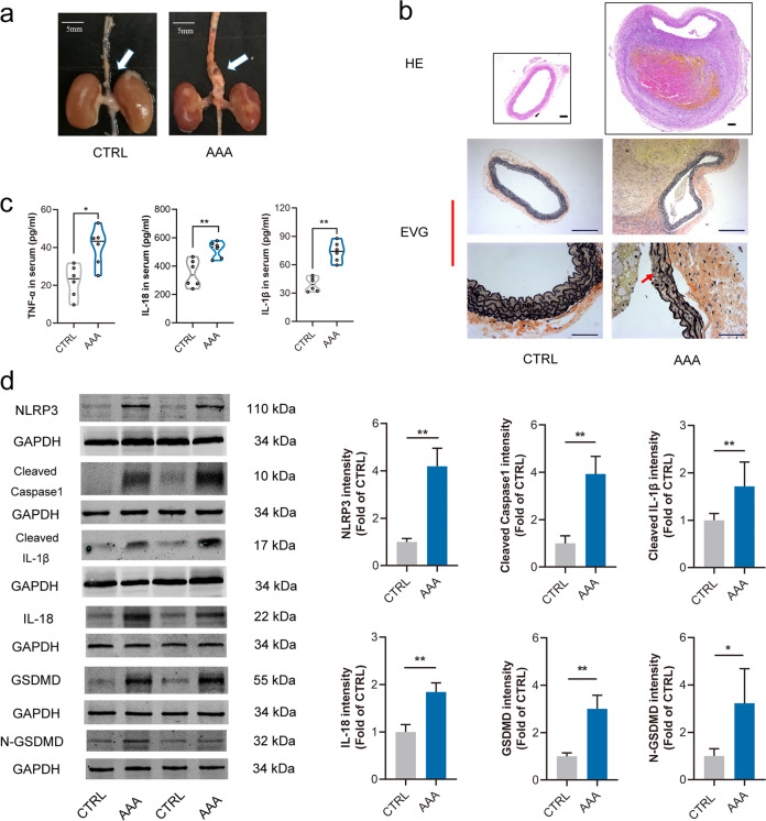
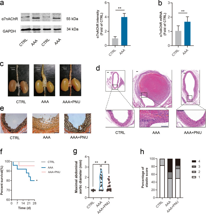
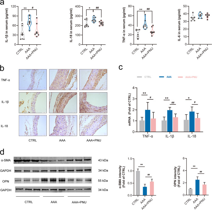
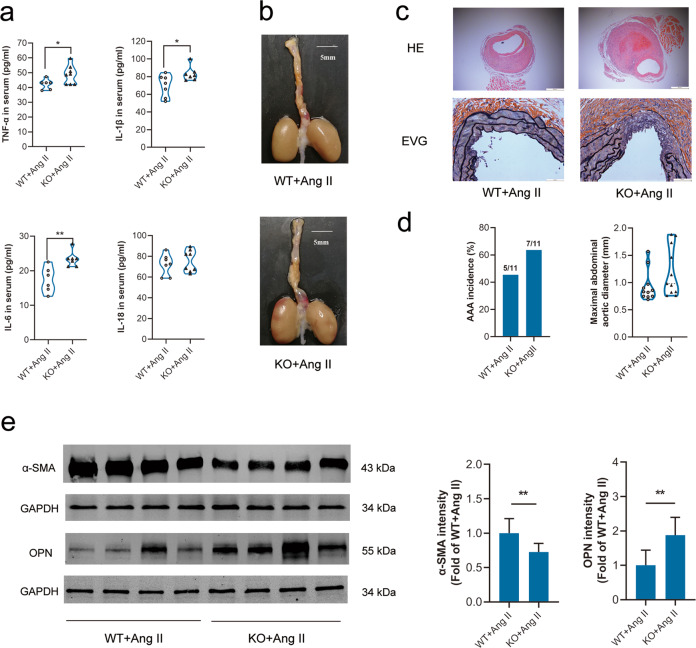
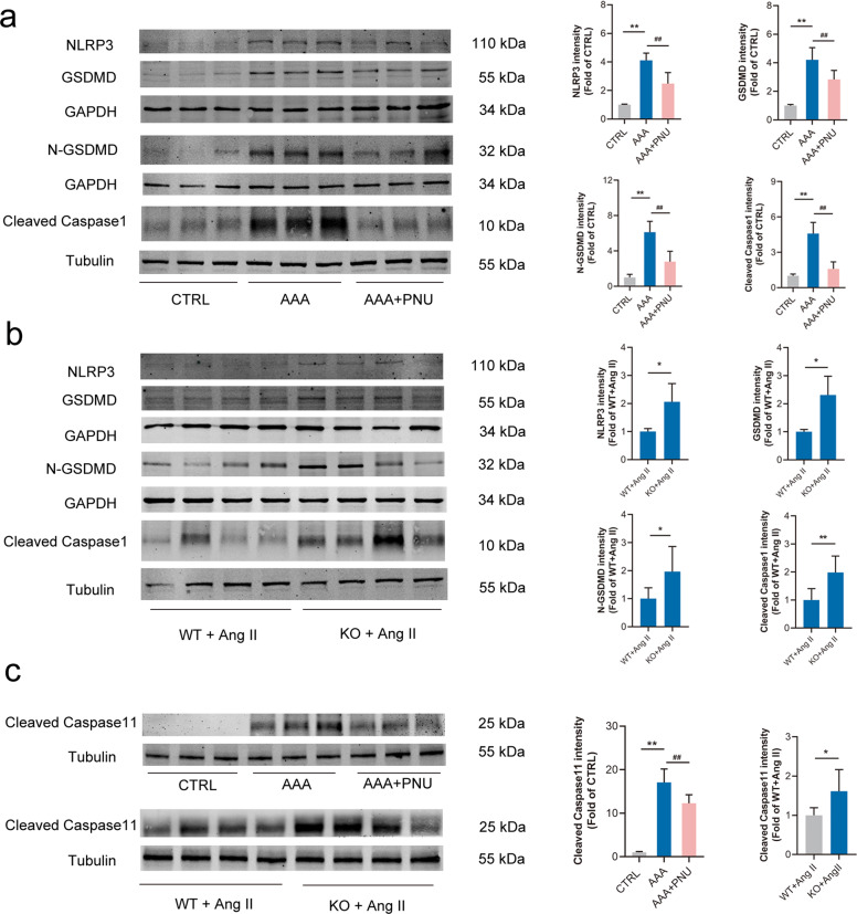
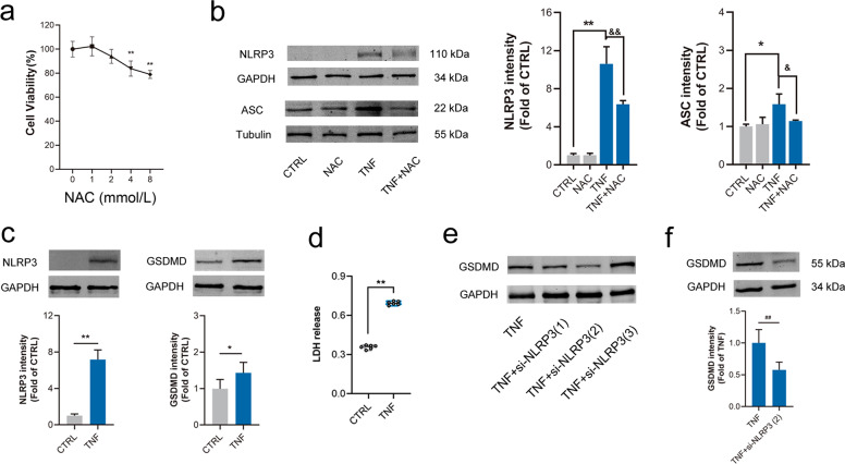
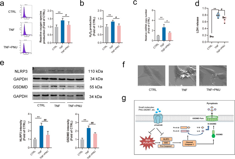
Abdominal aortic aneurysm (AAA) is defined as a dilated aorta in diameter at least 1.5 times of a normal aorta. Our previous studies found that activating α7 nicotinic acetylcholine receptor (α7nAChR) had a protective effect on vascular injury. This work was to investigate whether activating α7nAChR could influence AAA formation and explore its mechanisms. AAA models were established by angiotensin II (Ang II) infusion in ApoE-/- mice or in wild type and α7nAChR-/- mice. In vitro mouse aortic smooth muscle (MOVAS) cells were treated with tumor necrosis factor-α (TNF-α). PNU-282987 was chosen to activate α7nAChR. We found that cell pyroptosis effector GSDMD and NLRP3 inflammasome were activated in abdominal aorta, and inflammatory cytokines in serum were elevated in AAA models of ApoE-/- mice. Activating α7nAChR reduced maximal aortic diameters, preserved elastin integrity and decreased inflammatory responses in ApoE-/- mice with Ang II infusion. While α7nAChR-/- mice led to aggravated aortic injury and increased inflammatory cytokines with Ang II infusion when compared with wild type. Moreover, activating α7nAChR inhibited NLRP3/caspase-1/GSDMD pathway in AAA model of ApoE-/- mice, while α7nAChR deficiency promoted this pathway. In vitro, N-acetylcysteine (NAC) inhibited NLRP3 inflammasome activation and NLRP3 knockdown reduced GSDMD expression, in MOVAS cells treated with TNF-α. Furthermore, activating α7nAChR inhibited oxidative stress, reduced NLRP3/GSDMD expression, and decreased cell pyroptosis in MOVAS cells with TNF-α. In conclusion, our study found that activating α7nAChR retarded AAA through inhibiting pyroptosis mediated by NLRP3 inflammasome. These suggested that α7nAChR would be a potential pharmacological target for AAA.

Keywords: NLRP3 inflammasome; abdominal aortic aneurysm; cell pyroptosis; inflammation; α7nAChR.


Conflict of interest statement

The authors declare no competing interests.

Figures

Fig. 1
Fig. 1. Cell pyroptosis and NLRP3 inflammasome were involved in AAA formation.
Male ApoE−/− mice were infused with angiotensin II (Ang II, 1000 ng·kg−1·min−1) for 4 week to induce abdominal aortic aneurysm (AAA), the control (CTRL) was infused with normal saline. a Representative images of abdominal aortas in ApoE−/− mice, scale bar = 5 mm. b HE and EVG staining displayed intraluminal thrombus and disruption of the elastin in AAA mice, scale bar = 100 µm (up), or 200 µm (middle), or 50 µm (down). c Serum TNF-α, IL-18 and IL-1β were significantly increased in AAA mice, data were shown as each value of inflammatory cytokines. n = 6 mice per group, *P < 0.05, **P < 0.01 vs. CTRL. d Expressions of NLRP3, Cleaved caspase-1, Cleaved IL-1β, IL-18, GSDMD and N-GSDMD were increased in AAA tissues. n = 6 mice per group, data were shown as means ± SD. *P < 0.05, **P < 0.01 vs. CTRL.
Fig. 2
Fig. 2. Activating α7nAChR slowed down AAA formation.
Male ApoE−/− mice were infused with Ang II to induce AAA. PNU-282987 (PNU) was injected to activate α7nAChR. a, b Both protein and mRNA levels of α7nAChR were increased in aortas from AAA mice. n = 5–7 mice per group, data were shown as means ± SD, **P < 0.01 vs. CTRL. c Representative images of abdominal aortas in ApoE−/− mice, scale bar = 5 mm. d Representative images of HE staining in ApoE−/− mice, scale bar = 100 µm (up) or 50 µm (down). e Representative images of EVG staining in ApoE−/− mice. scale bar = 50 µm. f The survival curve in ApoE−/− mice. n = 20 mice for CTRL group; n = 26 mice for AAA group; and n = 22 mice for AAA + PNU group. g PNU treatment reduced maximal abdominal aortic diameters with Ang II infusion in ApoE−/− mice. n = 19–21 mice per group, data were shown as each value of per mouse. **P < 0.01 vs. CTRL; #P < 0.05 vs. AAA. h PNU treatment improved the elastin integrity, n = 12 mice per group
Fig. 3
Fig. 3. Activating α7nAChR inhibited inflammation and prevented the switch of VSMCs to a synthetic phenotype in AAA mice.
Male ApoE−/− mice were infused with Ang II to induce AAA. PNU-282987 (PNU) was injected to activate α7nAChR. a PNU inhibited inflammation in ApoE−/− mice with Ang II. n = 7–8 mice per group, data were shown as each inflammatory cytokine’s concentration. *P < 0.05, **P < 0.01 vs. CTRL; #P < 0.05, ##P < 0.01 vs. AAA. b Immunohistochemistry for TNF-α, IL-1β and IL-18 in aortas from ApoE−/− mice, scale bar = 50 µm. c Real-time PCR for TNF-α IL-1β and IL-18 expression in aortas from ApoE−/− mice. n = 6–7 mice per group, data were shown as means ± SD. *P < 0.05, **P < 0.01 vs. CTRL; #P < 0.05, ##P < 0.01 vs. AAA. d Activating α7nAChR prevented the decrease of α-SMA (a contractile phenotype marker of VSMCs) and increase of OPN (a synthetic phenotype marker of VSMCs) with Ang II infusion. n = 6 mice per group, data were shown as means ± SD. **P < 0.01 vs. CTRL; ##P < 0.01 vs. AAA.
Fig. 4
Fig. 4. α7nAChR deficiency promoted AAA formation and inflammation.
Age-matched male wild type (WT) and α7nAChR−/− (KO) mice with Ang II infusion for 4 week to induce AAA. a α7nAChR deficiency increased inflammatory cytokine expression in serum. n = 6–8 mice per group, data were shown as each cytokine’s concentration, *P < 0.05, **P < 0.01 vs. WT + Ang II. b, c α7nAChR deficiency accelerated the abdominal aortic dilation and displayed more serious disruption of elastin, scale bar = 5 mm (b), 500 µm (c up) or 50 µm (c down). d The AAA incidence and the maximal abdominal aortic diameter in WT and α7nAChR−/− mice infused with Ang II. n = 11 mice per group, the maximal abdominal aortic diameter was shown as each value. e VSMCs were more inclined to the synthetic phenotype in AAA tissues in α7nAChR−/− mice with Ang II infusion. n = 8 mice per group, data were shown as means ± SD, **P < 0.01 vs. WT + Ang II.
Fig. 5
Fig. 5. Activating α7nAChR inhibited cell pyroptosis in AAA mice.
Male ApoE−/− mice were infused with Ang II to induce AAA and PNU-282987 (PNU) was injected to activate α7nAChR in ApoE−/− mice. Age-matched male wild type (WT) and α7nAChR−/− (KO) mice were infused with Ang II to induce AAA. a PNU decreased the levels of NLRP3, cleaved caspase-1, GSDMD and N-GSDMD in aortas from ApoE−/− mice. n = 6 mice per group, data were shown as means ± SD, **P < 0.01 vs. CTRL; ##P < 0.01 vs. AAA. b α7nAChR deficiency promoted NLRP3, cleaved caspase-1, GSDMD and N-GSDMD expression in aortas from mice with Ang II infusion. n = 8 mice per group, data were shown as means ± SD, *P < 0.01, **P < 0.01 vs. WT+Ang II. c The expressions of Cleaved caspase-11 in aortas from ApoE−/− mice, or WT and KO mice with Ang II infusion. PNU decreased the levels of Cleaved caspase-11 in aortas from ApoE−/− mice. n = 6 mice per group, data were shown as means ± SD, **P < 0.01 vs. CTRL; ##P < 0.01 vs. AAA. α7nAChR deficiency promoted Cleaved caspase-11 expression in aortas from mice with Ang II infusion. n = 8 mice per group, data were shown as means ± SD, *P < 0.01 vs. WT+Ang II.
Fig. 6
Fig. 6. Knockdown NLRP3 inhibited GSDMD expression and LDH release in MOVAS by TNF-α.
Mouse aortic vascular smooth muscle cells (MOVAS) were treated with TNF-α for 24 h. a Different concentrations of N-acetylcysteine (NAC, a ROS scavenger) on cell viability. n = 6 per group, data were shown as means ± SD. **P < 0.01 vs. CTRL (0 mmol/L). b The increased expressions of NLRP3 and ASC by TNF-α were inhibited by NAC (2 mmol/L). n = 4 per group, data were shown as means ± SD. *P < 0.05, **P < 0.01 vs. CTRL; &P < 0.05, &&P < 0.01 vs. TNF. c, d NLRP3 and GSDMD expression, and LDH release (OD at 490 nm) were increased in MOVAS cells by TNF-α. n = 6 per group, data were shown as means ± SD or each value, *P < 0.05, **P < 0.01 vs. CTRL. e, f NLRP3 knockdown inhibited GSDMD expression in MOVAS cells treated with TNF-α. n = 6 per group, data were shown as means ± SD. ##P < 0.01 vs. TNF-α.
Fig. 7
Fig. 7. Activating α7nAChR inhibited oxidative stress, NLRP3 expression and cell pyroptosis.
Mouse aortic vascular smooth muscle cells (MOVAS) were treated with TNF-α with or without pretreatment with PNU-282987 (PNU) to activate α7nAChR. a, b PNU treatment inhibited the production of ROS and H2O2 in MOVAS by TNF-α. n = 4 per group, data were shown as means ± SD. **P < 0.01 vs. CTRL; #P < 0.05 vs. TNF. c PNU treatment reduced the relative mitochondrial DNA (mtDNA) level in MOVAS by TNF-α. n = 6 per group, data were shown as means ± SD. **P < 0.01 vs. CTRL; #P < 0.05 vs. TNF-α. d PNU treatment reduced the release of LDH in MOVAS cells by TNF-α (n = 5 per group), data were shown as each value. **P < 0.01 vs. CTRL; #P < 0.05 vs. TNF-α. e PNU treatment alleviated the expression of NLRP3/GSDMD in MOVAS by TNF-α. n = 6 per group, data were shown as means ± SD. **P < 0.01 vs. CTRL; ##P < 0.01 vs. TNF. f PNU treatment reduced bubble-like protrusions in MOVAS induced by TNF-α under scanning electron microscope (SEM) examination, scale bar = 20 µm. g The schematic representation for the mechanism of α7nAChR in AAA: Activating α7nAChR played a protective role in the pathophysiological process of AAA through inhibiting cell pyroptosis dependent on NLRP3/caspase-1/GSDMD pathway.

References

    1. Ma D, Zheng B, Suzuki T, Zhang R, Jiang C, Bai D, et al. Inhibition of KLF5-Myo9b-RhoA pathway-mediated podosome formation in macrophages ameliorates abdominal aortic aneurysm. Circ Res. 2017;120:799–815. doi: 10.1161/CIRCRESAHA.116.310367. - DOI - PubMed
    1. GBD 2013 Mortality and Causes of Death Collaborators. Global, regional, and national age-sex specific all-cause and cause-specific mortality for 240 causes of death, 1990-2013: a systematic analysis for the Global Burden of Disease Study 2013. Lancet. 2015;385:117–71. doi: 10.1016/S0140-6736(14)61682-2. - DOI - PMC - PubMed
    1. Sampson UK, Norman PE, Fowkes FG, Aboyans V, Song Y, Harrell FE, et al. Global and regional burden of aortic dissection and aneurysms: mortality trends in 21 world regions, 1990 to 2010. Glob Heart. 2014;9:171–80. - PubMed
    1. Nordon IM, Hinchliffe RJ, Loftus IM, Thompson MM. Pathophysiology and epidemiology of abdominal aortic aneurysms. Nat Rev Cardiol. 2011;8:92–102. doi: 10.1038/nrcardio.2010.180. - DOI - PubMed
    1. Quintana RA, Taylor WR. Cellular mechanisms of aortic aneurysm formation. Circ Res. 2019;124:607–18. doi: 10.1161/CIRCRESAHA.118.313187. - DOI - PMC - PubMed

MeSH terms