Review
. 2022 May;18(5):307-320.
doi: 10.1038/s41581-022-00538-3. Epub 2022 Feb 25.

The evolving story of apolipoprotein L1 nephropathy: the end of the beginning

Affiliations
Review

The evolving story of apolipoprotein L1 nephropathy: the end of the beginning

Parnaz Daneshpajouhnejad et al. Nat Rev Nephrol. 2022 May.

Abstract

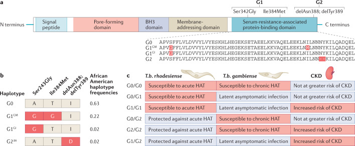
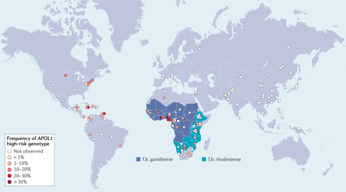
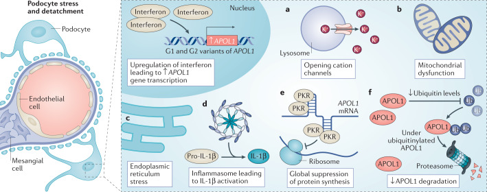
Genetic coding variants in APOL1, which encodes apolipoprotein L1 (APOL1), were identified in 2010 and are relatively common among individuals of sub-Saharan African ancestry. Approximately 13% of African Americans carry two APOL1 risk alleles. These variants, termed G1 and G2, are a frequent cause of kidney disease - termed APOL1 nephropathy - that typically manifests as focal segmental glomerulosclerosis and the clinical syndrome of hypertension and arterionephrosclerosis. Cell culture studies suggest that APOL1 variants cause cell dysfunction through several processes, including alterations in cation channel activity, inflammasome activation, increased endoplasmic reticulum stress, activation of protein kinase R, mitochondrial dysfunction and disruption of APOL1 ubiquitinylation. Risk of APOL1 nephropathy is mostly confined to individuals with two APOL1 risk variants. However, only a minority of individuals with two APOL1 risk alleles develop kidney disease, suggesting the need for a 'second hit'. The best recognized factor responsible for this 'second hit' is a chronic viral infection, particularly HIV-1, resulting in interferon-mediated activation of the APOL1 promoter, although most individuals with APOL1 nephropathy do not have an obvious cofactor. Current therapies for APOL1 nephropathies are not adequate to halt progression of chronic kidney disease, and new targeted molecular therapies are in clinical trials.


Conflict of interest statement

The authors declare no competing interests.

Figures

Fig. 1
Fig. 1. APOL1 domains and variants.
a | The APOL1 protein has four functional domains and a signal peptide, which is required for secretion of liver-produced APOL1 into plasma. Forms of APOL1 that lack the signal peptide owing to alternative splicing are retained as intracellular proteins. The G1 and G2 variant isoforms are the primary drivers of APOL1-mediated chronic kidney disease (CKD). The presence of two missense mutations (Ser342Gly and Ile384Met) in the nucleotide sequence encoding the serum resistance-associated protein-binding domain of APOL1 generates the G1 variant, whereas a 6 base-pair deletion that results in the loss of two amino acids (delAsn388 and delTry389) generates the G2 variant. b | The three kidney risk variants form only four observed haplotypes. Owing to the close physical proximity of the three disease-associated alleles, recombination events have not been observed and therefore the G1 and G2 alleles are mutually exclusive and do not occur together on the same chromosome. The G1 and G2 haplotypes are unique to individuals with sub-Saharan ancestry whereas the ancestral G0 haplotype is found in all global populations. Haplotype frequencies are shown for a healthy African American population. c | Susceptibility and resistance (indicated by red and blue shading, respectively) to acute human African trypanosomiasis (HAT) caused by Trypansoma brucei rhodesiense and chronic HAT caused by T.b. gambiense, and risk of APOL1-associated CKD, vary as a function of APOL1 haplotype. Heterozygous or homozygous carriers of the G0 allele are not at increased risk of kidney disease. Carriers of 1 or 2 copies of the G1 allele are susceptible to infection by T.b. gambiense but are less likely to develop symptoms of HAT; the mechanism of this protective association is unknown. The G2 variant protein efficiently lyses T.b. rhodesiense in vitro, thereby limiting infection in G2 carriers. People with G1/G1, G2/G2 and G1/G2 genotypes are at an increased risk of CKD. In certain uncommon settings (for example, in individuals with untreated HIV infection), G1/G0 individuals may also be at an increased risk of CKD.
Fig. 2
Fig. 2. Global distribution of APOL1 high-risk genotypes and endemic areas of Trypanosoma brucei gambiense and Trypansoma brucei rhodesiense.
The highest prevalence of APOL1 high-risk genotypes, defined as the presence of two risk alleles (i.e. G1/G1, G1/G2 or G2/G2), is in West Africa, but carriage of high-risk genotypes is found throughout sub-Saharan Africa and among those with African ancestry in the Americas and elsewhere. Allele frequency data are derived from Nadkarni et al. and Limou et al.) and unpublished data (C.A. Winkler, unpublished work).
Fig. 3
Fig. 3. Potential mechanisms of APOL1-associated kidney disease.
APOL1 is expressed in an interferon-dependent manner, and high-interferon states might result from the actions of a ‘second hit’, such as HIV infection. Several pathways have been implicated in the pathogenic response to APOL1 risk variants. For example, APOL1 variants may be associated with enhanced opening of cation channels, which compromises cell function (a), or may promote mitochondrial dysfunction through activation of the mitochondrial permeability transition pore (b). Alternatively, APOL1 variants may induce endoplasmic reticulum stress (c) and/or activate the NLRP3 inflammasome to generate active IL-1β (d). Other studies have demonstrated that APOL1 risk variants have stretches of double-stranded RNA, to which protein kinase R binds, inducing its autophosphorylation and suppressing of protein synthesis (e). Finally, APOL1 variants may reduce ubiquitin levels, prolonging the retention of intracellular proteins, including APOL1 itself (f). Although the relative importance of each of these injury pathways remains uncertain, the end result is damage to particular kidney cells, particularly the glomerular podocyte, resulting in dysfunction and in some cases, cell death and/or loss. IL-1β, interleukin-1β; PKR, protein kinase R; Ub, ubiquitin.

Similar articles

Cited by

References

    1. Genovese G, et al. Association of trypanolytic ApoL1 variants with kidney disease in African Americans. Science. 2010;329:841–845. doi: 10.1126/science.1193032. - DOI - PMC - PubMed
    1. Tzur S, et al. Missense mutations in the APOL1 gene are highly associated with end stage kidney disease risk previously attributed to the MYH9 gene. Hum. Genet. 2010;128:345–350. doi: 10.1007/s00439-010-0861-0. - DOI - PMC - PubMed
    1. The Churchill Society London. The Churchill Society http://www.churchill-society-london.org.uk/EndoBegn.html (1942).
    1. Nadkarni GN, et al. Worldwide frequencies of APOL1 renal risk variants. N. Engl. J. Med. 2018;379:2571–2572. doi: 10.1056/NEJMc1800748. - DOI - PMC - PubMed
    1. Micheletti SJ, et al. Genetic consequences of the transatlantic slave trade in the Americas. Am. J. Hum. Genet. 2020;107:265–277. doi: 10.1016/j.ajhg.2020.06.012. - DOI - PMC - PubMed

Publication types