. 2022 Mar 15;149(6):dev200472.
doi: 10.1242/dev.200472. Epub 2022 Mar 18.

The β8 integrin cytoplasmic domain activates extracellular matrix adhesion to promote brain neurovascular development

Affiliations

The β8 integrin cytoplasmic domain activates extracellular matrix adhesion to promote brain neurovascular development

Arpan De et al. Development. .

Abstract

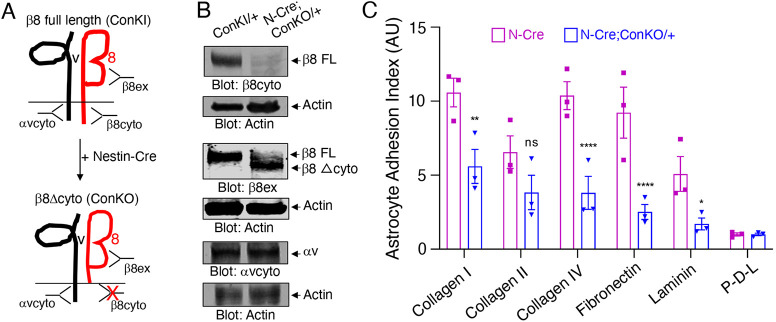
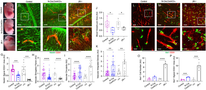
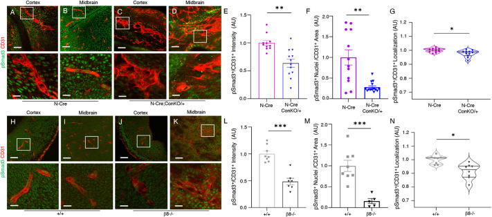
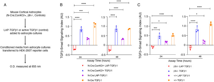
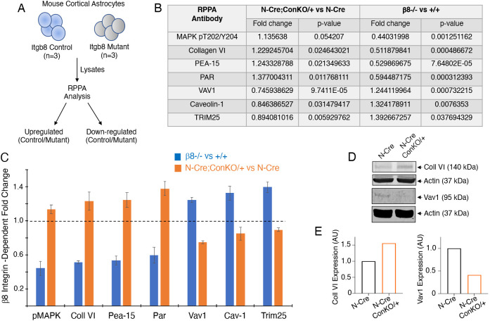
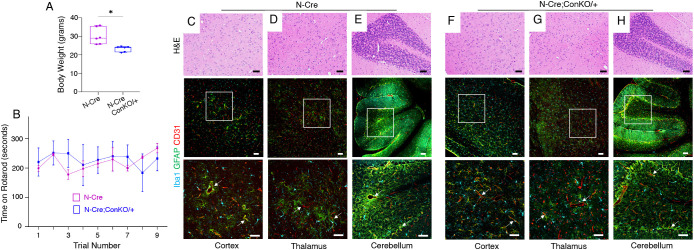
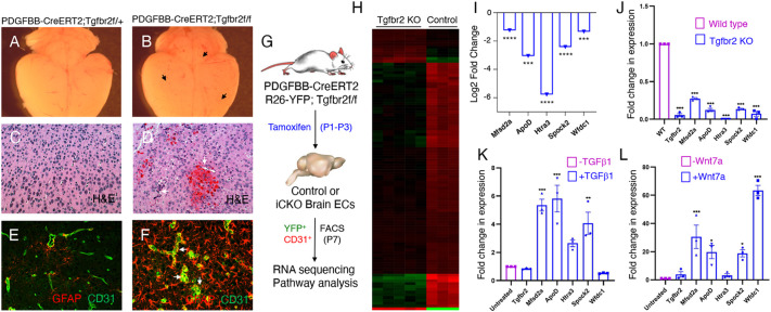
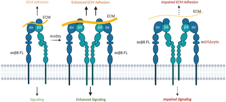
In the developing mammalian brain, neuroepithelial cells interact with blood vessels to regulate angiogenesis, blood-brain barrier maturation and other key neurovascular functions. Genetic studies in mice have shown that neurovascular development is controlled, in part, by Itgb8, which encodes the neuroepithelial cell-expressed integrin β8 subunit. However, these studies have involved complete loss-of-function Itgb8 mutations, and have not discerned the relative roles for the β8 integrin extracellular matrix (ECM) binding region versus the intracellular signaling tail. Here, Cre/lox strategies have been employed to selectively delete the cytoplasmic tail of murine Itgb8 without perturbing its transmembrane and extracellular domains. We report that the β8 integrin cytoplasmic domain is essential for inside-out modulation of adhesion, including activation of latent-TGFβs in the ECM. Quantitative sequencing of the brain endothelial cell transcriptome identifies TGFβ-regulated genes with putative links to blood vessel morphogenesis, including several genes linked to Wnt/β-catenin signaling. These results reveal that the β8 integrin cytoplasmic domain is essential for the regulation of TGFβ-dependent gene expression in endothelial cells and suggest that cross-talk between TGFβs and Wnt pathways is crucial for neurovascular development.

Keywords: Angiogenesis; Endothelial cell; Latent-TGFβ; Mouse; Neuroepithelial; Perivascular astrocyte; Wnt.


Conflict of interest statement

Competing interests The authors declare no competing or financial interests.

Figures

Fig. 1.
Fig. 1.
Development of a Cre/lox mouse model to selectively delete the Itgb8 cytoplasmic domain. (A) General strategy for targeting the murine Itgb8 gene to inhibit expression of the cytoplasmic domain. The 3′ region of the wild type Itgb8 gene is shown at top with the endogenous STOP codon in exon 14. A premature STOP codon was knocked into exon 13. A floxed ‘mini-gene’ comprising a cDNA containing exon 13 and the coding sequence of exon 14 as well as a 3′ poly-A (pA) sequence was inserted upstream of the knock-in exon to avoid potential dominant-negative effects of the engineered STOP (ConKI-Neo). The Neo cassette was subsequently removed using Flp recombinase to generate the conditional knock-in allele (ConKI). Cre-mediated deletion of the floxed mini-gene sequence results in premature termination of translation (via the engineered STOP in exon 13) and generation of the conditional knockout allele (ConKO), which encodes a truncated protein that lacks the cytoplasmic domain. (B) Southern blot of genomic DNA isolated from wild-type control of ConKI Neo/+ heterozygote mice, digested with BamHI and hybridized with a probe corresponding to the Neo cassette. (C) Genomic PCR analysis using DNA isolated from E14 brain and tail tissue samples confirms correct targeting and Cre-mediated recombination at the Itgb8 ConKI locus. Sample identities from brain and tail tissues in C are as follows: lane 1, Nestin-Cre−;+/+ (Cre-negative, wild type); lane 2, Nestin-Cre+;+/+ (Cre-positive, wild type); lane 3, Nestin-Cre+;ConKI/+ (Cre-positive, mutant). Forward and reverse DNA primer pairs used for PCR (05-02 and 04-02) are shown in the schematic (arrows in A). Note that Cre-mediated recombination occurs in the brain samples, but not in the tail samples. (D) Cortical astrocytes (Astro) cultured from mouse pups were infected with control adenovirus (Adeno) or Adeno-Cre and recombination at the engineered Itgb8 ConKI allele was analyzed with PCR. Alternatively, dissected brain tissue from E18 ConKI/+ control (N-Cre−) or Nestin-Cre;ConKO/+ mice (N-Cre+) was analyzed by genomic PCR. Primer pairs used for genomic PCR analyses are shown by red arrows in A. (E) Cre-mediated recombination excises the mini-gene (Ex13/14), as confirmed by RT-PCR using RNA isolated from ConKI/+ brain neurospheres infected with control Adeno or Adeno-Cre. (F) Schematic showing the amino acid sequences for the β8 integrin transmembrane domain (black) and cytoplasmic domain (green) and corresponding exon 13 and 14 coding sequences (cds) of the wild-type Itgb8 allele or the engineered Itgb8 ConKI allele containing the mini-gene insertion (β8 ConKI, upper panel). The black asterisk indicates the endogenous STOP codon. Cre-mediated deletion of the loxP-flanked mini-gene allows transcriptional read through to a STOP codon engineered in exon 13, leading to termination of the Itgb8 coding sequence and truncation of the β8 integrin cytoplasmic domain (ConKO, lower panel). The red asterisk indicates the inserted premature STOP codon shown in panel A.
Fig. 2.
Fig. 2.
The β8 integrin cytoplasmic domain promotes adhesion to the ECM. (A) Summary of antibodies directed against different regions (extracellular or cytoplasmic) of αv or β8 integrin proteins. Note that the β8cyto antibody cannot recognize the truncated β8 integrin protein (ConKO) owing to deletion of the cytoplasmic domain, whereas the β8ex antibody can recognize both the full-length (FL) and truncated β8 proteins. (B) Brain lysates from control (ConKI/+) or Nestin-Cre;ConKO/+ neonatal mice (P0) were analyzed by immunoblotting with antibodies directed against the β8 integrin extracellular domain (β8ex), the β8 integrin cytoplasmic domain (β8cyto) and the αv integrin cytoplasmic domain (αvcyto). (C) Quantification of β8 integrin cytoplasmic domain-dependent adhesion to various ECM proteins. Astrocyte progenitors from Nestin-Cre control or Nestin-Cre;ConKO/+ brains were added to wells coated with the indicated ECM proteins (x-axis) and cell adhesion was quantified after 16 h. Note that loss of the β8 integrin cytoplasmic tail leads to defects in ECM adhesion. Differences between groups were analyzed using two-way ANOVA and Tukey post-hoc analysis (n=3, mean±s.e.m., *P<0.05, **P<0.01, ****P<0.0001). ns, not significant.
Fig. 3.
Fig. 3.
Deletion of the β8 integrin cytoplasmic domain leads to defective cell adhesion and aberrant blood vessel morphogenesis during brain development. (A-C) Representative images of E14 Nestin-Cre control (A), Nestin-Cre;ConKO/+ (B) and β8−/− (C) embryos. Note that unlike β8−/− embryonic brains, which are hemorrhagic and hydrocephalic, there is a lack of grossly obvious hemorrhage in Nestin-Cre;ConKO/+ embryos. (D-F) Sagittal sections through the ganglionic eminence regions of E14 Nestin-Cre control (D), Nestin-Cre;ConKO/+ (E) or β8−/− embryos (F) were analyzed by double immunofluorescence using anti-CD31 antibody (red) to detect vascular endothelial cells combined with anti-nestin (green) antibody to detect neuroepithelial cells. Note that, in comparison to blood vessels in control mice, microscopic analysis of blood vessels in the Nestin-Cre;ConKO/+ and β8−/− brains reveal abnormal, distended morphologies as well as disrupted contacts with perivascular neuroepithelial cells. The lower panels are higher magnified images of boxed areas in upper panels. (G) Quantitation of neuroepithelial-endothelial cell juxtaposition in the ganglionic eminences of E14 Nestin-Cre control versus Nestin-Cre;ConKO/+ mutant embryos as well as wild-type control versus β8−/− mutant embryos. (H,I) Quantitation of blood vessel morphologies in the ganglionic eminences of E14 Nestin-Cre control versus Nestin-Cre;ConKO/+ mutant embryos and wild-type control versus β8−/− mutant embryos. Note the significant decrease in vascular elongation along the neuroepithelium (H) and a corresponding increase in circularity indicative of shape defects (I). The β8−/− mutant mice brains revealed similar alterations in cerebral blood vessel morphology. (J) Quantitation of blood vessels per area in the ganglionic eminences of control and mutant mice. Note the decrease in the number of CD31+ blood vessels per area in Nestin-Cre;ConKO/+ and β8−/− mutant embryos compared with the controls. (K) Quantitation of blood vessel shapes within the ganglionic eminences of control, Nestin-Cre;ConKO/+ and β8−/− mutant embryos. In comparison with controls, CD31+ blood vessels in mutant brains were more dilated. Aberrantly shaped blood vessels covered larger areas and thus showed enhanced anti-CD31 fluorescence intensity, as characterized by higher integrated density values. (L-N) Sagittal sections through midbrain regions of E14 Nestin-Cre control (L), Nestin-Cre;ConKO/+ (M) or β8−/− embryos (N) were analyzed by double immunofluorescence using anti-CD31 (red) antibody to detect vascular endothelial cells combined with anti-Iba1 (green) antibody to detect microglial cells. Note that in comparison to blood vessels in control mice, the blood vessels in the Nestin-Cre;ConKO/+ and β8−/− brains have abnormal morphologies and show increased juxtaposition with microglia. The lower panels are higher magnified images of boxed areas in upper panels. (O,P) Quantitation of perivascular microglial cells in the ganglionic eminences of E14 control and mutant embryos. In comparison to control mice, a higher number of Iba1+ cells was identified near blood vessels in Nestin-Cre;ConKO/+ mutant mice brains. The β8−/− mutant brain sections contained higher numbers of total microglia and perivascular microglia. Differences between groups were analyzed using unpaired two-tailed Student's t-test (n=3, mean±s.e.m., *P<0.05, **P<0.01, ***P<0.001, ****P<0.0001). Scale bars: 20 µm (D-F,L-N, upper panels); 5 µm (D-F,L-N, lower panels).
Fig. 4.
Fig. 4.
Deletion of β8 integrin cytoplasmic domain in brain neuroepithelial cells leads to reduced TGFβ-Smad signaling in vascular endothelial cells. (A-D) Analysis of Smad3 phosphorylation in brains from E14 Nestin-Cre control (A,B) and Nestin-Cre;ConKO/+ embryos (C,D). Shown are sagittal sections through the forebrains/cerebral cortices (A,C) and the ganglionic eminence regions of the midbrain (B,D). Note the abnormal CD31+ blood vessel morphologies in Nestin-Cre;ConKO/+ brains with reduced levels of pSmad3. The lower panels are higher magnification images of boxed areas in upper panels. (E-G) Quantification of pSmad3 levels in E14 Nestin-Cre control and Nestin-Cre;ConKO/+ midbrain regions as plotted in a bar graph measuring fluorescence intensity of pSmad3 relative to CD31 (E), numbers of pSmad3+ nuclei normalized to total CD31+ area (F) or by violin plots showing pSmad3+/CD31+ co-localization (G). Note the reduced levels of pSmad3 in Nestin-Cre;ConKO/+ brain samples. (H-K) Analysis of Smad3 phosphorylation in brains from E14 wild-type control (+/+; H,I) and β8−/− embryos (J,K). Shown are sagittal sections through the developing cerebral cortices (H,J) and the ganglionic eminence regions of the midbrain (I,K). Note the abnormal CD31+ blood vessel morphologies in β8−/− brains with defects in levels of pSmad3. The lower panels are higher magnification images of boxed areas in upper panels. (L-N) Quantification of pSmad3 levels in E14 wild-type control and β8−/− midbrain regions as plotted in a bar graph measuring fluorescence intensity of pSmad3 relative to CD31 (L), numbers of pSmad3+ nuclei normalized to total CD31+ area (M) or by violin plots showing pSmad3+/CD31+ co-localization (N). Note the reduced levels of pSmad3 in β8−/− brain samples. Differences between groups were determined using unpaired two-tailed Student's t-test (n=3, mean±s.e.m., *P<0.05, **P<0.01, ***P<0.001). Violin plots show distribution of statistically comparable mean values by analyzing differences between control and mutant brains (n=3 per group). Scale bars: 20 µm (A-D,H-K, upper panels); 5 µm (A-D,H-K, lower panels).
Fig. 5.
Fig. 5.
The β8 integrin cytoplasmic domain promotes latent-TGFβ activation in vitro. (A) Experimental strategy for quantifying β8 integrin-dependent latent-TGFβ activation and signaling in vitro using control and mutant astrocytes and HEK-293 cells expressing SBE-iSEAP reporter constructs. (B,C) Quantification of β8 integrin-mediated activation of latent-TGFβ1 using Nestin-Cre control and Nestin-Cre;ConKO/+ astrocytes (B) as well as wild-type and β8−/− astrocytes (C) in cell-based SBE-iSEAP reporter assays. Astrocyte conditioned media (+/− exogenous LAP-TGFβ1 or bioactive TGFβ1) was transferred to HEK-SBE Blue cells followed by quantification of reporter activities at 24 and 48 h. Note that both Nestin-Cre;ConKO/+ and β8−/− astrocytes show reduced latent-TGFβ activation and signaling at 24 and 48 h in comparison with control astrocytes. Differences between groups were analyzed using unpaired two-tailed Student's t-test (n=3, mean±s.e.m., *P<0.05, ***P<0.001, ****P<0.0001).
Fig. 6.
Fig. 6.
The β8 integrin cytoplasmic domain regulates signal transduction in cultured glial cells. (A) Strategy to identify β8 integrin-dependent changes in protein expression or phosphorylation in mouse astrocytes using reverse phase protein array (RPPA) platforms. (B) Table summarizing the seven most differentially expressed and/or phosphorylated proteins (with relevant P-values) in mutant versus control astrocytes identified by RPPA. (C) Bar graph summarizing select proteins that show statistically significant differences in expression and/or phosphorylation in control versus β8−/− astrocytes or Nestin-Cre control versus Nestin-Cre;ConKO/+ astrocytes. Shown are the top seven proteins displaying differential expression and/or phosphorylation in mutant cells. Note the inverse correlation between expression and/or phosphorylation of the seven proteins in astrocyte progenitors cultured from Nestin-Cre;ConKO/+ versus β8−/− pups. Data are mean±s.d. (D) Detergent-soluble lysates from Nestin-Cre control and Nestin-Cre;ConKO/+ mutant astrocytes were immunoblotted using anti-collagen VI and anti-Vav1 antibodies. Note the integrin-dependent changes in expression, which correlates with the expression differences identified in the RPPA screen (B,C). (E) Densitometry-based quantification of collagen VI (left) and Vav1 (right) protein levels based on immunoblot data in D.
Fig. 7.
Fig. 7.
Absence of brain vascular pathologies and neurodegenerative phenotypes in adult Nestin-Cre;ConKO/+ mice. (A) Body weights were recorded for 7 month old male Nestin-Cre control and Nestin-Cre;ConKO/+ mice. Unpaired two-tailed Student's t-test was used to compare differences between groups (n=3, mean±s.e.m., *P<0.05). Box and whisker plot shows distribution of statistically comparable minimum and maximum values (boxes) around mean (middle bars) as determined by quantifying differences between control and mutant groups (n=3 mice per group). (B) Rotarod analyses of 7 month old male Nestin-Cre control (n=3) and Nestin-Cre;ConKO/+ mutant mice (n=3). The time (y-axis) the mice remained on the rod rotating at an increasing speed versus the trial number (x-axis) is plotted. Note the lack of statistically significant differences in time spent on the rod for mutant versus control mice. (C-H) Sagittal brain sections from 7 month old Nestin-Cre control (C-E) or Nestin-Cre;ConKO/+ mutant mice (F-H) through the cerebral cortex (C,F), thalamus (D,G) and cerebellum (E,H) were stained with Hematoxylin and Eosin (upper panels) or fluorescently labeled with anti-Iba1 (cyan), anti-GFAP (green) and anti-CD31 (red) antibodies (lower panels) to reveal microglia, astrocytes and vascular endothelial cells, respectively. Note the absence of obvious neurovascular pathologies in Nestin-Cre;ConKO/+ mutant brain sections (F-H). White arrows in the fluorescent panels indicate blood vessels with closely juxtaposed astrocytes and/or microglial cells. The lower panels are higher magnification images of the boxed areas within the middle panels. Scale bars: 50 µm.
Fig. 8.
Fig. 8.
β8 integrin-activated TGFβ signaling regulates gene expression in brain endothelial cells. (A-F) Images of brains and fixed sections from tamoxifen-injected Pdgfbb-CreERT2 control (A) or Pdgfbb-CreERT2;Tgfbr2f/f conditional knockout (B) mice stained with Hematoxylin and Eosin (C,D) or anti-CD31 and anti-GFAP antibodies (E,F). Note that Pdgfbb-CreERT2;Tgfbr2f/f mutant mice develop focal brain hemorrhages and angiogenesis defects (arrows in B,D) as well as higher numbers of perivascular astrocytes (arrows in F). Mice received three consecutive intragastric tamoxifen injections between P1 and P3 and were analyzed at P7. (G) Strategy to identify TGFβ-regulated genes involved in cerebral angiogenesis and neurovascular development. Bulk transcriptome sequencing was performed on YFP+/CD31+ brain endothelial cells (ECs) isolated from P7 PDGFBB-CreERT2f/+;R26-YFP/+ control or PDGFBB-CreERT2;Tgfbr2f/f;R26-YFP/+ inducible knockout (iCKO) mice. (H) Heat map listing the top differentially expressed genes in control Pdgfbb-CreERT2;Tgfbr2f/+;R26-YFP/+ (n=3) and mutant Pdgfbb-CreERT2;Tgfbr2f/f;R26-YFP/+ (n=5) brain endothelial cells. (I) Decreased relative expression of five select genes (Mfsd2a, Apod, Htra3, Spock2, Wfdc1) as measured by quantitative mRNA sequencing from isolated control and Tgfbr2−/− brain endothelial cells. (J) Quantitative RT-PCR analysis validates differential expression in control and Tgfbr2−/− brain endothelial cell samples for five genes identified from transcriptome sequencing. (K) Brain endothelial cells (bEnd.3) were treated with TGFβ1 for 12 h and expression of Tgfbr2, Mfsd2a, ApoD, Htra3, Spock2 and Wfdc1 was quantified by RT-PCR. (L) Brain endothelial cells were treated with Wnt7a for 24 h and expression levels of Tgfbr2, Mfsd2a, ApoD, Htra3, Spock2 and Wfdc1 mRNAs were quantified by RT-PCR. Statistical differences of means were determined by two-way ANOVA (n=3, mean±s.e.m.). *P<0.05, **P<0.01, ***P<0.001, ****P<0.0001.
Fig. 9.
Fig. 9.
Model for the β8 integrin cytoplasmic tail in control of ECM adhesion and intracellular signaling. Activated full-length αvβ8 integrin promotes ECM engagement and clustering, leading to enhanced avidity to protein ligands and robust intracellular signaling. Genetic deletion of the β8 cytoplasmic domain impairs intracellular signaling and cytoskeletal linkages. αvβ8Δcyto is shown in a clustered complex with full-length αvβ8 integrin (ConKO/+). This figure was created with BioRender.com.

Similar articles

Cited by

References

    1. Allinson, K. R., Lee, H. S., Fruttiger, M., McCarty, J. H. and Arthur, H. M. (2012). Endothelial expression of TGFβ type II receptor is required to maintain vascular integrity during postnatal development of the central nervous system. PLoS ONE 7, e39336. 10.1371/journal.pone.0039336 - DOI - PMC - PubMed
    1. Anders, S., Pyl, P. T. and Huber, W. (2015). HTSeq--a Python framework to work with high-throughput sequencing data. Bioinformatics 31, 166-169. 10.1093/bioinformatics/btu638 - DOI - PMC - PubMed
    1. Arnold, T. D., Ferrero, G. M., Qiu, H., Phan, I. T., Akhurst, R. J., Huang, E. J. and Reichardt, L. F. (2012). Defective retinal vascular endothelial cell development as a consequence of impaired integrin αVβ8-mediated activation of transforming growth factor-β. J. Neurosci. 32, 1197-1206. 10.1523/JNEUROSCI.5648-11.2012 - DOI - PMC - PubMed
    1. Arnold, T. D., Niaudet, C., Pang, M.-F., Siegenthaler, J., Gaengel, K., Jung, B., Ferrero, G. M., Mukouyama, Y.-S., Fuxe, J., Akhurst, R.et al. (2014). Excessive vascular sprouting underlies cerebral hemorrhage in mice lacking αVβ8-TGFβ signaling in the brain. Development 141, 4489-4499. 10.1242/dev.107193 - DOI - PMC - PubMed
    1. Arnold, T. D., Lizama, C. O., Cautivo, K. M., Santander, N., Lin, L., Qiu, H., Huang, E. J., Liu, C., Mukouyama, Y.-S., Reichardt, L. F.et al. (2019). Impaired alphaVbeta8 and TGFbeta signaling lead to microglial dysmaturation and neuromotor dysfunction. J. Exp. Med. 216, 900-915. 10.1084/jem.20181290 - DOI - PMC - PubMed

Publication types