. 2022 Apr;9(12):e2102220.
doi: 10.1002/advs.202102220. Epub 2022 Feb 26.

Bioactive NIR-II Light-Responsive Shape Memory Composite Based on Cuprorivaite Nanosheets for Endometrial Regeneration

Affiliations

Bioactive NIR-II Light-Responsive Shape Memory Composite Based on Cuprorivaite Nanosheets for Endometrial Regeneration

Chenle Dong et al. Adv Sci (Weinh). 2022 Apr.

Abstract

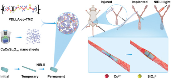
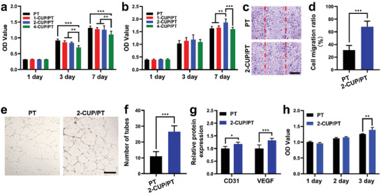
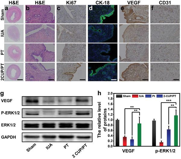
Intrauterine adhesions (IUAs) caused by mechanical damage or infection increase the risk of infertility in women. Although numerous physical barriers such as balloon or hydrogel are developed for the prevention of IUAs, the therapeutic efficacy is barely satisfactory due to limited endometrial healing, which may lead to recurrence. Herein, a second near-infrared (NIR-II) light-responsive shape memory composite based on the combination of cuprorivaite (CaCuSi4 O10 ) nanosheets (CUP NSs) as photothermal conversion agents and polymer poly(d,l-lactide-co-trimethylene carbonate) (PT) as shape memory building blocks is developed. The as-prepared CUP/PT composite possesses excellent shape memory performance under NIR-II light, and the improved operational feasibility as an antiadhesion barrier for the treatment of IUAs. Moreover, the released ions (Cu, Si) can stimulate the endometrial regeneration due to the angiogenic bioactivity. This study provides a new strategy to prevent IUA and restore the injured endometrium relied on shape memory composite with enhanced tissues reconstruction ability.

Keywords: NIR-II light-responsive; cuprorivaite nanosheets; endometrial regeneration; intrauterine adhesions; shape memory.


Conflict of interest statement

The authors declare no conflict of interest.

Figures

Figure 1
Figure 1
Schematic illustration of NIR‐II light‐responsive CUP/PT composites for IUA prevention and endometrial regeneration.
Figure 2
Figure 2
Characterization of 2D CUP NSs. a,b) SEM and TEM images of bulk CUP. c) AFM image and the corresponding height profile d) of exfoliated 2D CUP NSs. e) UV–vis/NIR absorption spectra of aqueous suspensions of CUP NSs with indicated concentrations. f) Concentration‐dependent photothermal property of CUP NSs aqueous dispersions under the mentioned irradiation conditions. g) NIR‐II laser power‐dependent photothermal property of CUP NSs (400 ppm).
Figure 3
Figure 3
Characterization of CUP/PT composites. a) Photographs of tubular CUP/PT composites. b) NIR‐II photothermal properties of PT, 1‐CUP/PT, 2‐CUP/PT, and 4‐CUP/PT under 1064 nm laser (0.5 W cm−2) excitation for 2 min. c) Photothermal stability of the 2‐CUP/PT under six repetitive NIR‐II laser on/off cycles at a power density of 0.5 W cm−2. d) Compressive Young's moduli of different CUP/PT composites (n = 3). e) Degradation profile of the as‐mentioned CUP/PT composites in PBS buffer (n = 3). Cumulative release of f) Cu and g) Si ions from indicated CUP/PT composites (n = 3). All data are presented as mean ± SD. Statistical analysis was performed using one‐way ANOVA analysis, **p < 0.01, ***p < 0.001.
Figure 4
Figure 4
Shape memory performance of CUP/PT composites. a) T g of PT and different CUP/PT composites from the DSC test (n = 3). b,c) 2D (b) and 3D (c) shape memory stress–strain–temperature curves of a representative 2‐CUP/PT composite from DMA testing. d) Shape recovery of a stretched tubular 2‐CUP/PT composite placed on a desk. e,f) Photoacoustic imaging of 2‐CUP/PT composite implanted underneath the skin (e), and into the isolated uterine lumen of a rat (f) under irradiation of NIR‐II light (0.5 W cm−2), respectively. Scale bar: 1.5 mm in (e) and 2.5 mm in (f). All data are presented as mean ± SD.
Figure 5
Figure 5
Bioactivities of different composites on HEECs and HUVECs. a,b) Proliferation of HEECs (a) and HUVECs (b) after culture on different CUP/PT composites for 1, 3, and 7 days (n = 4). c) Cell migration analysis using scratch method after cocultured with PT and 2‐CUP/PT composite for 16 h. d) The corresponding cell migration ratio (n = 3). e) The tube formation assessment of HUVECs after cocultured with PT and 2‐CUP/PT composite for 12 h. f) The corresponding number of formed tubes (n = 3). g) ELISA analysis of CD31 and VEGF from HUVECs after cocultured with PT and 2‐CUP/PT composite for 48 h (n = 5). h) Proliferation of HEECs after culture with medium collected from HUVECs study (n = 5). Scale bar: 500 µm. All data are presented as mean ± SD. Statistical analysis was performed using one‐way ANOVA analysis, **p < 0.01, ***p < 0.001.
Figure 6
Figure 6
In vivo endometrium regeneration. a) Representative H&E staining images of uterine cavity in different treatments at day 14. b) The corresponding high magnification H&E images for the analysis of glands in each group. c,d) The immunohistochemistry staining targeting Ki67 (c) and immunofluorescence staining targeting CK‐18 (d) in different treatments at day 14. e,f) Representative images of immunohistochemistry staining targeting CD31 (e) and VEGF (f) in new‐formed tissues in different treatments at day 14. g) Western blotting analysis of the protein expressions of VEGF and P‐ERK1/2 in each group. h) The corresponding optical density analysis for western blotting assay (n = 5). Scale bar: 1 mm in (a), 50 µm in (b,c,e,f), and 10 µm in (d). All data are presented as mean ± SD. Statistical analysis was performed using one‐way ANOVA analysis, **p < 0.01, ***p < 0.001.

Similar articles

Cited by

References

    1. Evans J., Salamonsen L. A., Winship A., Menkhorst E., Nie G. Y., Gargett C. E., Dimitriadis E., Nat. Rev. Endocrinol. 2016, 12, 654. - PubMed
    1. a) Deans R., Abbott J., J. Minim. Invasive Gynecol. 2010, 17, 555; - PubMed
    2. b) Zhang H., Zhang Q., Zhang J., Sheng F., Wu S., Yang F., Li W., Biomater. Sci. 2020, 8, 988. - PubMed
    1. Wei C., Pan Y. B., Zhang Y. L., Dai Y. D., Jiang L. L., Shi L. B., Yang W. J., Xu S. Q., Zhang Y. Y., Xu W. Z., Zhang Y. L., Lin X. N., Zhang S. Y., Cell Death Dis. 2020, 11, 755. - PMC - PubMed
    1. a) Sardo A. D., Calagna G., Scognamiglio M., O'Donovan P., Campo R., De Wilde R. L., Eur. J. Obstet. Gynecol. Reprod. Biol. 2016, 203, 182; - PubMed
    2. b) Leprince S., Huberlant S., Allegre L., Warembourg S., Leteuff I., Bethry A., Paniagua C., Taillades H., De Tayrac R., Coudane J., Letouzey V., Garric X., Commun. Biol. 2019, 2, 196. - PMC - PubMed
    1. Kou L., Jiang X., Xiao S., Zhao Y.‐Z., Yao Q., Chen R., J. Controlled Release 2020, 318, 25. - PubMed