. 2022 May:93:110294.
doi: 10.1016/j.cellsig.2022.110294. Epub 2022 Feb 24.

IBD-associated G protein-coupled receptor 65 variant compromises signalling and impairs key functions involved in inflammation

Affiliations

IBD-associated G protein-coupled receptor 65 variant compromises signalling and impairs key functions involved in inflammation

Virginie Mercier et al. Cell Signal. 2022 May.

Abstract

Background and aims: Inflammatory bowel diseases (IBD) result in chronic inflammation of the gastrointestinal tract. Genetic studies have shown that the GPR65 gene, as well as its missense coding variant, GPR65*Ile231Leu, is associated with IBD. We aimed to define the signalling and biological pathways downstream of GPR65 activation and evaluate the impact of GPR65*231Leu on these.

Methods: We used HEK 293 cells stably expressing GPR65 and deficient for either Gαs, Gαq/11 or Gα12/13, to define GPR65 signalling pathways, IBD patient biopsies and a panel of human tissues, primary immune cells and cell lines to determine biologic context, and genetic modulation of human THP-1-derived macrophages to examine the impact of GPR65 in bacterial phagocytosis and NLRP3 inflammasome activation.

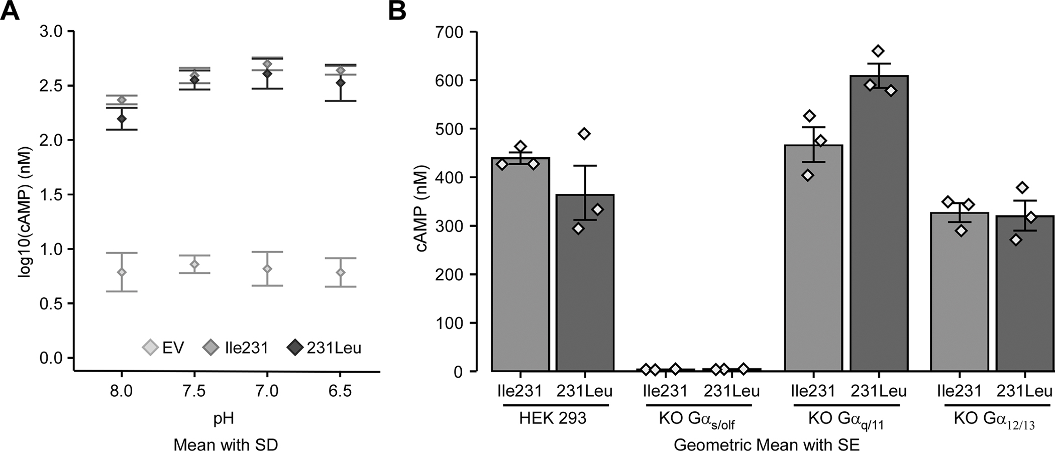
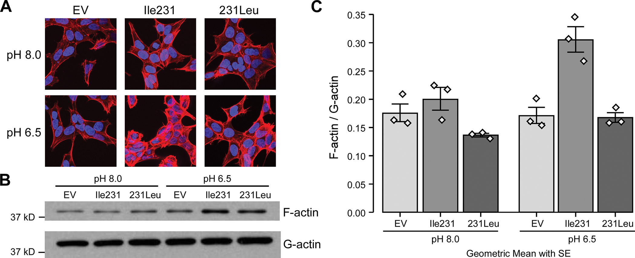
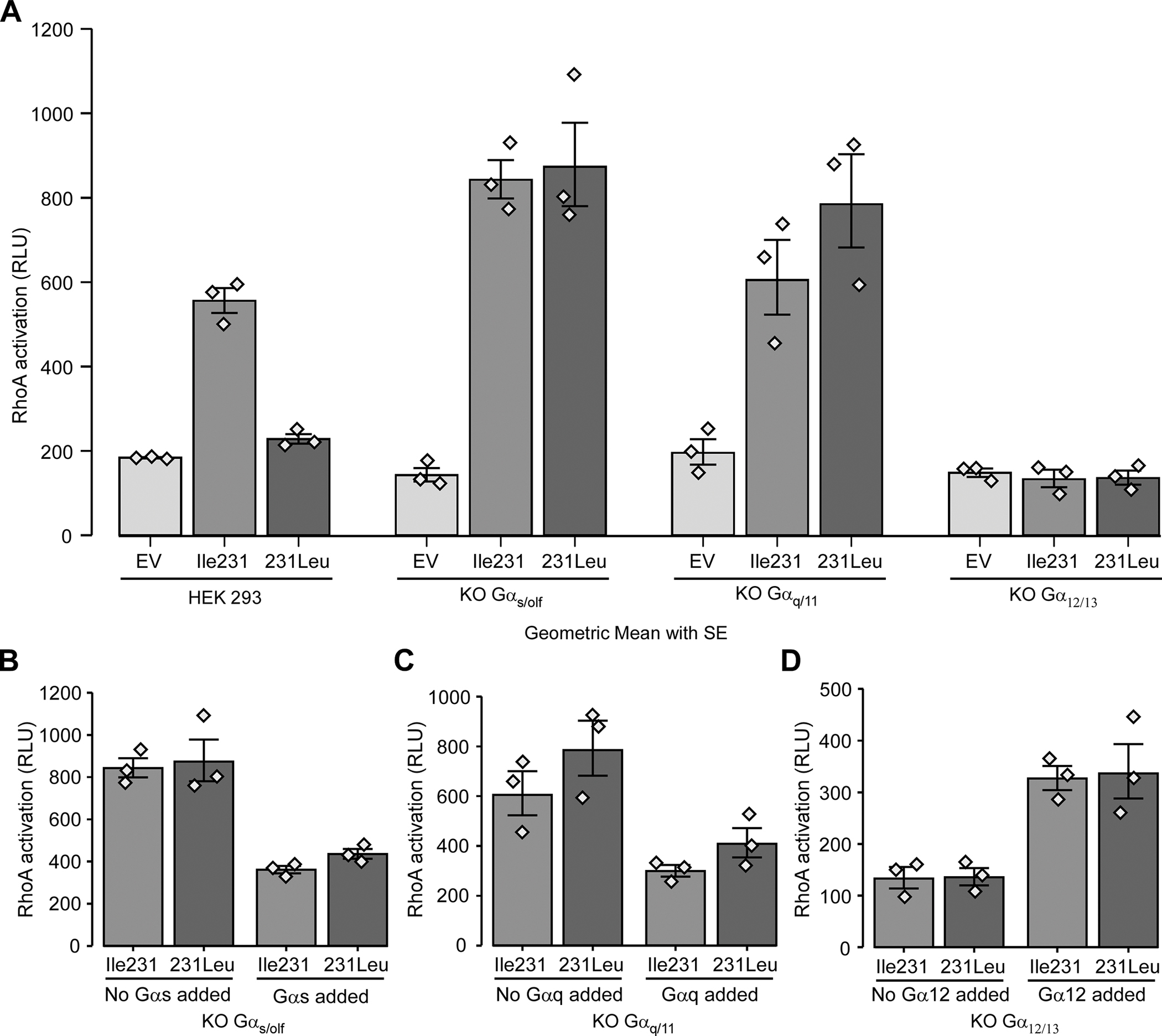
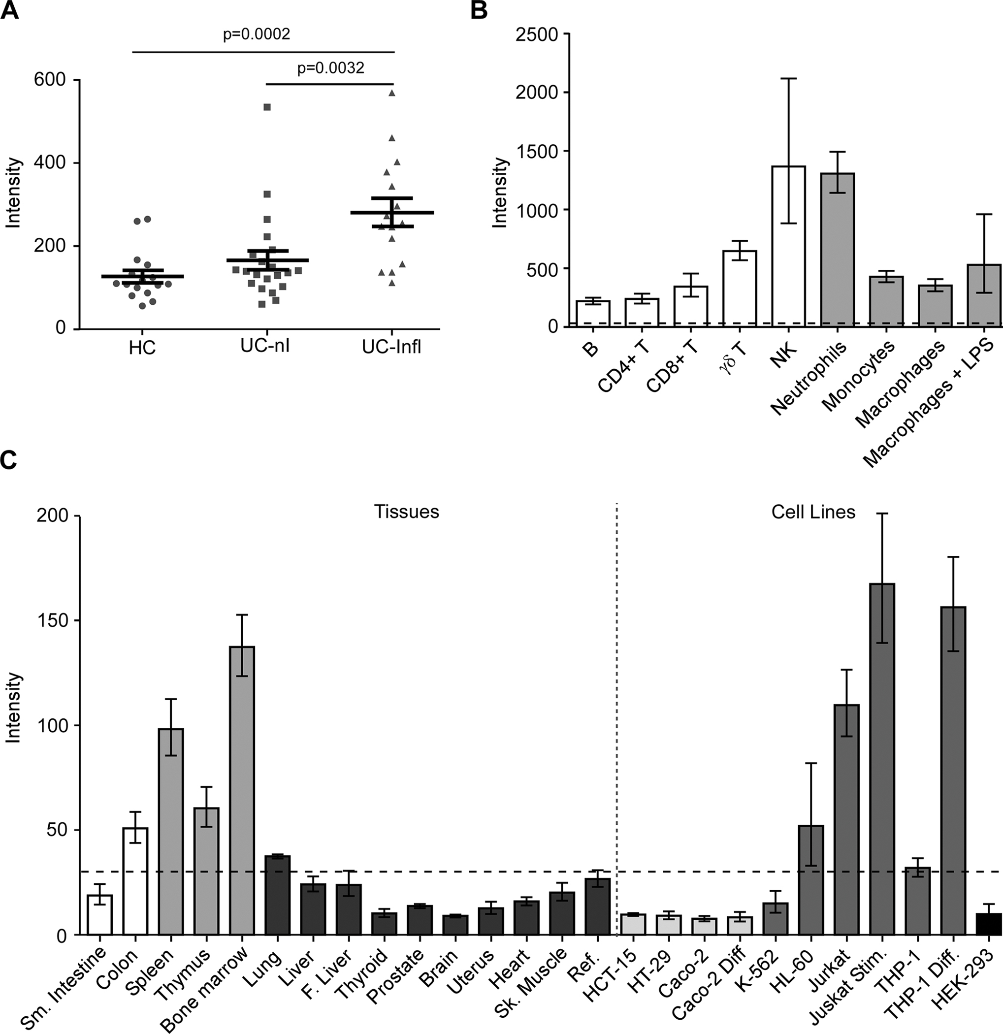
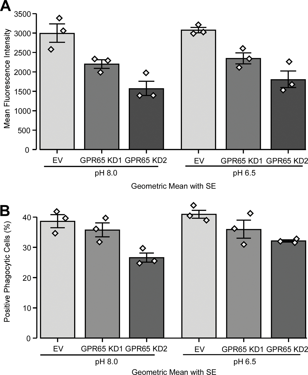
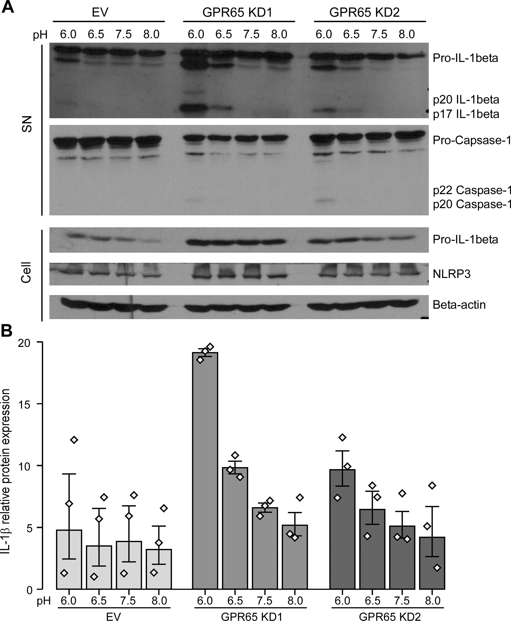
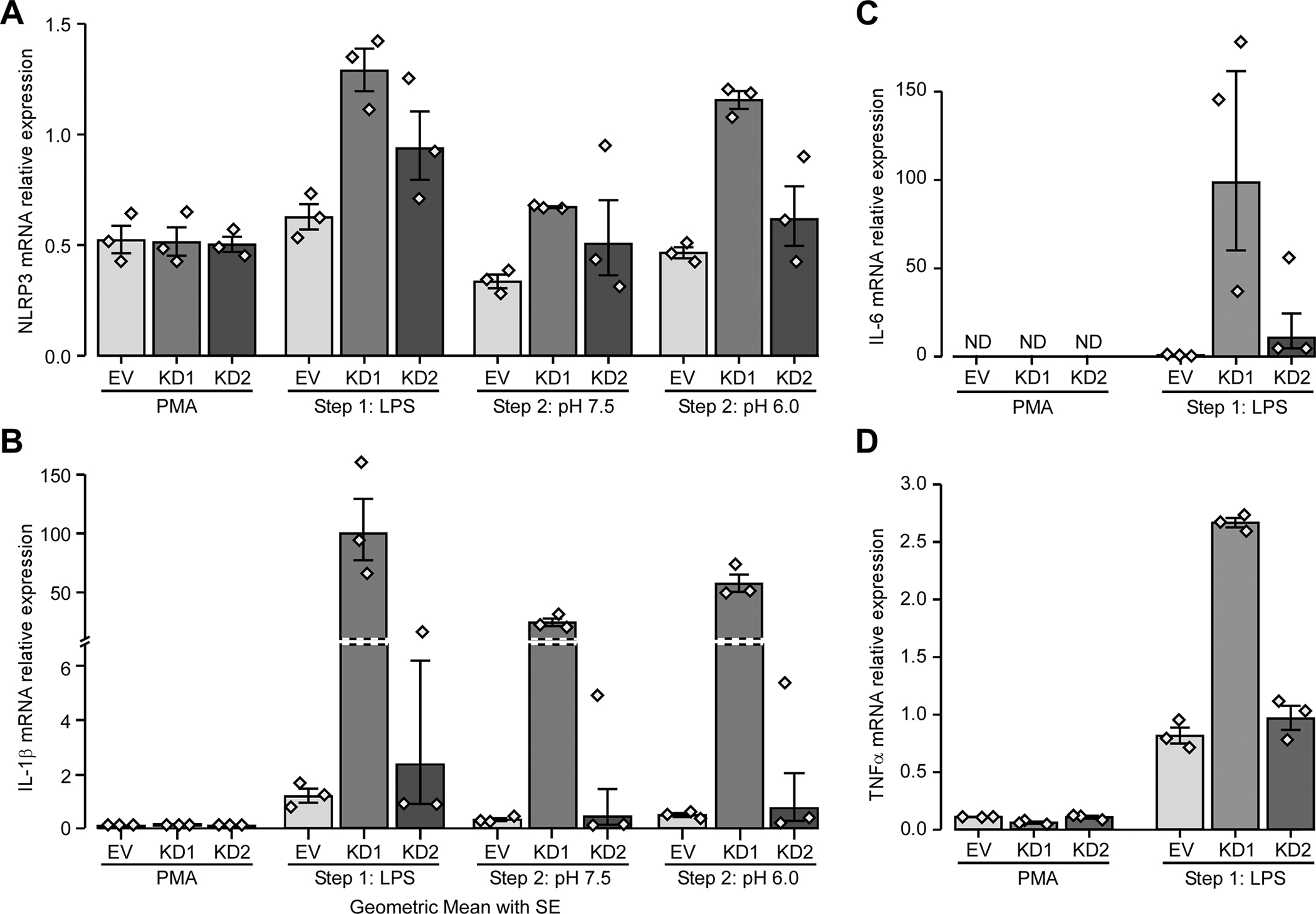
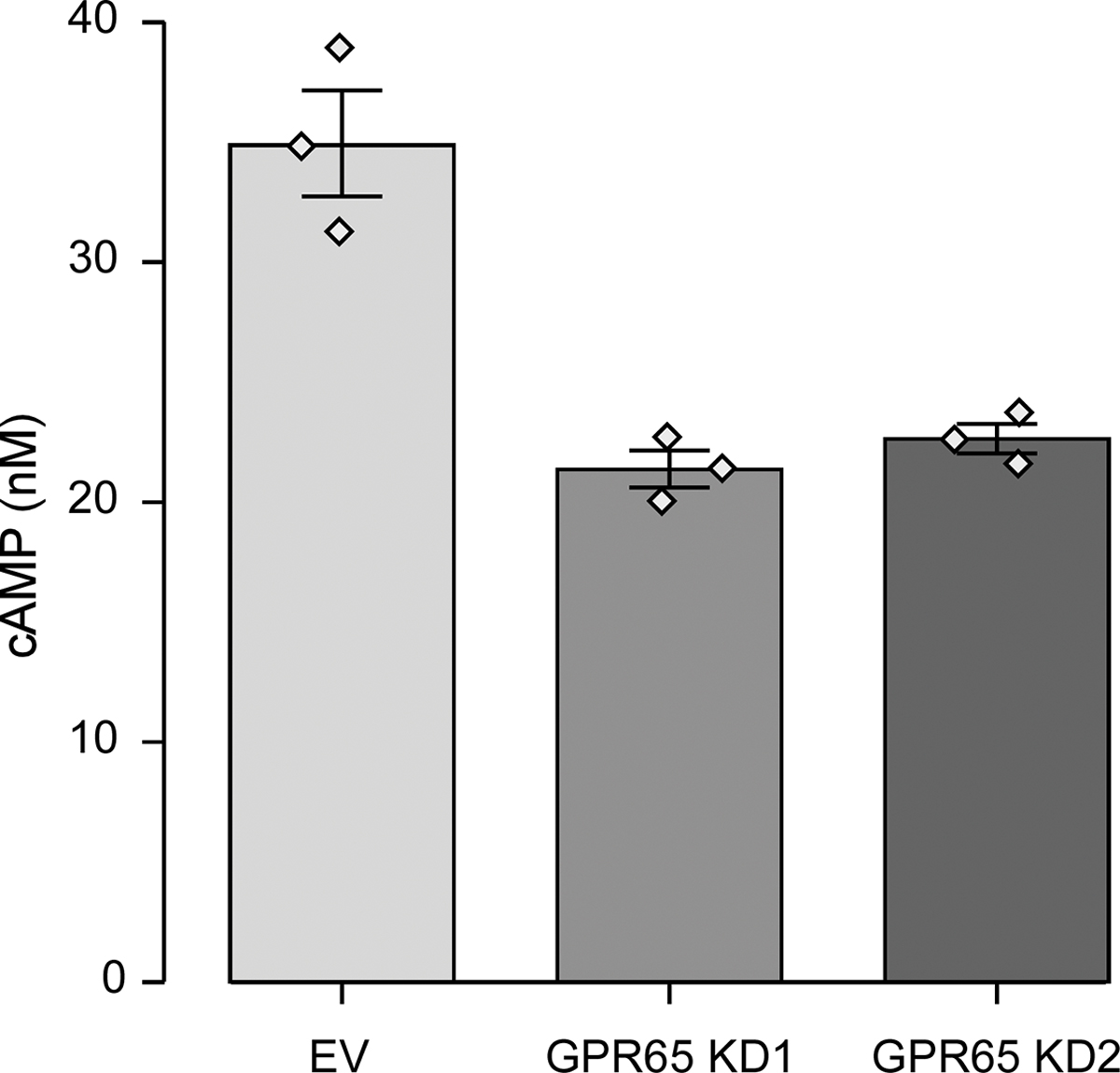
Results: We confirmed that GPR65 signals via the Gαs pathway, leading to cAMP accumulation. GPR65 can also signal via the Gα12/13 pathway leading to formation of stress fibers, actin remodeling and RhoA activation; all impaired by the IBD-associated GPR65*231Leu allele. Gene expression profiling revealed greater expression of GPR65 in biopsies from inflamed compared to non-inflamed tissues from IBD patients or control individuals, potentially explained by infiltration of inflammatory immune cells. Decreased GPR65 expression in THP-1-derived macrophages leads to impaired bacterial phagocytosis, increased NLRP3 inflammasome activation and IL-1β secretion in response to an inflammatory stimulus.

Conclusions: We demonstrate that GPR65 exerts its effects through Gαs- and Gα12/13-mediated pathways, that the IBD-associated GPR65*231Leu allele has compromised interactions with Gα12/13 and that KD of GPR65 leads to impaired bacterial phagocytosis and increased inflammatory signalling via the NLRP3 inflammasome. This work identifies a target for development of small molecule therapies.

Keywords: Actin remodeling; Bacterial phagocytosis; G proteins; GPR65; NLRP3 inflammasome.


Conflict of interest statement

CONFLICT OF INTEREST

The authors declare that they have no conflicts of interest related to the contents of this article.

Figures

Figure 1 -
Figure 1 -. IBD-associated allele GPR65*231Leu impairs cAMP response following pH-mediated GPR65 activation
Parental HEK 293 cells and three different Gα-KO HEK 293 cell lines, stably transfected with the pcDNA3.1 expression vector containing the different GPR65*Ile231Leu alleles (Ile231 or 231Leu) or with empty vector (EV), were assessed for cAMP accumulation. cAMP accumulation was measured after a 30 minute incubation in HEM-PSS in the presence of IBMX at pH 8.0, 7.5, 7.0 and 6.5 in parental HEK 293 cells (A) and the G protein-KO cell lines (B). Detailed effect sizes analyses from ANOVA are presented in Table S1.
Figure 2 -
Figure 2 -. IBD-associated allele GPR65*231Leu impairs stress fiber formation
(A) HEK 293 cells stably expressing the different GPR65*Ile231Leu alleles (Ile231 or 231Leu) or empty vector (EV) were evaluated for stress fiber formation after 30 minutes incubation in HEM-PSS at pH 8.0 or 6.5 using AlexaFluor555-phalloidin detection by immunofluorescence on confocal microscopy. Representative images of 3 independent experiments are shown. (B) HEK 293 cells stably expressing the different GPR65*Ile231Leu alleles or empty vector were evaluated for F-actin accumulation through the ratio of cellular filamentous actin (F-actin) and globular actin (G-actin) levels detected via western blot. (C) Quantification of band density was performed. Barplots represent geometric mean with SE. Detailed effect sizes analyses from ANOVA are presented in Table S2.
Figure 3 -
Figure 3 -. IBD-associated allele GPR65*231Leu impairs RhoA activation and is mediated via Gα12/13
(A) Parental HEK 293 and three different Gα-KO HEK 293 cell lines stably expressing the different GPR65*Ile231Leu alleles (Ile231 or 231Leu) or empty vector (EV) were assessed for RhoA activation after two minutes incubation in HEM-PSS at pH 6.5. (B-D) Three different Gα-KO HEK 293 cell lines stably expressing the different GPR65*Ile231Leu alleles or empty vector were transiently transfected with 200ng of Gα expressing plasmid in order to rescue the expression their respective knocked-out Gα subunit and then assessed for RhoA activation after two minutes incubation in HEM-PSS at pH 6.5. A dose-response experiment was performed before-hand (Suppl. Fig. S3) and 200ng of plasmid was chosen as it was the lowest dose of plasmid that led to an effect. Barplots represent geometric mean with SE. Detailed effect sizes analyses from ANOVA are presented in Table S3.
Fig. 4 -
Fig. 4 -. Expression profiling of GPR65 suggests that its role in IBD is primarily mediated by immune cells
(A) GPR65 expression in biopsies from healthy controls (HC, ●), UC patients in non-inflamed tissues (UC-nI, ■) or inflamed tissues (UC-Infl, ▲) measured using the Human Genome U133 Plus 2.0 Array (Affymetrix). The lines in panel A represent the mean and SEM of each group. P values (p) were calculated using a Student’s t test. For panel B and C, GPR65 expression was evaluated with a custom-made expression microarray (Agilent). (B) Gene expression in primary immune cells, cells were isolated from peripheral blood of healthy donors (n=6). Macrophages were generated in vitro following a seven-day treatment of monocytes with M-CSF (Millipore, Cedarlane, 50 ng/mL). Macrophage activation was achieved by a one-day treatment with 1 μg/mL LPS (macrophages + LPS) for a total incubation period of eight days. Barplots represent geometric mean with SE. (C) Expression levels of GPR65 in a panel of human tissues (bone marrow, heart, skeletal muscle, uterus, liver, fetal liver, spleen, thymus, thyroid, prostate, brain, lung, small intestine, and colon). Cell lines represent models of human T lymphocytes (Jurkat), monocytes (THP-1), erythroleukemia cells (K562), promyelocytic cells (HL-60), colonic epithelial cells (HCT-15, HT-29, Caco-2), and cells from embryonic kidney (HEK 293). For gene expression in immortalized cell lines, a differentiation model of colonic epithelium was obtained by culture of Caco-2 cells after 21 days of confluence. Jurkat cells were stimulated using 40 ng/mL PMA and 1 μg/mL ionomycin (Millipore Sigma) for 6 hours (Jurkat Stim) and THP-1 cells were differentiated using 10 ng/mL TNFα and 400 U/mL IFNγ (Biolegend, Cedarlane) for 24 hours (THP-1 Diff). Intensity values for each tissue/cell line represent the geometric mean with geometric standard deviation of 3 independent (technical) measurements; each measurement represents the geometric mean of all probes (one per exon) for each gene followed by a median normalization across all genes on the array. Dotted line indicates the threshold level for detection of basal expression. The reference sample is composed of a mixture of RNAs derived from 10 different human tissues.
Figure 5 -
Figure 5 -. GPR65 knockdown in macrophages alters phagocytosis
THP-1 cells were transduced with two different GPR65 shRNAs or empty vector (EV) control. PMA-differentiated THP-1 cells were placed in two different pHs (6.5 and 8.0) for four hours, with cytochalasin D or DMSO for the last hour. Cells were then incubated for one hour with pH sensitive pHRodo E. coli (A) GPR65 KD and pH effect on the mean fluorescence intensity (MFI) of positive cells. (B) GPR65 KD and pH effect on the percentage of positive phagocytic cells. Barplots represent geometric mean with SE. Detailed effect sizes analyses from ANOVA are presented in Table S4.
Figure 6 -
Figure 6 -. GPR65-knockdown macrophages secrete more IL-1β in response to acidic pH
THP-1 cells were transduced with two different GPR65 shRNAs or empty vector (EV) control. PMA-differentiated THP-1 cells were incubated with LPS and then in custom RPMI media at different pHs for 6 hours. (A) Supernatants (SN) and cell lysates (Cell) were assessed by western blot for active IL-1β and caspase-1 and pro-IL-1β, NLRP3 and β-actin respectively. Representative blots of three experiments are shown. (B) Band density for active p17 IL-1β was obtained with ImageJ. Barplots represent geometric mean with SE. Detailed effect sizes analyses from ANOVA are presented in Table S5.
Figure 7 -
Figure 7 -. GPR65 affects the inflammasome pathway, at least partly, at the transcriptional level
THP-1 cells were transduced with two different GPR65 shRNAs or empty vector (EV) control. RNA samples were taken after each of the three steps (PMA, LPS and incubation in different pH) and (A) NLRP3, (B) pro-IL1β, (C) IL-6 and (D) TNFα and expression were evaluated by RT-qPCR. Barplots represent geometric mean with SE. Detailed effect sizes analyses from ANOVA are presented in Table S6.
Figure 8 –
Figure 8 –. GPR65 is at least partly responsible for cAMP accumulation in THP-1-derived macrophages
THP-1 cells were transduced with two different GPR65 shRNAs or empty vector (EV) control. PMA-differentiated THP-1 cells were assessed for cAMP accumulation after an incubation at pH 6.0 with IBMX for 30 minutes. Barplots represent geometric mean with SE. Detailed effect sizes analyses from ANOVA are presented in Table S7.

References

    1. Jostins L, et al., Host-microbe interactions have shaped the genetic architecture of inflammatory bowel disease. Nature, 2012. 491(7422): p. 119–24. - PMC - PubMed
    1. Liu JZ, et al., Association analyses identify 38 susceptibility loci for inflammatory bowel disease and highlight shared genetic risk across populations. Nat Genet, 2015. 47(9): p. 979–86. - PMC - PubMed
    1. Rivas MA, et al., Deep resequencing of GWAS loci identifies independent rare variants associated with inflammatory bowel disease. Nat Genet, 2011. 43(11): p. 1066–73. - PMC - PubMed
    1. Mohanan V, et al., C1orf106 is a colitis risk gene that regulates stability of epithelial adherens junctions. Science, 2018. 359(6380): p. 1161–1166. - PMC - PubMed
    1. Rioux JD, et al., Genome-wide association study identifies new susceptibility loci for Crohn disease and implicates autophagy in disease pathogenesis. Nat Genet, 2007. 39(5): p. 596–604. - PMC - PubMed

Publication types

MeSH terms

Substances

LinkOut - more resources