Observational Study
. 2022 Mar:77:103893.
doi: 10.1016/j.ebiom.2022.103893. Epub 2022 Feb 23.

Oxidative stress-induced endothelial dysfunction and decreased vascular nitric oxide in COVID-19 patients

Affiliations
Observational Study

Oxidative stress-induced endothelial dysfunction and decreased vascular nitric oxide in COVID-19 patients

Virginie Montiel et al. EBioMedicine. 2022 Mar.

Abstract

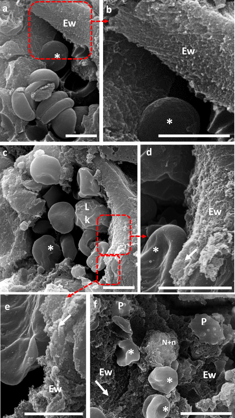
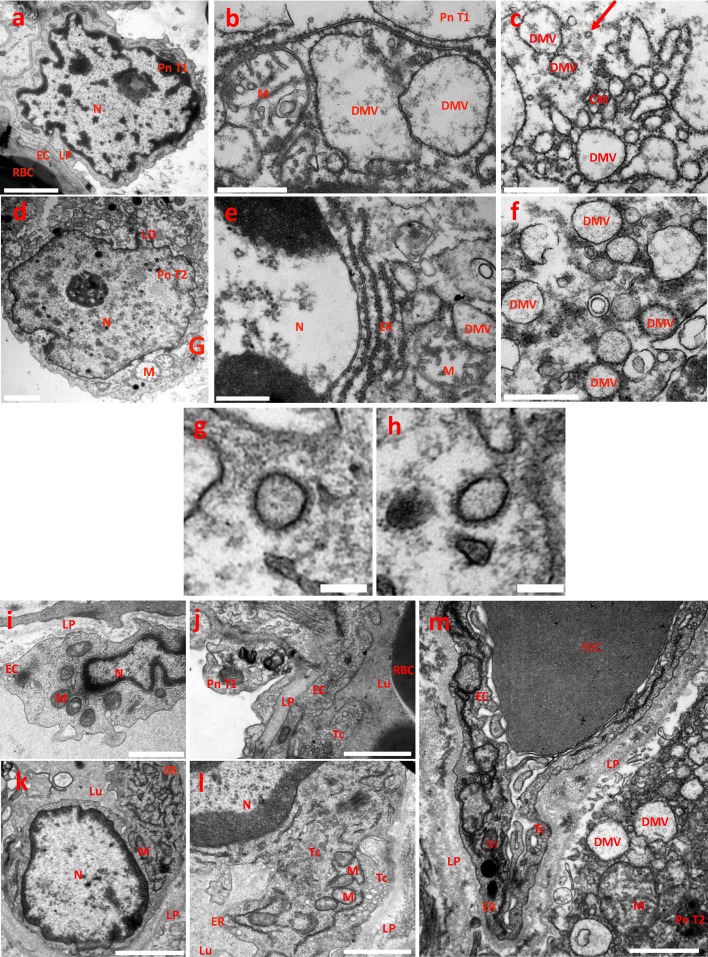
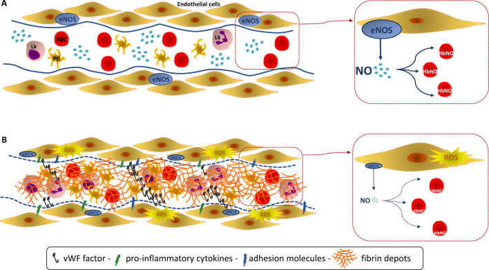
Background: SARS-CoV-2 targets endothelial cells through the angiotensin-converting enzyme 2 receptor. The resulting endothelial injury induces widespread thrombosis and microangiopathy. Nevertheless, early specific markers of endothelial dysfunction and vascular redox status in COVID-19 patients are currently missing.

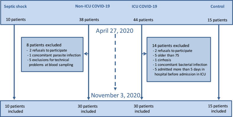
Methods: Observational study including ICU and non-ICU adult COVID-19 patients admitted in hospital for acute respiratory failure, compared with control subjects matched for cardiovascular risk factors similar to ICU COVID-19 patients, and ICU septic shock patients unrelated to COVID-19.

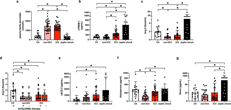
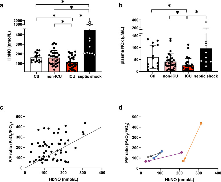
Findings: Early SARS-CoV-2 infection was associated with an imbalance between an exacerbated oxidative stress (plasma peroxides levels in ICU patients vs. controls: 1456.0 ± 400.2 vs 436 ± 272.1 mmol/L; P < 0.05) and a reduced nitric oxide bioavailability proportional to disease severity (5-α-nitrosyl-hemoglobin, HbNO in ICU patients vs. controls: 116.1 ± 62.1 vs. 163.3 ± 46.7 nmol/L; P < 0.05). HbNO levels correlated with oxygenation parameters (PaO2/FiO2 ratio) in COVID-19 patients (R2 = 0.13; P < 0.05). Plasma levels of angiotensin II, aldosterone, renin or serum level of TREM-1 ruled out any hyper-activation of the renin-angiotensin-aldosterone system or leucocyte respiratory burst in ICU COVID-19 patients, contrary to septic patients.

Interpretation: Endothelial oxidative stress with ensuing decreased NO bioavailability appears as a likely pathogenic factor of endothelial dysfunction in ICU COVID-19 patients. A correlation between NO bioavailability and oxygenation parameters is observed in hospitalized COVID-19 patients. These results highlight an urgent need for oriented research leading to a better understanding of the specific endothelial oxidative stress that occurs during SARS-CoV-2.

Funding: Stated in the acknowledgments section.

Keywords: Angiotensin II; Endothelial dysfunction; Microvascular thrombosis; Nitric oxide; Oxidative stress; SARS-CoV-2.


Conflict of interest statement

Declaration of interests Marc Derive is a co-founder and employee of Inotrem Company, a drug development company that is developing anti-TREM-1 approaches in septic shock and COVID-19. The other authors have disclosed that they do not have any potential conflicts of interest.

Figures

Fig 1
Figure 1
Flow chart of the ENDOCOVID observational study.
Figure 2.
Figure 2
Oxidative stress in ICU versus non-ICU COVID-19 and septic shock patients. Datapoints indicate individual measurements whereas horizontal bars show geometric mean (SD) for plasma lipid peroxides (2a), soluble TREM-1 (2b), plasma Ang II (2c), plasma Ang II ± ACEs and ARBs therapy (2d), soluble ACE-2 (2e), Aldosterone (2f) and Renin (2g). * P < 0.05 by one-way ANOVA followed by Tukey's correction for multiple comparisons, normal distribution except plasma Ang II and Aldosterone performed by Kruskal-Wallis corrected by Dunn's correction for multiple comparisons. Number of subjects for each analysis. 2a: Controls n = 15, non-ICU COVID-19 n = 30, ICU COVID-19 n = 30, Septic shock n = 7 (related to an interference with hyperbilirubinemia in 3 patients). 2b: Controls n = 15, non-ICU COVID-19 n = 30, ICU COVID-19 n = 29, Septic shock n = 10. 2c-d: Controls n = 15, non-ICU COVID-19 n = 29, ICU COVID-19 n = 28, Septic shock n = 9. 2e: Controls n = 14, non-ICU COVID-19 n = 27, ICU COVID-19 n = 28, Septic shock n = 10. 2f: Controls n = 14, non-ICU COVID-19 n = 29, ICU COVID-19 n = 29, Septic shock n = 9. 2g: Controls n = 15, non-ICU COVID-19 n = 28, ICU COVID-19 n = 29, Septic shock n = 10.
Figure 3.
Figure 3
NO bioavailability and plasma NOx in ICU versus non-ICU COVID-19 and septic shock patients. Datapoints indicate individual measurements whereas horizontal bars show geometric mean (SD) for HbNO (3a) and plasma NOx (3b). * P < 0.05 by one-way ANOVA followed by Tukey's correction for multiple comparisons, normal distribution. Linear regression analysis between PaO2/FiO2 ratio and HbNO (R2 = 0.13; P = 0.005) (3c). Decreased HbNO levels and PaO2/FiO2 ratio in 4 COVID-19 patients requiring secondarily ICU support (3d). Number of subjects for each analysis. 3a: Controls n = 15, non-ICU COVID-19 n = 30, ICU COVID-19 n = 29, Septic shock n = 10. 3b: Controls n = 14, non-ICU COVID-19 n = 30, ICU COVID-19 n = 29, Septic shock n = 10. 3c: non-ICU COVID-19 n = 30, ICU COVID-19 n = 30. 3d: ICU COVID-19 initially admitted to the general ward requiring secondarily ICU support n = 4.
Figure 4.
Figure 4
SARS-CoV-2-induced endothelial alterations in lung blood vessels of COVID-19 patients. 4. A, B: Scanning Electron Microscopy (SEM) of a cross section of healthy lung inner vessel with red blood cells (*). The luminal layer is a thin layer, composed of a single continuous layer of endothelial cells, the endothelial wall (Ew) (4A). Zoom of the boxed region showing the smooth surface of this wall (4B). 4. C–F: Cross-section of lung inner vessels of ICU COVID-19 patients. Blood vessel showing the presence of several leukocytes (Lk) and red blood cells (*) with irregular aspect of the endothelial wall (4C). Zooms of the boxed region highlighting the irregular aspect of the endothelial wall (Ew) due to a fibrillar network (arrow) of fibrin depots (4D-E). Large view of a vessel showing activated platelets (P), red blood cells (*), neutrophils and associated NETs and the dramatic fibrillar aspect of the endothelial wall contributed mainly by fibrin depots (4F). Digital zoom; Bars, A, B, C, D: 5 µm; E: 2 µm: F: 10 µm. *: red blood cells; Lk: leukocytes; Ew: Endothelial wall; P: platelets; arrow heads: fibrin; N+n: Neutrophils and NET.
Figure 5.
Figure 5
SARS-CoV-2 replication compartments in lung samples of ICU COVID-19 patients. 5. A–F. Pneumocytes. Transmission electron Microscopy (TEM) image of a control sample showing the normal morphology of a pneumocyte type 1 (5A). TEM images of damaged pneumocytes type 1 and type 2 (Pn T1, PnT2, respectively), from patients infected by SARS-CoV-2 coronavirus and showing virus replication compartments (5B to 5F). Pneumocytes contained abnormal swollen mitochondria with loss of cristae (M) (5B-D-E) and lipid droplets (LD) accumulation (5D). Pneumocytes contained typical membranous structures associated to coronavirus replication, such as double-membranes spherical vesicles (DMV), containing fibrous material (dsRNA) in their lumen and attached ribosomes at their external surface; due to their rough-endoplasmic reticulum (ER) origin and intensive genome virus replication (5B-C-E-F). DMVs were bridged by convoluted ER-derived membranes (CM) characteristic of the above-mentioned SARS-CoV-2 replication compartments (5B-C-E-F). Arrow, SARS-CoV-2 virion. Bars, A, D: 2 µm; B, C, E, F: 500 nm. 5. G, H: Mature SARS-CoV-2 virions. Typical morphology of coronaviruses with its crown of spike trimers. Diameters including the crowns: G, 191.4 nm; H, 189.3 nm. Bar, 150 nm. 5. I–M: Endothelial cells. TEM image of a control sample showing the normal morphology of a lung endothelial cells (5I, J). TEM images of lung endothelial cells from patients infected by SARS-CoV-2 coronavirus (5K–M). Despite the cell death of subjacent pneumocytes type I and type 2, endothelial cells (EC) retain swollen but active mitochondria with decrease in the electron density of their matrix (M) and transcytosis (Tc) activity, as evidenced by the massive caveolae and vesicle intracellular trafficking. Endothelial cells expressed abundant ribosomes expression associated with a large surface area of rough endoplasmic reticulum and Golgi apparatus (5K-L-M). Virus replication compartments in a type 2 pneumocyte subjacent to an EC (5M); note the detachment of the Pn T2 from the LP and the abnormal mitochondria. Bars, I, J, L, M: 1 µm; K: 500 nm. LD, lipid droplets; DMV, Double membrane vesicles surrounded by ribosomes; CM, convoluted membranes; M, mitochondrion; N, nucleus; RBC, red blood cell; ER, rough endoplasmic reticulum; LP, lamina propria; Tc, transcytosis; Lu, blood vessel lumen. Pn T1, pneumocytes type 1; Pn T2, pneumocytes type 2; EC, endothelial cells.
Figure 6.
Figure 6
Endothelial function reflected by the level of the 5-α-nitrosyl-hemoglobin (HbNO). Endothelial nitric oxide (NO) is the pivotal endothelium-derived substance that reduces platelet aggression and adhesion, inhibits adhesion of leukocytes and expression of pro-inflammatory cytokine genes. 5-α-nitrosyl-hemoglobin (or HbNO) is measured in venous erythrocytes but is mainly influenced by the vascular NO sources (despite expression of an erythrocyte NOS) and is used as a surrogate for the NO-dependent endothelial function. (A, Upper) A healthy endothelium is represented with high venous erythrocyte levels of HbNO. (B, lower) Classically, NO-dependent endothelial dysfunction is defined as an imbalance between the Nitric Oxide (NO) bioavailability and reactive oxidant species (ROS). A decrease of the endothelium-derived NO is observed in response to an endothelial “aggression” related to the SARS-CoV-2 invasion with a pronounced oxidative stress leading to a decreased NO bioavailability. This severe NO-dependent endothelial dysfunction is reflected by decreased venous erythrocyte levels of HbNO. Such alteration could participate to the microvascular dysfunction observed in COVID-19 patients characterized by a hypercoagulable and a pro-inflammatory state with expression of pro-inflammatory cytokines and adhesion molecules, but without DIC as such. RBC: red blood cells - erythrocyte, Lk: leukocyte, Ptl: platelet.

Comment in

References

    1. Xiong X., Chi J., Gao Q. Prevalence and risk factors of thrombotic events on patients with COVID-19: a systematic review and meta-analysis. Thromb J. 2021;19(1):32. - PMC - PubMed
    1. Malas M.B., Naazie I.N., Elsayed N., Mathlouthi A., Marmor R., Clary B. Thromboembolism risk of COVID-19 is high and associated with a higher risk of mortality: a systematic review and meta-analysis. EClinicalMedicine. 2020;29 - PMC - PubMed
    1. Lemos A.C.B., do Espirito Santo D.A., Salvetti M.C., et al. Therapeutic versus prophylactic anticoagulation for severe COVID-19: a randomized phase II clinical trial (HESACOVID) Thromb Res. 2020;196:359–366. - PMC - PubMed
    1. Libby P., Luscher T. COVID-19 is, in the end, an endothelial disease. Eur Heart J. 2020;41(32):3038–3044. - PMC - PubMed
    1. Ackermann M., Verleden S.E., Kuehnel M., et al. Pulmonary vascular endothelialitis, thrombosis, and angiogenesis in Covid-19. N Engl J Med. 2020;383(2):120–128. - PMC - PubMed

Publication types