

Photodermatitis and ocular changes in nine horses after ingestion of wild parsnip (pastinaca sativa)
- PMID: 35219345
- PMCID: PMC8881838
- DOI: 10.1186/s12917-022-03162-2
Photodermatitis and ocular changes in nine horses after ingestion of wild parsnip (pastinaca sativa)
Abstract
Background: Primary photosensitization rarely occurs in horses and can easily be misinterpreted. Descriptions of the disease in horses after ingestion of parsnip are lacking. The aim of this case series was to describe the dermatological and ocular changes due to photosensitization and to raise awareness of parsnip being a possible aetiologic agent.
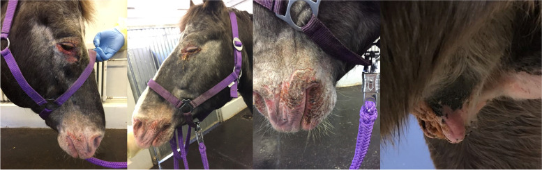
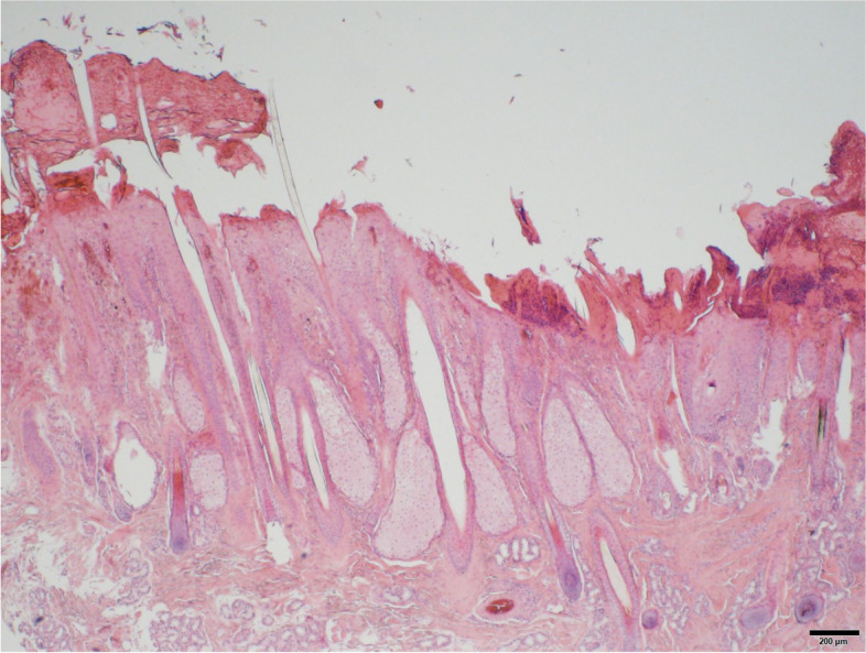
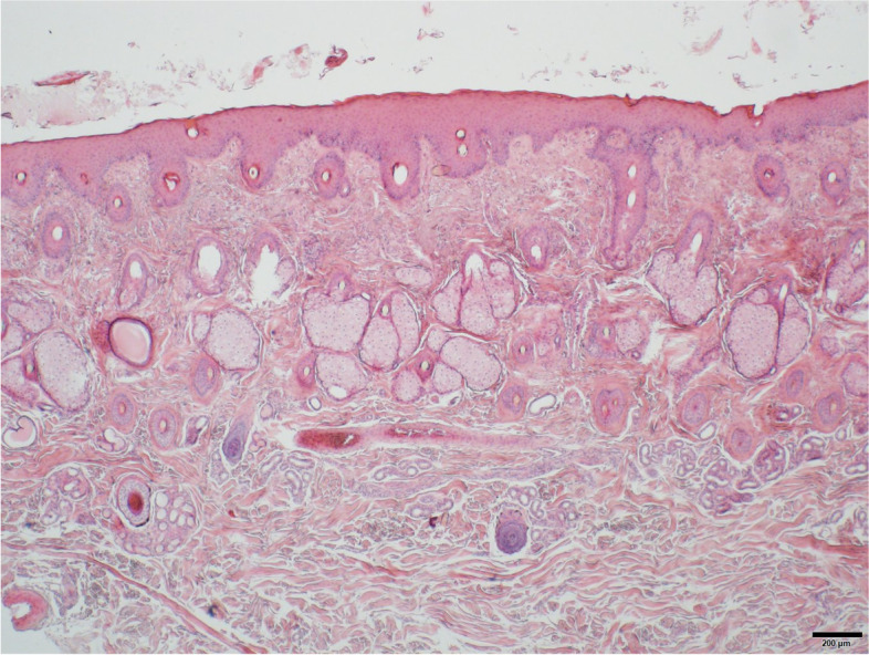
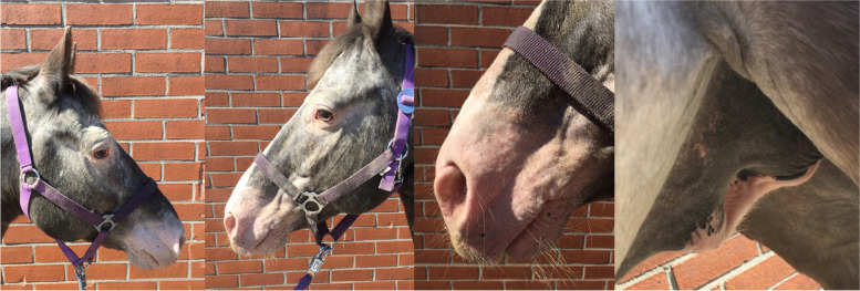
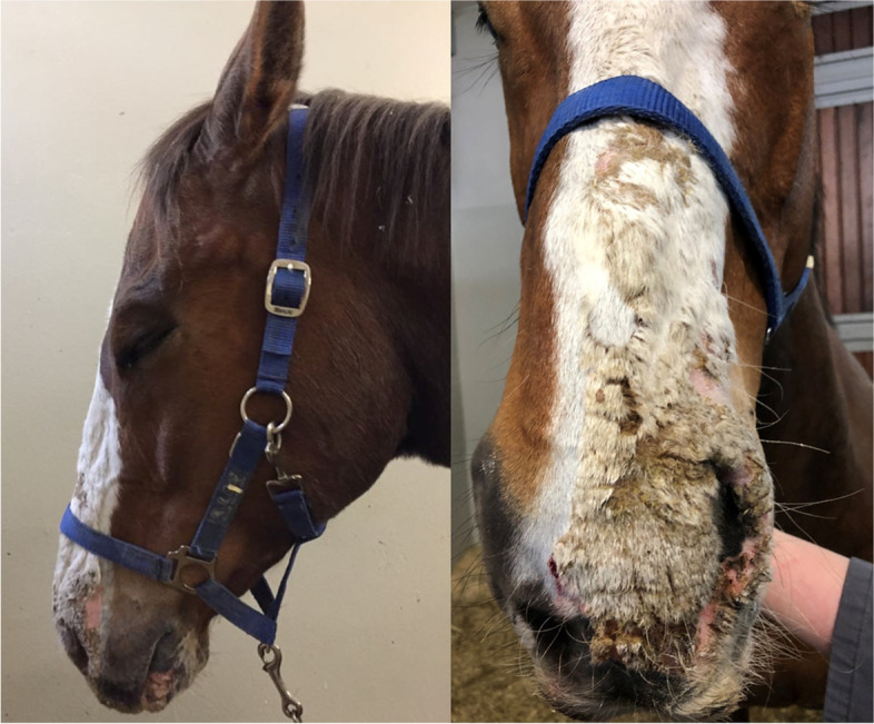
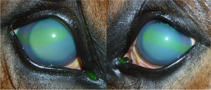
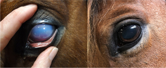
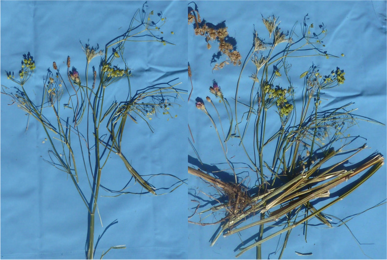
Case presentation: Nine horses from three different stables in Berlin and Brandenburg, Germany, presented variable degrees of erythema, scaling, crusting and necrosis of unpigmented skin at the head and prepuce. Horses were of different breeds with a median age of 15 ± 5.9 years. A mild leukocytosis was diagnosed in 1/9 horses at admission. Analyzed liver enzymes were within the reference ranges in all horses. Ocular changes were diagnosed as follows: blepharitis (3/9), conjunctivitis (7/9), corneal edema without additional signs of keratitis and/or uveitis (2/9), corneal edema with signs of uveitis (1/9) and photophobia (4/9). One horse developed a fluorescein positive corneal erosion. Skin biopsy (1/9) revealed a moderate to severe acute, eosinophilic and lymphocytic dermatitis with dermal edema and vasculitis. All stables housing these patients fed hay from the same distributer. Analyzed hay samples showed high contents of wild parsnip (plants, seeds, roots). Wild parsnip is widespread in Europe and contains furocoumarins, a family of photodynamic pigments, which may cause primary photodermatitis, keratoconjunctivitis and uveitis. Horses were treated according to severity of clinical symptoms systemically with flunixine meglumine (1.1 mg/kg BW 1-2x/day) or prednisolone (1 mg/kg BW 1x/day). Topically, either gentamicin (3x/day), dexamethasone (2-3x/day) and/or atropine (1x/day) were used. Skin care was provided with almond oil or dexpanthenol (2x/day). All horses were kept in a dark environment or were treated with sunscreen and facemasks. Duration of treatment varied from 6-30 days (median 11.3 days).
Conclusion: Ingestion of wild parsnip (Pastinaca sativa) can induce primary photosensitization with dermatitis and ocular injury in horses. In times of extreme weather, hay may alter in botanical composition, resulting in high amounts of uncharacteristic plants causing novel problems.
Keywords: Bergapten; Cornea; Edema; Erythema; Furocoumarins; Intoxication; Ocular changes; Parsnip; Photosensitization; Sunburn.
© 2022. The Author(s).
Conflict of interest statement
The authors declare no conflict of interest.
Figures
Similar articles
-
Wild parsnip (Pastinaca sativa)-induced photosensitization.Toxicon. 2019 Sep;167:60-66. doi: 10.1016/j.toxicon.2019.06.007. Epub 2019 Jun 4. Toxicon. 2019. PMID: 31173794
-
Alfalfa hay induced primary photosensitization in horses.Vet J. 2016 May;211:32-8. doi: 10.1016/j.tvjl.2016.03.004. Epub 2016 Mar 17. Vet J. 2016. PMID: 27040919
-
Evaluation of furanocoumarins from seeds of the wild parsnip (Pastinaca sativa L. s.l.).J Chromatogr B Analyt Technol Biomed Life Sci. 2019 Jan 15;1105:54-66. doi: 10.1016/j.jchromb.2018.12.012. Epub 2018 Dec 12. J Chromatogr B Analyt Technol Biomed Life Sci. 2019. PMID: 30562630
-
Review of Pharmacological Properties and Chemical Constituents of Pastinaca sativa.J Pharmacopuncture. 2021 Mar 31;24(1):14-23. doi: 10.3831/KPI.2021.24.1.14. J Pharmacopuncture. 2021. PMID: 33833896 Free PMC article. Review.
-
The Parsnip (Pastinaca sativa L), A Proposed Remedy as to a Fertile Agent in the Viewpoint of Iranian Traditional Medicine.Curr Drug Discov Technol. 2020;17(5):711-715. doi: 10.2174/1570163816666190820143052. Curr Drug Discov Technol. 2020. PMID: 31429690 Review.
Cited by
-
Medicinal Plants of the Flora of Kazakhstan Used in the Treatment of Skin Diseases.Molecules. 2023 May 19;28(10):4192. doi: 10.3390/molecules28104192. Molecules. 2023. PMID: 37241933 Free PMC article. Review.
References
-
- Barrington GM. Integumentary System In: Kahn CM, editor. The Merck Veterinary Manual. 10. London, United Kingdom Wiley; 2010. p. 665 - 800
-
- Roth L, Daunderer M, Kormann K. Giftpflanzen-Pflanzengifte. 4. Hamburg: Nikol Verlagsgesellschaft mbH & Co KG; 1994.
Publication types
MeSH terms
Substances
LinkOut - more resources
Full Text Sources
