. 2022 Apr;102(4):115636.
doi: 10.1016/j.diagmicrobio.2022.115636. Epub 2022 Jan 16.

Y380Q novel mutation in receptor-binding domain of SARS-CoV-2 spike protein together with C379W interfere in the neutralizing antibodies interaction

Affiliations

Y380Q novel mutation in receptor-binding domain of SARS-CoV-2 spike protein together with C379W interfere in the neutralizing antibodies interaction

Ivaine Tais Sauthier Sartor et al. Diagn Microbiol Infect Dis. 2022 Apr.

Abstract

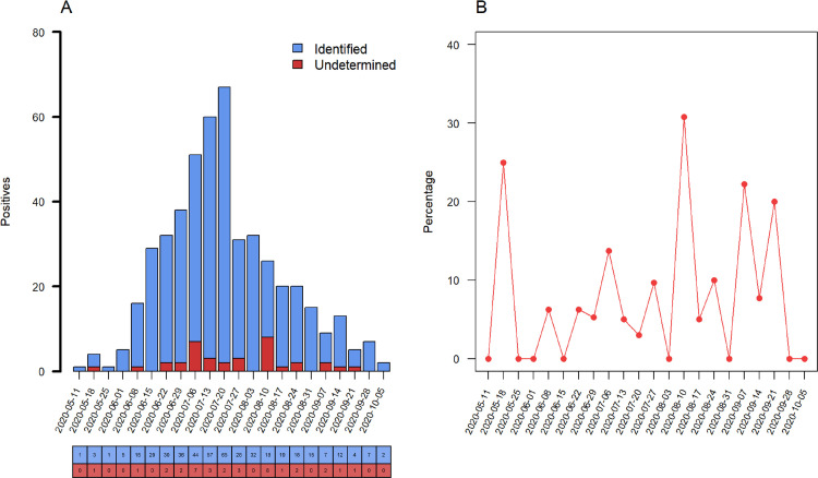
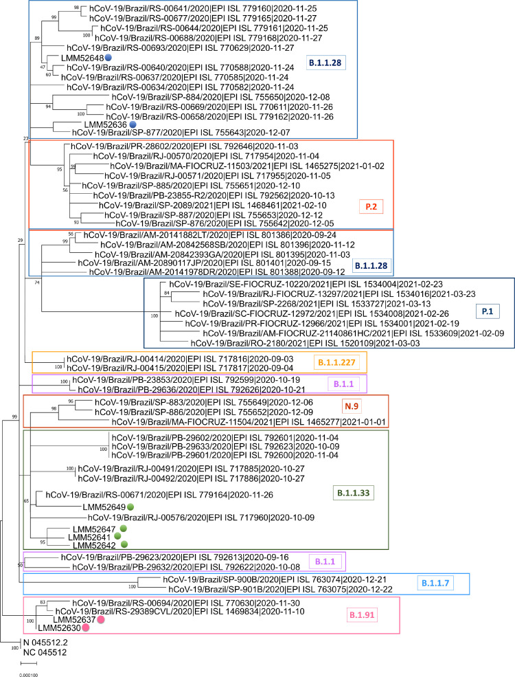
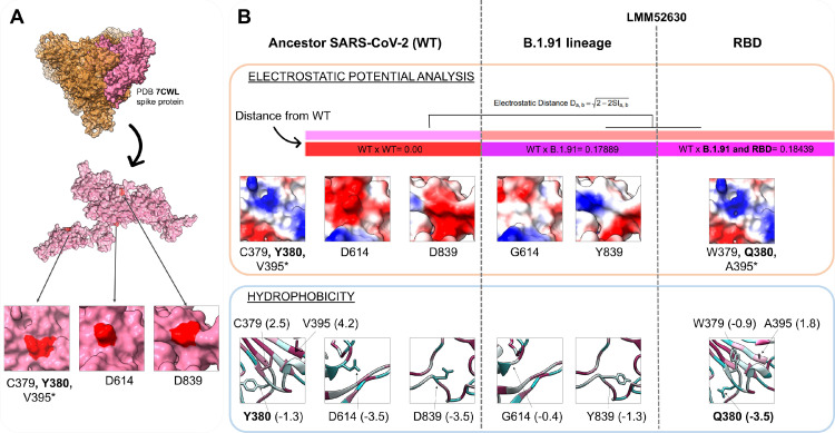
We aimed to describe the SARS-CoV-2 lineages circulating early pandemic among samples with S gene dropout and characterize the receptor-binding domain (RBD) of viral spike protein. Adults and children older than 2 months with signs and symptoms of COVID-19 were prospectively enrolled from May to October in Porto Alegre, Brazil. All participants performed RT-PCR assay, and samples with S gene dropout and cycle threshold < 30 were submitted to high-throughput sequencing (HTS). 484 out of 1,557 participants tested positive for SARS-CoV-2. The S gene dropout was detected in 7.4% (36/484) and a peak was observed in August. The B.1.1.28, B.1.91 and B.1.1.33 lineages were circulating in early pandemic. The RBD novel mutation (Y380Q) was found in one sample occurring simultaneously with C379W and V395A, and the B.1.91 lineage in the spike protein. The Y380Q and C379W may interfere with the binding of neutralizing antibodies (CR3022, EY6A, H014, S304).

Keywords: COVID-19; Novel mutation; RBD; SARS-CoV-2; Variants.


Figures

Fig 1
Fig. 1
Prevalence of S dropout profile from May to October 2020. (A) Number of positive RT-PCR assays, blue columns represent the raw counts of positive assays identified by S gene target and red columns the raw counts of S dropout (characterized as undetected RT-PCR by S, and detected by ORF1ab and N gene targets). (B) Percentage of S dropout over epidemiological week with an increase of undetermined results of the S gene target on the 10th August week, (8/26, 30.8%, P = 0.007). (Color version of figure is available online.)
Fig 2
Fig. 2
SARS-CoV-2 complete genome phylogenetic tree. The Maximum Likelihood phylogenetic analysis under General Time Reversible allows a proportion of invariable sites, and the substitution rates were inferred empirically in MEGAX web server applying 200 replicates and 1000 bootstrap.
Fig 3
Fig. 3
Physicochemical modifications of mutations in the B.1.91 lineage and RBD region (C379W, Y380Q, V395A). (A) The PDB 7CWL crystal with the model generated for the spike protein monomer is detached in pink. Variants' positions at the tridimensional structure surface are highlighted in red. The neighbor residues C379 and Y380 are depicted together, while the V395* is buried and not perceptible at the surface. Both regions (D614 and D839) of the B.1.91 lineage are in exposure regions of the spike protein surface. (B) The SARS-COV-2 ancestor lineage (represented by C379, Y380, V395, D614 and D839 residues) was compared to the G614 and Y839 residues of the B.1.91 variant of concern (VOC), and to the mutations presented by the LMM52630 subject (namely: G614 and Y839 of B.1.91 lineage, in addition to W379, Q380 and A395 from RBD residues) considering the electrostatic potential (EP) (orange rectangle) and hydrophobicity (blue rectangle) properties. The epogram (color bars) presents the electrostatic distances (ED) of the mutated models compared to the wild type (ancestor). Both models (B.1.91 lineage and RBD mutations) are similar with ED = 0.18, and diverging from the ancestor. Below of the epogram, are shown the divergences on electrostatic surface distribution, considering the wild and mutated residues. The color scale is represented by a variation from red (more electronegative residues, -5) to blue (more electropositive, +5) passing through neutral (white, 0). The hydrophobicity scale varies from more hydrophobic residues (represented in magenta, positive values) to more hydrophilic (in blue, negative values). PDB = Protein Data Bank; RBD = Receptor binding domain; WT = Wild type. (Color version of figure is available online.)
Fig 4
Fig. 4
RBD mutation regions of the LMM52630 subject in contact with neutralizing antibodies. (A) The comparison of the mutated sequences with the wild type. Below, the structural model with the selected residues, depicted (red color) for wild and mutated RBD region. The model of the monomer was obtained from the 7CWL PDB structure. (B) The contact region among the RBD (pink), the Fab CR3022 neutralizing antibody (light and heavy chains, represented in blue and green colors, respectively) and the residues C379 and Y380 (red). (C) The same RBD region contacting the neutralizing antibody EY6A (the light chain in purple and heavy in grey). RBD = Receptor binding domain; WT = Wild type. (Color version of figure is available online.)

References

    1. Anichini G, Terrosi C, Gori Savellini G, Gandolfo C, Franchi F, Cusi MG. Neutralizing antibody response of vaccines to SARS-CoV-2 variants. Vaccines. 2021;9:517. doi: 10.3390/vaccines9050517. - DOI - PMC - PubMed
    1. Awadasseid A, Wu Y, Tanaka Y, Zhang W. SARS-CoV-2 variants evolved during the early stage of the pandemic and effects of mutations on adaptation in Wuhan populations. Int J Biol Sci. 2021;17:97–106. doi: 10.7150/ijbs.47827. - DOI - PMC - PubMed
    1. Barnes CO, Jette CA, Abernathy ME, Dam K-MA, Esswein SR, Gristick HB, et al. SARS-CoV-2 neutralizing antibody structures inform therapeutic strategies. Nature. 2020;588:682–687. doi: 10.1038/s41586-020-2852-1. - DOI - PMC - PubMed
    1. Benkert P, Biasini M, Schwede T. Toward the estimation of the absolute quality of individual protein structure models. Bioinformatics. 2011;27:343–350. doi: 10.1093/bioinformatics/btq662. - DOI - PMC - PubMed
    1. cov-lineages/pangolin. CoV-lineages; 2021.