. 2022 Jun;162(7):2032-2046.e12.
doi: 10.1053/j.gastro.2022.02.032. Epub 2022 Feb 24.

Mucin 5AC-Mediated CD44/ITGB1 Clustering Mobilizes Adipose-Derived Mesenchymal Stem Cells to Modulate Pancreatic Cancer Stromal Heterogeneity

Affiliations

Mucin 5AC-Mediated CD44/ITGB1 Clustering Mobilizes Adipose-Derived Mesenchymal Stem Cells to Modulate Pancreatic Cancer Stromal Heterogeneity

Koelina Ganguly et al. Gastroenterology. 2022 Jun.

Abstract

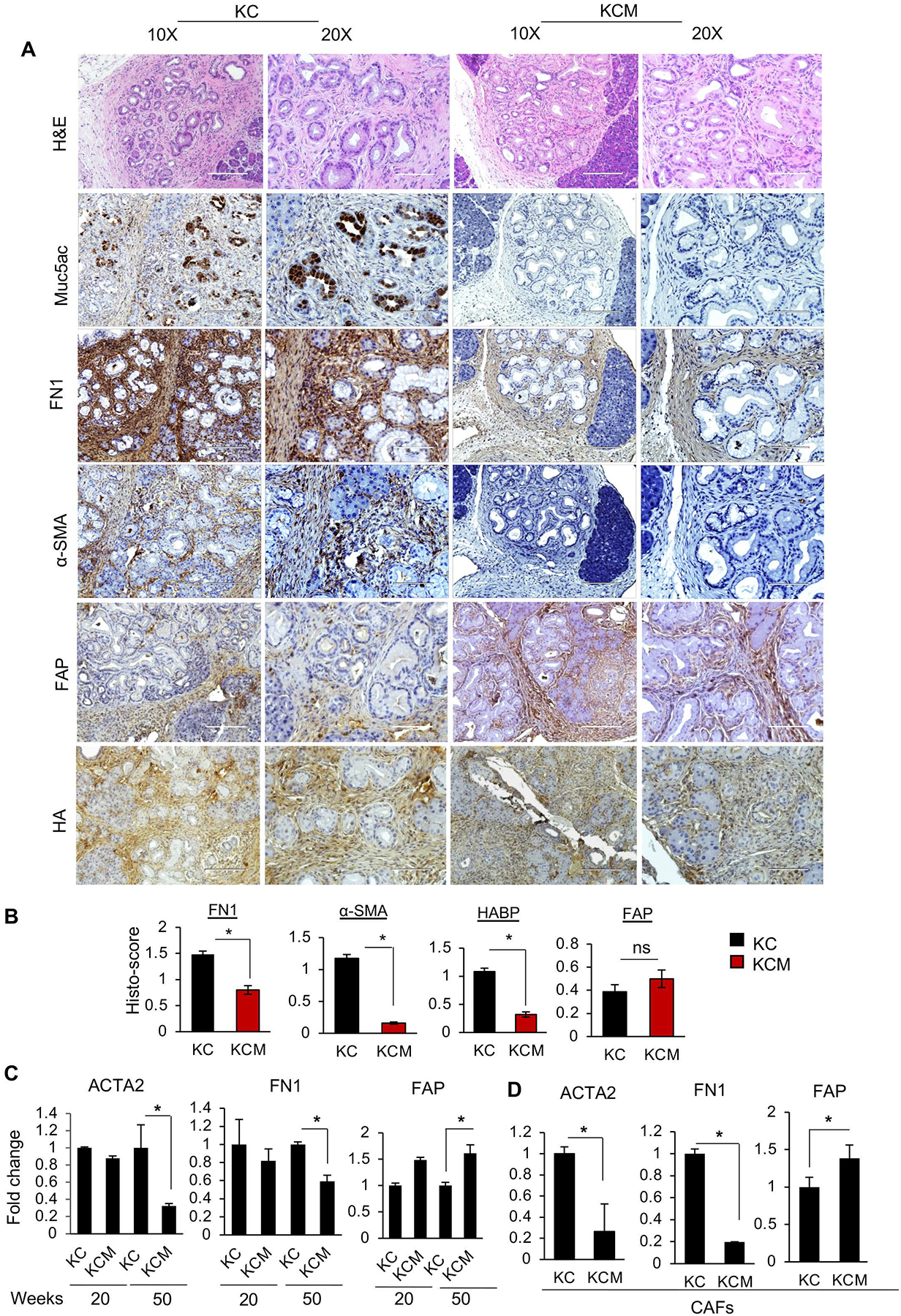
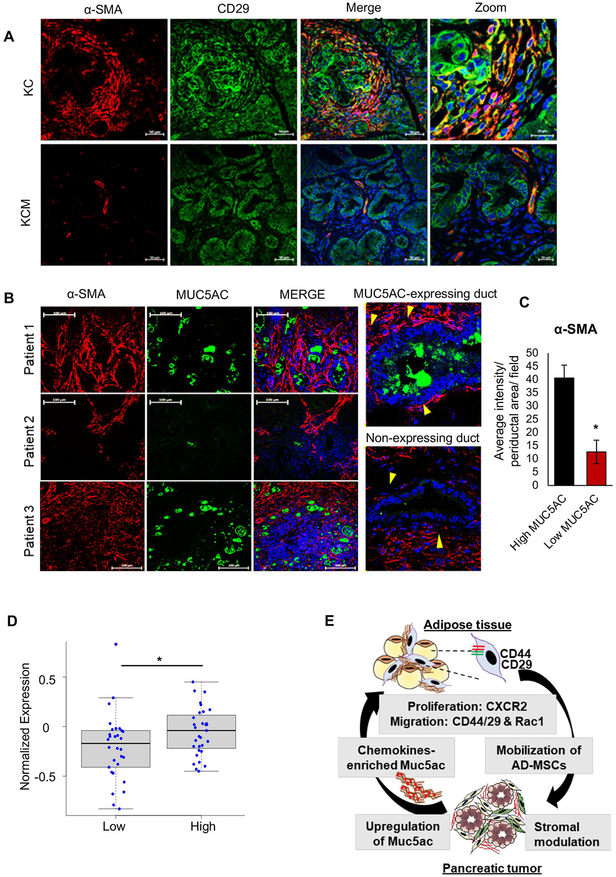
Background & aims: Secreted mucin 5AC (MUC5AC) promotes pancreatic cancer (PC) progression and chemoresistance, suggesting its clinical association with poor prognosis. RNA sequencing analysis from the autochthonous pancreatic tumors showed a significant stromal alteration on genetic ablation of Muc5ac. Previously, depletion or targeting the stromal fibroblasts showed an ambiguous effect on PC pathogenesis. Hence, identifying the molecular players and mechanisms driving fibroblast heterogeneity is critical for improved clinical outcomes.

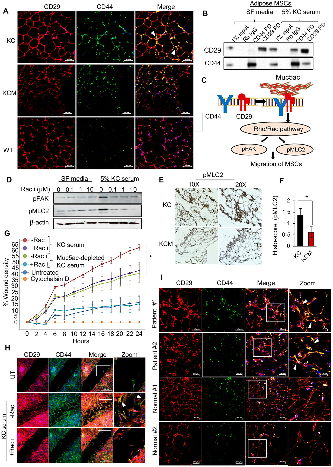
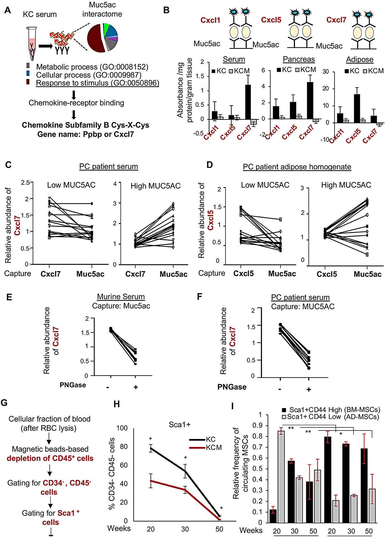
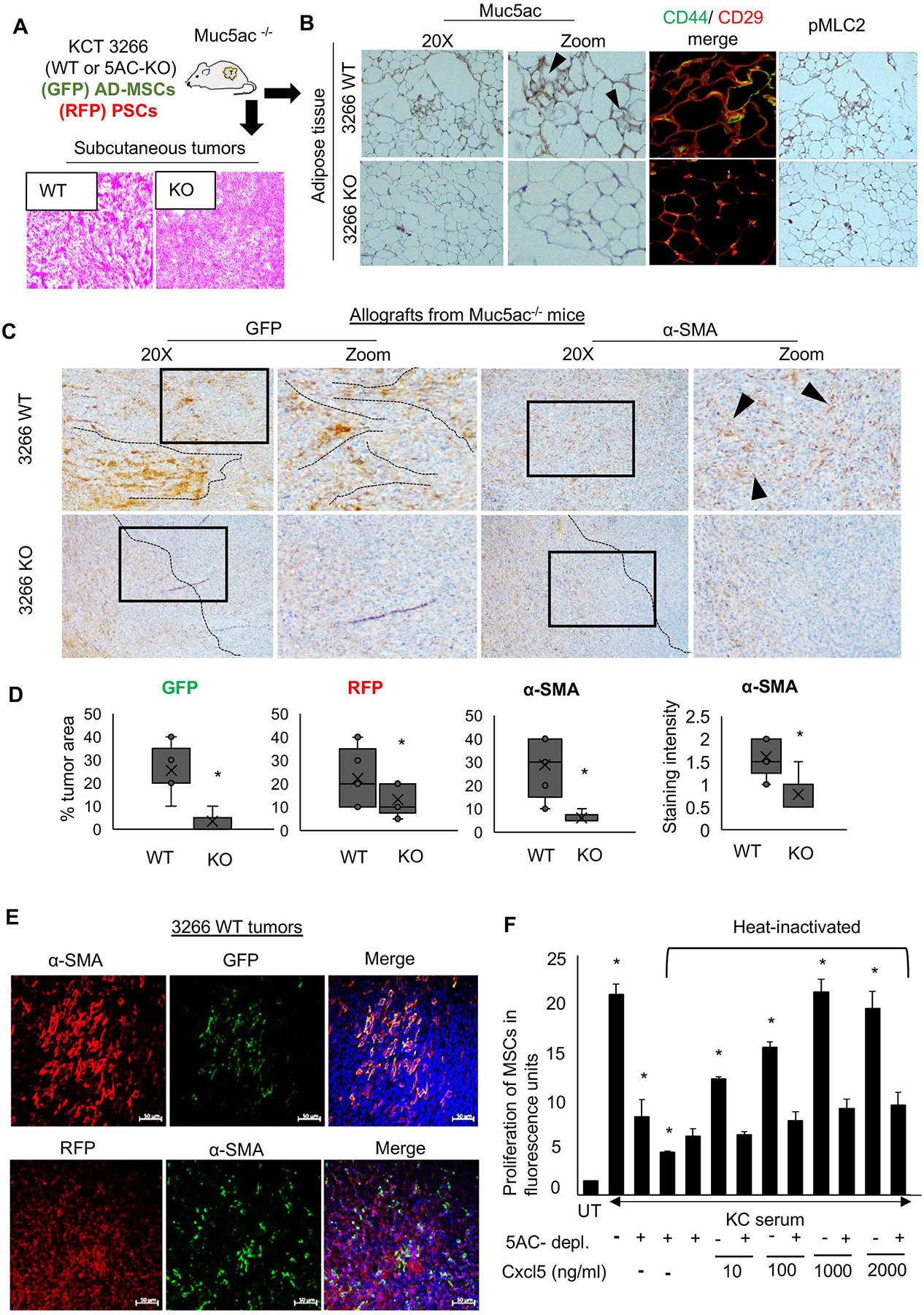
Methods: Autochthonous murine models of PC (KrasG12D, Pdx1-Cre [KC] and KrasG12D, Pdx1-Cre, Muc5ac-/- [KCM]) and co-implanted allografts of murine PC cell lines (Muc5ac wild-type and CRISPR/Cas knockout) with adipose-derived mesenchymal stem cells (AD-MSCs) were used to assess the role of Muc5ac in stromal heterogeneity. Proliferation, migration, and surface expression of cell-adhesion markers on AD-MSCs were measured using live-cell imaging and flow cytometry. MUC5AC-interactome was investigated using mass-spectrometry and enzyme-linked immunosorbent assay.

Results: The KCM tumors showed a significant decrease in the expression of α-smooth muscle actin and fibronectin compared with histology-matched KC tumors. Our study showed that MUC5AC, carrying tumor secretome, gets enriched in the adipose tissues of tumor-bearing mice and patients with PC, promoting CD44/CD29 (integrin-β1) clustering that leads to Rac1 activation and migration of AD-MSCs. Furthermore, treatment with KC-derived serum enhanced proliferation and migration of AD-MSCs, which was abolished on Muc5ac-depletion or pharmacologic inhibition of CXCR2 and Rac1, respectively. The AD-MSCs significantly contribute toward α-smooth muscle actin-positive cancer-associated fibroblasts population in Muc5ac-dependent manner, as suggested by autochthonous tumors, co-implantation xenografts, and patient tumors.

Conclusion: MUC5AC, secreted during PC progression, enriches in adipose and enhances the mobilization of AD-MSCs. On recruitment to pancreatic tumors, AD-MSCs proliferate and contribute towards stromal heterogeneity.

Keywords: Cancer-Associated Fibroblast; Chemokines; MUC5AC; Mesenchymal Stem Cells; Pancreatic Cancer; Stroma.


Conflict of interest statement

Conflicts of Interest: Authors have no conflicts of interest to declare.

Figures

Figure 1.
Figure 1.. Muc5ac is associated with stromal modulation in PC.
(A) Representative images and (B) quantitative analyses of immunohistochemistry (IHC) for stromal markers on tumor sections from KC and KCM mice at 50 weeks of age (n=4/ group). Expression of α-SMA, FN1, and HA was significantly reduced, accompanied by a nonsignificant increase in expression of FAP in KCM tumors, as compared to KC tumors. (C) Quantitative PCR analysis on RNA isolated from KC/ KCM tumors (n=4/ group) and (D) CAFs (isolated from 50 weeks KC and KCM tumors, n=4/ group) demonstrate heterogeneity in stromal markers. t-test *p<0.05.
Figure 2.
Figure 2.. Secreted Muc5ac enriches in AT and modulates AD-MSCs of tumor-bearing mice.
(A) ELISA analysis of the tissue homogenates from KC mice (n=4) demonstrates the abundant expression of Muc5ac in the pancreatic tumor and adipose tissue. The stomach serves as a positive control. (B) Immunoblot from WT (n=2) and KC mice (n=4) serum and tissue homogenates. Muc5ac was absent in WT mouse adipose and pancreas; Liver and stomach homogenates served as negative and positive controls, respectively. Arrows indicate MUC5AC protein bands. (C) Representative images of IHC for Muc5ac demonstrating its spatial localization proximal to the AD-MSCs in the KC AT, with no trace of Muc5ac in KCM and WT adipose. (D) Representative histograms and quantitative analyses from flow cytometry demonstrate significant reductions in the expression of CD44 and CD29 on the AD-MSCs isolated from KCM adipose, as compared to KC. (I) Representative histograms and (F) Quantitative analyses from flow cytometry demonstrate a significant increase in the expression of CD44 and CD29 on the AD-MSCs after treatment with KC serum with significant reductions in the Muc5ac-depleted KC serum-treated AD-MSCs. t-test *p<0.05
Figure 3.
Figure 3.. Muc5ac promotes AD-MSCs’ migration via CD44/CD29 clustering and Rac1 activation.
(A) Immunofluorescence images demonstrate the colocalization of CD44 and CD29 (focal clusters marked by white arrowhead) in KC, absent in KCM and WT ATs. (B) Direct and reciprocal co-immunoprecipitation demonstrate the interaction of CD44 and CD29 on AD-MSCs treated with KC serum. Rabbit IgG serves as a negative control, and 1% input demonstrates endogenous expression of the target molecules. (C) Schematic working model of Muc5ac-mediated clustering of CD44 and CD29 on AD-MSCs leading to the activation of Rac 1 pathway, manifested by the enhanced expression of pMLC2 and pFAK. (D) Representative images and (E) quantitative analysis from immunohistochemistry show a significant decline in the expression of pMLC2 in KCM adipose compared to KC. (F) Immunoblot demonstrate upregulation of pMLC2 and pFAK in AD-MSCs treated with 5% KC serum and subsequent decline in these markers upon Rac1 inhibition in a dose-dependent manner. (G) Live-cell imaging-based kinetic wound-healing assay demonstrates a significant decline in migratory propensity (manifested by a lower percentage of wound density) of AD-MSCs treated with Muc5ac-depleted KC serum and Rac1 inhibitor, as compared to those treated with 5% KC serum. Treatment with Cytochalasin D, an F-actin blocker, was used as a negative control. (H) Immunofluorescence images from the migration assay at the endpoint (24 hours) demonstrate co-localization of CD44 and CD29 (white arrowheads) on the migratory AD-MSCs treated with KC serum. t-test *p<0.05.
Figure 4.
Figure 4.. Muc5ac scaffolds chemokines and promotes AD-MSCs’ mobilization in the systemic circulation.
(A) The peptide hits from mass spectrometry-analysis on Muc5ac immunoprecipitate from the KC serum were subjected to pathway analysis using Panther webtool. “Response to stimulus” was the top-most enriched pathway that led to “chemokine-receptor binding” upon functional classification. Subsequent candidate-based analysis led to the identification of Cxcl7 in the secreted Muc5ac interactome. (B) Sandwich-ELISA analysis demonstrates a significant abundance of CXCR2 ligands bound with Muc5ac captured from KC pancreas, serum, and adipose tissues. (C, D) Correlation plots demonstrate enrichments in the Muc5ac-bound form of Cxcl7 and Cxcl5, compared to their total (free and bound) forms in the serum and adipose of human PC patients with high MUC5AC expression; without any enrichments in the low MUC5AC-expressing group. (E, F) Correlation plot demonstrating a significant decline in the abundance of Muc5ac-bound Cxcl7 in the murine and human PC serum upon incubation with PNGase. (G) Workflow of isolation and flow cytometry-based detection of circulating MSCs. (H) Line graph demonstrates significantly higher Sca1+ MSCs in the blood of KC mice at 20 and 30 weeks, compared to age-matched KCM group (n=3/time-point/group). Subsequent analysis shows a significant decline in the CD44+CD29+ MSCs in KC blood, compared to KCM, at 30 and 50 weeks. t-test *p<0.05.
Figure 5.
Figure 5.. Muc5ac promotes AD-MSC expansion and maturation towards α-SMA-expressing CAFs.
(A) Schematic experimental plan for the co-implantation allograft model in Muc5ac−/− mice, using murine PC cell line KCT 3266 (WT and MUC5AC-KO) admixed with AD-MSCs isolated from GFP mice and RFP-labelled pancreatic stellate cells. Representative H&E images demonstrating the histology of WT and KO tumors (n=10/group). (B) Immunohistochemistry and immunofluorescence images from adipose tissue isolated from Muc5ac−/− animals demonstrate the presence of Muc5ac, with higher CD44/CD29 colocalization and pMLC2 expression in the mice grafted with Muc5ac-expressing cells, as compared to those receiving Muc5ac-KO cells. (C) Representative images and (D) quantitative analysis (n=10/group) from immunohistochemistry demonstrate the expansion of AD-MSCs (GFP), pancreatic stellate cells (RFP), and α-SMA+ CAFs with increased α-SMA staining in 3266 WT tumors, as compared to the KO group. Black dotted lines and arrowheads demonstrate the pattern of GFP-MSCs and α-SMA-CAFs, localized near the malignant cells in the WT tumors but dispersed towards the periphery of the KO tumors. (E) Immunofluorescence images show co-expression of GFP and α-SMA in the WT tumors, with no significant co-expression of RFP and α-SMA, suggesting AD-MSCs contribute more than the resident stellate cells towards the α-SMA+ stromal population in the Muc5ac-expressing pancreatic tumors. (F) The bar graph demonstrates that the proliferation of AD-MSCs significantly increased when treated with KC serum, which subsequently declined upon Muc5ac depletion and heat inactivation of KC serum. Ectopic addition of Cxcl5 at increasing concentrations of 10, 100, 1000, and 2000 ng/ml in heat-inactivated KC serum rescued the proliferation of AD-MSCs in a dose-dependent manner. t-test *p<0.05
Figure 6.
Figure 6.. MUC5AC expression significantly correlates with peri-ductal α-SMA+-expressing CAFs in murine and human pancreatic tumors.
(A) Immunofluorescence images show co-expression of CD29 and periductal α-SMA expressing population in the KC tumor stroma, but not in KCM tumors. (B) Representative immunofluorescence images from PC patient tumor sections (n=6) demonstrate the correlation of MUC5AC expression in the malignant ducts and α-SMA expression in the periductal CAFs. Yellow arrowheads demonstrate the α-SMA+ CAF clusters lying in the juxtacrine position of the malignant cells in MUC5AC-expressing ducts, unlike in non-MUC5AC- expressing ducts. (C) ImageJ-based quantitation demonstrates an increased intensity of α-SMA+ expression in the periductal area/field (n=10 areas/ field, n=3 fields, n=4 patients) in high MUC5AC-expressing ducts, as compared to low-expressing ducts. t-test *p<0.05. (D) Boxplot from PC patients’ proteomic data demonstrate a significant association of MUC5AChighCXCL5high expression with α-SMA+ expression. p=0.03 (E) Schematic workflow of MUC5AC-mediated crosstalk between adipose tissue and pancreatic tumors. Abundantly expressed MUC5AC, with entrapped chemokines, is secreted in the systemic circulation from the pancreatic tumors. The Muc5ac-entrapped tumor secretome reaches the adipose tissue, where it causes the expansion and migration of AD-MSCs via the CXCR2 and Rac1 axis. Muc5ac-mediated CD44/CD29 clustering mobilizes the AD-MSCs into circulation, from where they home to the pancreatic tumors and phenotypically mature into α-SMA+ CAF subtypes.

References

    1. Neoptolemos JP, Kleeff J, Michl P, et al. Therapeutic developments in pancreatic cancer: current and future perspectives. Nature reviews Gastroenterology & hepatology 2018;15:333–348. - PubMed
    1. Patra KC, Bardeesy N, Mizukami Y. Diversity of precursor lesions for pancreatic cancer: the genetics and biology of intraductal papillary mucinous neoplasm. Clinical and translational gastroenterology 2017;8:e86. - PMC - PubMed
    1. Sahai E, Astsaturov I, Cukierman E, et al. A framework for advancing our understanding of cancer-associated fibroblasts. Nature Reviews Cancer 2020;20:174–186. - PMC - PubMed
    1. Gore J, Korc M. Pancreatic cancer stroma: friend or foe? Cancer cell 2014;25:711–712. - PMC - PubMed
    1. Neesse A, Bauer CA, Öhlund D, et al. Stromal biology and therapy in pancreatic cancer: ready for clinical translation? Gut 2019;68:159–171. - PubMed

Publication types