. 2022 Mar:77:103891.
doi: 10.1016/j.ebiom.2022.103891. Epub 2022 Feb 24.

Short-chain fatty acid acetate triggers antiviral response mediated by RIG-I in cells from infants with respiratory syncytial virus bronchiolitis

Affiliations

Short-chain fatty acid acetate triggers antiviral response mediated by RIG-I in cells from infants with respiratory syncytial virus bronchiolitis

Krist H Antunes et al. EBioMedicine. 2022 Mar.

Abstract

Background: Gut microbiota-derived short-chain fatty-acid (SFCA) acetate protects mice against RSV A2 strain infection by increasing interferon-β production and expression of interferon-stimulated genes (ISGs). However, the role of SFCA in RSV infection using strains isolated from patients is unknown.

Methods: We first used RSV clinical strains isolated from infants hospitalized with RSV bronchiolitis to investigate the effects of in vitro SCFA-acetate treatment of human pulmonary epithelial cells. We next examined whether SCFA-acetate treatment is beneficial in a mouse model of RSV infection using clinical isolates. We sought to investigate the relationship of gut microbiota and fecal acetate with disease severity among infants hospitalized with RSV bronchiolitis, and whether treating their respiratory epithelial cells with SCFA-acetate ex-vivo impacts viral load and ISG expression. We further treated epithelial cells from SARS-CoV-2 infected patients with SCFA-acetate.

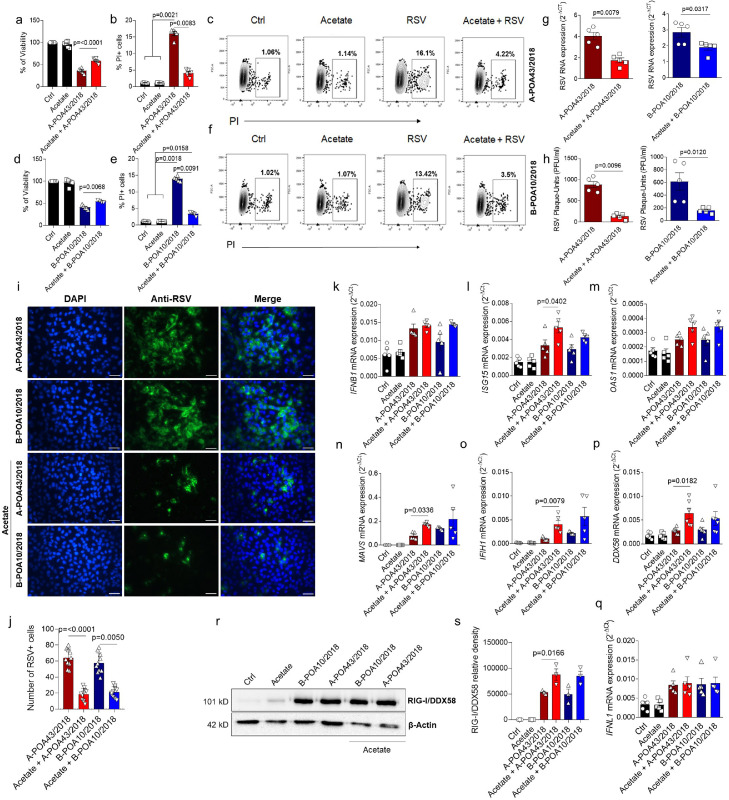
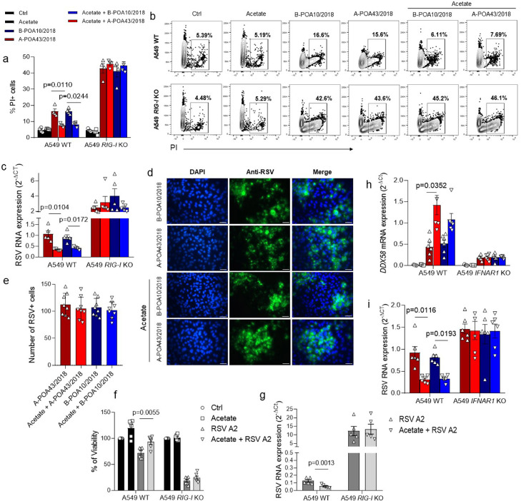
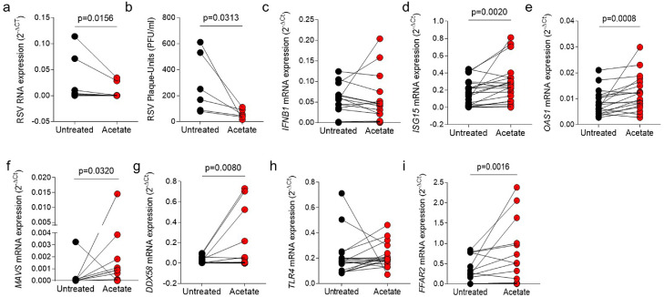
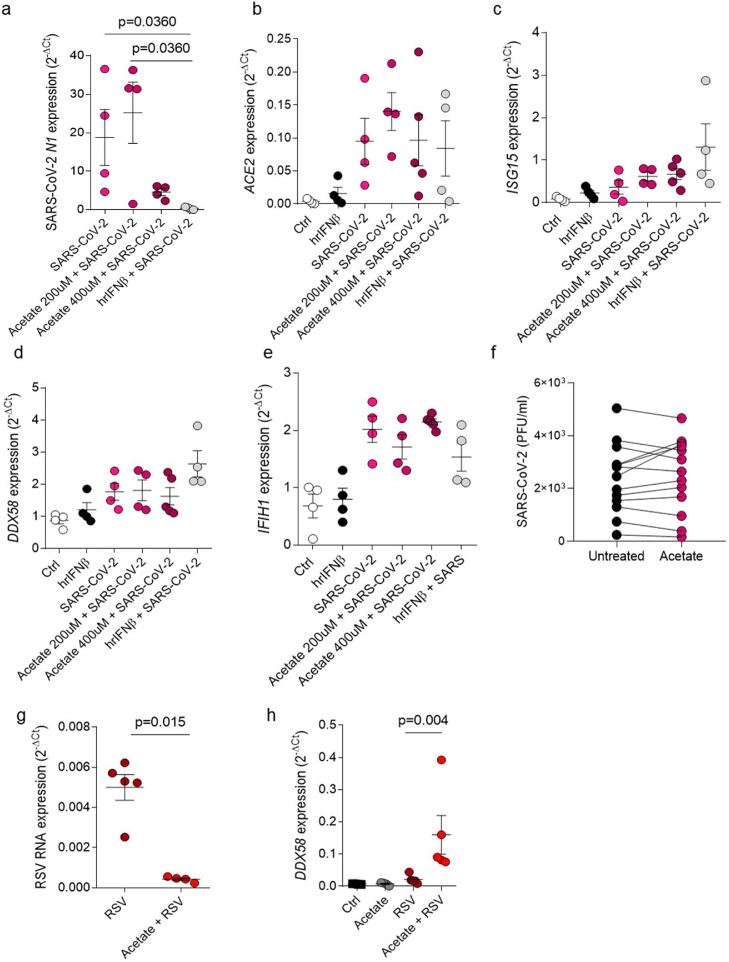
Findings: In vitro pre-treatment of A549 cells with SCFA-acetate reduced RSV infection with clinical isolates and increased the expression of RIG-I and ISG15. Animals treated with SCFA-acetate intranasally recovered significantly faster, with reduction in the RSV clinical isolates viral load, and increased lung expression of IFNB1 and the RIG-I. Experiments in RIG-I knockout A549 cells demonstrated that the protection relies on RIG-I presence. Gut microbial profile was associated with bronchiolitis severity and with acetate in stool. Increased SCFA-acetate levels were associated with increasing oxygen saturation at admission, and shorter duration of fever. Ex-vivo treatment of patients' respiratory cells with SCFA-acetate reduced RSV load and increased expression of ISGs OAS1 and ISG15, and virus recognition receptors MAVS and RIG-I, but not IFNB1. These SCFA-acetate effects were not found on cells from SARS-CoV-2 infected patients.

Interpretation: SCFA-acetate reduces the severity of RSV infection and RSV viral load through modulation of RIG-I expression.

Funding: FAPERGS (FAPERGS/MS/CNPq/SESRS no. 03/2017 - PPSUS 17/2551-0001380-8 and COVID-19 20/2551-0000258-6); CNPq 312504/2017-9; CAPES) - Finance Code 001.

Keywords: Acetate; Clinical isolates; RIG-I; Respiratory syncytial virus.


Conflict of interest statement

Declaration of interests The authors declare no competing interests.

Figures

Fig 1
Figure 1
Acetate protects against RSV clinical isolate infection in human pulmonary cells. a, A549 cells were pre-treated with 260 µM of acetate for 24 h and infected with 104 PFU/ml of clinical isolate RSV A-POA43/2018 or B-POA10/2018 for 96 h (a–j) or 24 h (q–s). a and c, Percent of cell viability measured by MTT assay (using untreated/uninfected cells as viability control). b and e, Percent of PI (propidium iodide) positive cells detected by flow cytometry. c and f, Representative FACS profile of PI positive cells. Gating strategy is shown in Supplementary Fig. E4 3. g, RSV F protein RNA levels detected using real-time PCR (2−ΔCt). h, Viral load measured by Plaque-forming unit (PFU) using specific RSV antibody. i, Fluorescence images of RSV (green) and cell nuclei using Hoescht (blue). Third panel shows data co-localization (merge). Scale bars = 60 µm. j, Immunofluorescence quantification measured by the number of RSV positive (green) cells. k-q, IFNB1, ISG15, OAS1, MAVS, IFIH1, DDX58 (RIG-I) and IFNL1 gene expression detected by real-time PCR (2−ΔCt). r, Western blot analysis of RIG-I/DDX58 and β-actin in the whole cell. The whole blot image is available on Supplementary Fig. 5. s, Protein bands quantification. Graphic shows data normalized by β-actin. Data are presented as mean ± SEM. All the experiments were performed in quadruplicates. Data are representative of 3 independent experiments, except in r and s, where 2 independent experiments were performed. Statistical significance between groups was determined with Kruskal–Wallis, except in G and H, in which Mann–Whitney was used. A p-value lower than 0.05 was considered as significative.
Fig 2
Figure 2
Acetate protection against RSV clinical isolates depends on RIG-I presence in human pulmonary cells. a–d, CRISPR/Cas9-generated RIG-I knockout (RIG-I KO) or wild-type (WT) A549 cells were pre-treated with 260 µM of acetate for 24 h and infected with 104 PFU/ml (0.5 MOI) of clinical isolate RSV A-POA43/2018, B-POA10/2018 for 96 h or laboratory RSV strain A2 (f and g plots). a, Percent of PI (propidium iodide) positive cells detected by flow cytometry. b, Representative FACS profile of PI positive cells. Gating strategy is shown in appendix Figure 4. c, RSV F protein RNA levels detected using real-time PCR (2−ΔCt). d, Fluorescence images of RSV (green) and cell nuclei using Hoescht (blue). Third panel shows data co-localization (merge). Scale bars = 60 µm. e, Immunofluorescence quantification measured by the number of RSV positive (green) cells. f, Percent of cell viability measured by MTT assay (using untreated/uninfected cells as viability control) in cells infected with RSA A2 strain. g, RSV F protein RNA levels detected using real-time PCR (2−ΔCt) in cells infected with RSA A2 strain. h and i, CRISPR/Cas9-generated IFNAR1 knockout (IFNAR1 KO) or WT A549 cells were pre-treated with 260 µM of acetate for 24 h and infected with 104 PFU/ml of clinical isolate RSV A-POA43/2018 or B-POA10/2018 for 96 h or 24 h (h plot). h, DDX58 gene expression calculated using real-time PCR (2−ΔCt). i, RSV F protein RNA levels calculated using real-time PCR (2−ΔCt). Data are presented as mean ± SEM. All the experiments were performed in quadruplicates. The data are representative of 3 independent experiments, except in f to i, where 2 independent experiments were performed. Statistical significance between groups was determined with Kruskal–Wallis. A p-value lower than 0.05 was considered as significative.
Fig 3
Figure 3
Intranasal acetate treatment protects against RSV clinical isolate infection in vivo. a, Experimental scheme. Female BALB/cJ mice were infected with clinical isolate RSV A-POA43/2018 (107 PFU/ml) and 24 h post infection treatment with 20 mM (40 μl) of acetate started through the intranasal route and PBS was administered as vehicle control. Treatment was performed daily up to 5 days after infection. b, Percentage body weight loss post infection relative to initial weight (day 0) (n = 5). c, Total cell number and differential cell counts in bronchoalveolar lavage fluid. d, Representative images of lung tissue section stained with hematoxylin and eosin (H&E). Scale bars = 100 μm. e, H&E respective inflammation scores (n = 3). f, Viral titre in the lung (PFUs/g lung tissue). g–k, Ifnb1, Isg15, Oas1, Ddx58 and Mavs gene expression in the lung detected by real-time PCR (2−ΔCt). The data are representative of 2 independent experiments. Data are presented as mean ± SEM. Statistical significance between groups was determined with Kruskal–Wallis, except in F, in which Mann–Whitney was used. A p-value lower than 0.05 was considered as significative.
Fig 4
Figure 4
Gut microbiota composition and production of SCFA are associated with the severity of RSV-infected infants. a, microbiota composition analysis at phylum level using 16S rRNA gene sequencing (relative abundance). b, SCFA quantification in feces. c, Heatmap showing associations between the bacteria family and clinical parameters. Correlations were made through Spearman's rank correlation coefficient. Data in b is presented as mean ± SEM. The data are from 13 patients collected throughout the study. In b statistical significance between groups was determined with Kruskal–Wallis. A p-value lower than 0.05 was considered as significative.
Fig 5
Figure 5
SCFA-acetate treatment reduces viral load and increases expression of antiviral molecules in cells from nasal washes of patients with RSV-bronchiolitis. d–i, Cells from nasopharyngeal washes of infants with RSV bronchiolitis were treated with 260 µM of SCFA-acetate for 24 h and then harvested for analysis. a, Real-time PCR analysis of RSV protein F gene expression detected by real-time PCR (2−ΔCt). b, Viral plaque-assay (PFU/ml) performed using the supernatant. c-g, IFNB1, ISG15, OAS1, DDX58 (RIG-I gene), and MAVS expression detected by real-time PCR (2-ΔCt). h and i, TLR4 and FFAR2 (GPR43) expression detected by real-time PCR (2-ΔCt) Data are presented as mean ± SEM. The data are from 7 to 30 patients collected throughout the study. From a to i the difference between the treatment and its untreated control was accessed by Wilcoxon matched-pairs signed rank test. A p-value lower than 0.05 was considered as significative.
Fig 6
Figure 6
Acetate treatment does not have influence on SARS-CoV-2 infection. a–d, Calu-3 cells were pre-treated with 200 µM or 400 µM of acetate for 24 h or pre-treated with 100 UI of human recombinant IFNβ for 6 h. Cells were then infected with 0.1 MOI (multiplicity of infection) of SARS-CoV-2 and left in culture for 2 days. a, SARS-CoV-2 N1 (virus nucleocapsid) gene expression detected by real-time PCR (2−ΔCt). b-e, ACE2, ISG15, DDX58 (RIG-I) gene expression detected by real-time PCR (2−ΔCt). f, viral plaque-assay (PFU) of cells from nasopharyngeal aspirate samples from patients with SARS-CoV-2 treated with 260 µM of acetate for 24 h (PFU/ml). g,h, Calu-3 cells were pre-treated with 260 µM of acetate for 24 h and infected with 104 PFU/ml (0.5 MOI) of clinical isolate RSV A-POA43/2018 for 96 h (g) or 24 h (h). g, RSV F protein RNA levels calculated using real-time PCR (2−ΔCt). h, DDX58 gene expression assessed using real-time PCR (2−ΔCt). Data are presented as mean ± SEM. All the experiments were performed in quadruplicates. The data are representative of 2 independent experiments. Data in e is from 13 patients. Statistical significance between groups was determined with Kruskal–Wallis, except in f, where the difference between the treatment and its untreated control was accessed by Wilcoxon matched-pairs signed rank test. In g Mann–Whitney was used to determine the difference between the groups. A p-value lower than 0.05 was considered as significative.

References

    1. Nair H., Nokes D.J., Gessner B.D., et al. Global burden of acute lower respiratory infections due to respiratory syncytial virus in young children: a systematic review and meta-analysis. Lancet. 2010;375(9725):1545–1555. - PMC - PubMed
    1. Shi T., McAllister D.A., O'Brien K.L., et al. Global, regional, and national disease burden estimates of acute lower respiratory infections due to respiratory syncytial virus in young children in 2015: a systematic review and modelling study. Lancet. 2017;390(10098):946–958. - PMC - PubMed
    1. Foley D.A., Phuong L.K., Englund J.A. Respiratory syncytial virus immunisation overview. J Paediatr Child Health. 2020;56(12):1865–1867. - PubMed
    1. Mazur N.I., Higgins D., Nunes M.C., et al. The respiratory syncytial virus vaccine landscape: lessons from the graveyard and promising candidates. Lancet Infect Dis. 2018;18(10):e295–e311. - PubMed
    1. Ruckwardt T.J., Morabito K.M., Graham B.S. Immunological lessons from respiratory syncytial virus vaccine development. Immunity. 2019;51(3):429–442. - PubMed