. 2022 Feb 28;15(1):95-103.
doi: 10.15283/ijsc21195.

A Simple Method for Generating Cerebral Organoids from Human Pluripotent Stem Cells

Affiliations

A Simple Method for Generating Cerebral Organoids from Human Pluripotent Stem Cells

Yean Ju Hong et al. Int J Stem Cells. .

Abstract

Background and objectives: In recent years, brain organoid technologies have been the most innovative advance in neural differentiation research. In line with this, we optimized a method to establish cerebral organoids from feeder-free cultured human pluripotent stem cells. In this study, we focused on the consistent and robust production of cerebral organoids comprising neural progenitor cells and neurons. We propose an optimal protocol for cerebral organoid generation that is applicable to both human embryonic stem cells and human induced pluripotent stem cells.

Methods and results: We investigated formation of neuroepithelium, neural tube, and neural folding by observing the morphology of embryoid bodies at each stage during the cerebral organoid differentiation process. Furthermore, we characterized the cerebral organoids via immunocytochemical staining of sectioned organoid samples, which were prepared using a Cryostat and Vibratome. Finally, we established a routine method to generate early cerebral organoids comprising a cortical layer and a neural progenitor zone.

Conclusions: We developed an optimized methodology for the generation of cerebral organoids using hESCs and hiPSCs. Using this protocol, consistent and efficient cerebral organoids could be obtained from hiPSCs as well as hESCs. Further, the morphology of brain organoids could be analyzed through 2D monitoring via immunostaining and tissue sectioning, or through 3D monitoring by whole tissue staining after clarification.

Keywords: Brain organoid; Differentiation; Human pluripotent stem cells; Organoid; Pluripotency.


Conflict of interest statement

Potential Conflict of Interest

The authors have no conflicting financial interest.

Figures

Fig. 1
Fig. 1
Characterization of human embryonic stem cells (hESCs) and human induced pluripotent stem cells (hiPSCs). (A) Phase contrast images of hESCs and hiPSCs in feeder-free conditions. Scale bar=200 μm. (B) Immunocytochemical staining of the pluripotency markers OCT4, SOX2, and TRA-1-81 in hESCs (upper) and hiPSCs (lower). Scale bar=200 μm in the upper panels and 100 μm in the lower panels. (C) Growth rate of hESCs from 2 to 4 days after seeding. Growth rate was measured as an increase in colony size (μm2). Error bars represent standard error of the mean.
Fig. 2
Fig. 2
Brain organoid differentiation protocol using human pluripotent stem cells (hPSCs). (A) Schematic illustration of the generation of brain organoids. Representative images characterizing each stage are shown. Scale bar=200 μm. (B) Morphology of embryoid bodies (EBs) in EB formation medium, based on TeSR medium (upper left), and their enlarged images (upper right). EBs committed to the neural lineage showed a transparent neuroepithelium-like region at the periphery of the structure (lower left), which is depicted with a white dotted circle (lower right). Scale bar=500 μm (lower left), 200 μm (lower right). (C) Comparison of koSR-containing medium (hESC medium) and Aggrewell EB medium for the size of EBs differentiated from hESCs. Scale bars=200 μm.
Fig. 3
Fig. 3
Matrigel embedding culture and morphology of early-stage brain organoids. (A) Matrigel embedding process for small or large amount of embryoid bodies (EBs) and the spin culture system. (B) The neural folding stage of hESC-derived EBs at day 6 after Matrigel embedding with single (small amount) or bulk (large amount) EBs. Scale bar=200 μm (left and right) and 500 μm (middle). (C) Brain organoids with multiple neural rosette-like structures derived from human embryonic stem cells and (day 19) and human induced pluripotent stem cells (day 20). Scale bar=500 μm.
Fig. 4
Fig. 4
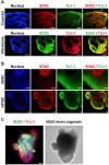
Characterization of human embryonic stem cell (hESC)- and human induced pluripotent stem cell (hiPSC)-derived brain organoids. (A) 2D immunocytochemistry staining of the early neuron marker TUJ-1 and the neural progenitor marker SOX2 in hPSC-derived organoids. A Cryostat (upper) and Vibratome (lower) were used for tissue sectioning. Scale bar=100 μm (upper panels) and 200 μm (lower panels). Green arrow indicates cortical plate, red arrow indicates ventricular zone, and white arrow indicates ventricle (Cryostat sample). Green arrow indicates ventricular zone, red arrow indicates cortical plate, and white arrow indicates ventricle (Vibratome sample). (B) Confocal images of 3D immunocytochemistry for the early neuron marker TUJ-1 and the neural progenitor marker SOX2 in hESC- and hiPSC-derived organoids. Scale bar=50 μm (upper panels) and 20 μm (lower panels). (C) Representative 3D image of whole brain organoids, which were stained with TUJ-1 and SOX2 (at day 12 hESC-derived). The image was captured using high contents screening (HCS). Scale bars=200 μm.

References

    1. Fatehullah A, Tan SH, Barker N. Organoids as an in vitro model of human development and disease. Nat Cell Biol. 2016;18:246–254. doi: 10.1038/ncb3312. - DOI - PubMed
    1. Kelava I, Lancaster MA. Dishing out mini-brains: current progress and future prospects in brain organoid research. Dev Biol. 2016;420:199–209. doi: 10.1016/j.ydbio.2016.06.037. - DOI - PMC - PubMed
    1. Di Lullo E, Kriegstein AR. The use of brain organoids to investigate neural development and disease. Nat Rev Neurosci. 2017;18:573–584. doi: 10.1038/nrn.2017.107. - DOI - PMC - PubMed
    1. Quadrato G, Arlotta P. Present and future of modeling human brain development in 3D organoids. Curr Opin Cell Biol. 2017;49:47–52. doi: 10.1016/j.ceb.2017.11.010. - DOI - PubMed
    1. Lancaster MA, Renner M, Martin CA, Wenzel D, Bicknell LS, Hurles ME, Homfray T, Penninger JM, Jackson AP, Knoblich JA. Cerebral organoids model human brain development and microcephaly. Nature. 2013;501:373–379. doi: 10.1038/nature12517. - DOI - PMC - PubMed

LinkOut - more resources