

Comparison of Morphological and Histological Characteristics of Human and Sheep: Sheep as a Potential Model for Testing Midurethral Slings in vivo
- PMID: 35220315
- PMCID: PMC10123539
- DOI: 10.1159/000522138
Comparison of Morphological and Histological Characteristics of Human and Sheep: Sheep as a Potential Model for Testing Midurethral Slings in vivo
Abstract
Introduction: The sheep was evaluated as a potential model for preclinical evaluation of urethral slings in vivo based on: (1) anatomical measurements of the sheep vagina and (2) histological tissue integration and host response to polypropylene (PP) slings.
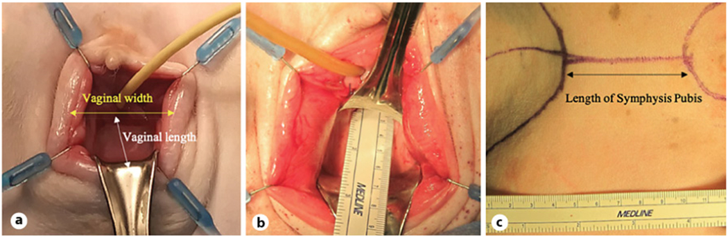
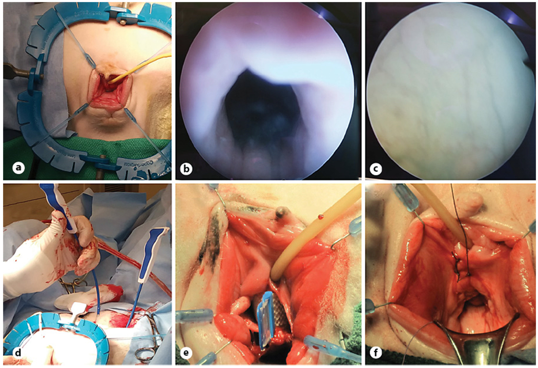
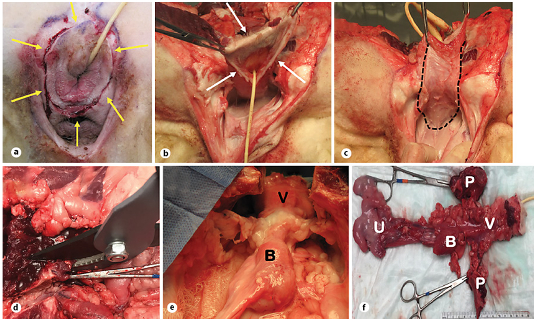
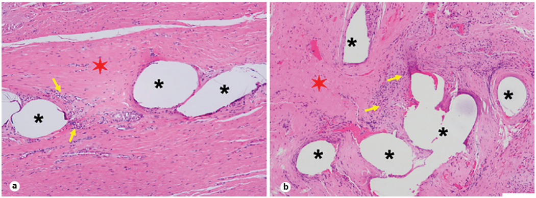
Methods: Eight female, multiparous sheep were utilized. Three of 8 animals underwent surgery mimicking human tension-free vaginal tape protocols for midurethral slings and were euthanized at 6 months. The following measurements were obtained: vaginal length, maximum vaginal width with retraction, symphysis pubis length, and distance from the pubic bone to incision. Explanted sling samples from sheep and human were stained with hematoxylin and eosin for host reaction assessment.
Results: Geometric measurements were similar between humans and sheep. Sheep vaginal anatomy allowed sling placement similar to procedures in human surgeries, and all sheep recovered without problems. Comparative histology between the sheep and human indicated similar host reaction and collagen deposition around implants, confirming suitability of the sheep model for biomaterial response assessment.
Conclusion: Sheep vaginal length is comparable to humans. Tissue integration and host response to PP slings showed chronic inflammation with rich collagen deposition around the material in both sheep and human specimens, highlighting the sheep as a potential animal model for preclinical testing of midurethral slings.
Keywords: Midurethral sling; Preclinical testing; Sheep model; Stress urinary incontinence.
© 2022 S. Karger AG, Basel.
Conflict of interest statement
Conflict of Interest Statement
Subbakrishna Shankar is the chief technology officer and holds equity in CollaMedix Inc. Ozan Akkus is the chief scientific officer of and holds equity in CollaMedix Inc. Adonis Hijaz is the chief medical officer and holds equity in CollaMedix Inc. The other authors declare no conflicts of interest.
Figures
Similar articles
-
Comparison of 2 single incision slings on the vagina in an ovine model.Am J Obstet Gynecol. 2021 Jan;224(1):78.e1-78.e7. doi: 10.1016/j.ajog.2020.07.005. Epub 2020 Jul 21. Am J Obstet Gynecol. 2021. PMID: 32707267
-
A preliminary evaluation of in vivo response to a filament-wound macroporous collagen midurethral sling in an ovine model.J Biomed Mater Res B Appl Biomater. 2022 Dec;110(12):2676-2685. doi: 10.1002/jbm.b.35120. Epub 2022 Jul 2. J Biomed Mater Res B Appl Biomater. 2022. PMID: 35779040 Free PMC article.
-
Comparing the vaginal wall sling with autologous rectus fascia and polypropylene sling: Short term outcomes and patient satisfaction.Eur J Obstet Gynecol Reprod Biol. 2018 Dec;231:98-103. doi: 10.1016/j.ejogrb.2018.10.012. Epub 2018 Oct 11. Eur J Obstet Gynecol Reprod Biol. 2018. PMID: 30340120 Review.
-
The evolution of midurethral slings.Nat Clin Pract Urol. 2008 Apr;5(4):194-201. doi: 10.1038/ncpuro1052. Epub 2008 Mar 4. Nat Clin Pract Urol. 2008. PMID: 18317496 Review.
-
Development of a Clinically Relevant Preclinical Animal Model to Mimic Suburethral Implantation of Support Materials for Stress Urinary Incontinence.Neurourol Urodyn. 2025 Feb;44(2):489-495. doi: 10.1002/nau.25630. Epub 2024 Nov 25. Neurourol Urodyn. 2025. PMID: 39584320
Cited by
-
Advances in vaginal bioengineering: Applications, techniques, and needs.Curr Res Physiol. 2023 Oct 18;6:100111. doi: 10.1016/j.crphys.2023.100111. eCollection 2023. Curr Res Physiol. 2023. PMID: 38107786 Free PMC article. Review. No abstract available.
-
Ex-vivo functional and mechanical assessment of human endopelvic fascia in men undergoing radical prostatectomy.World J Urol. 2025 Apr 3;43(1):209. doi: 10.1007/s00345-025-05578-5. World J Urol. 2025. PMID: 40178628
References
-
- Isali I, Mahran A, Khalifa AO, Sheyn D, Neudecker M, Qureshi A, et al. Gene expression in stress urinary incontinence: a systematic review. Int Urogynecol J. 2020. Jan;31(1):1–14. - PubMed
-
- Colaco M, Mettu J, Badlani G. The scientific basis for the use of biomaterials in stress urinary incontinence (SUI) and pelvic organ prolapse (POP). BJU Int. 2015. Jun;115(6):859–66. - PubMed
MeSH terms
Substances
Grants and funding
LinkOut - more resources
Full Text Sources
Medical
Miscellaneous
