. 2022 Apr;41(15):2210-2224.
doi: 10.1038/s41388-022-02241-w. Epub 2022 Feb 26.

Targeted siRNA nanocarrier: a platform technology for cancer treatment

Affiliations

Targeted siRNA nanocarrier: a platform technology for cancer treatment

Nicole Bäumer et al. Oncogene. 2022 Apr.

Abstract

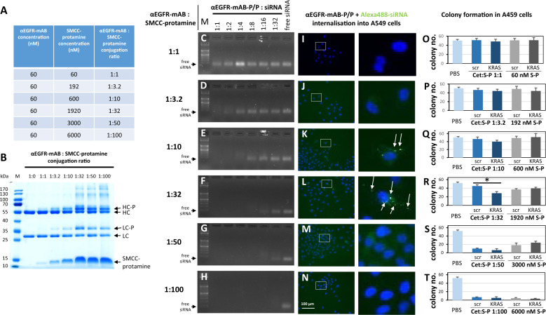
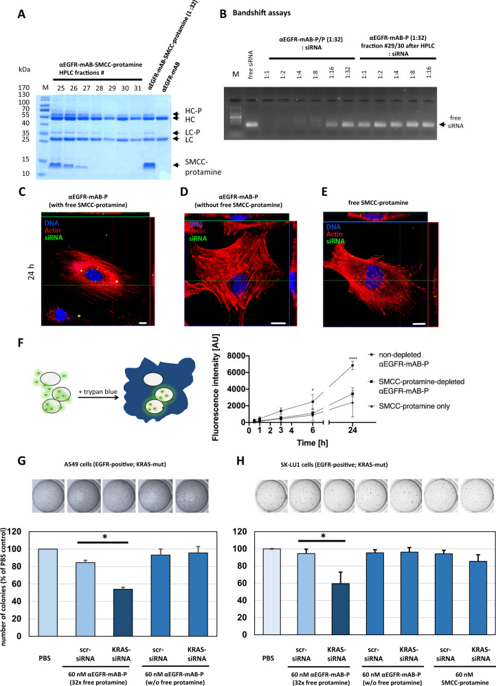
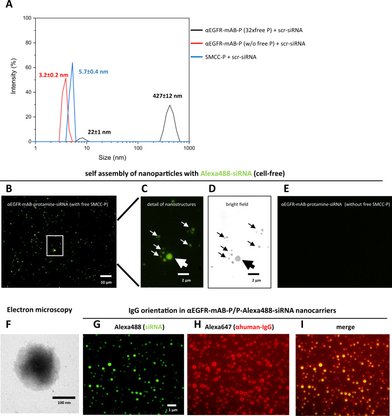
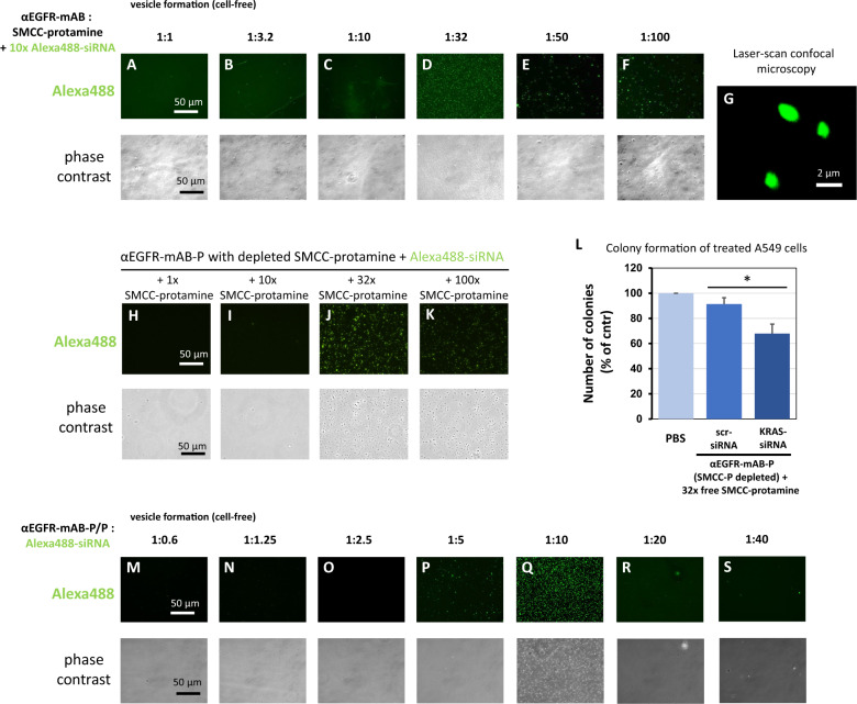
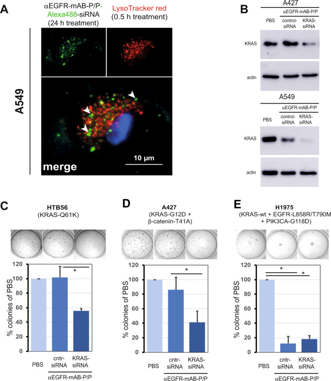
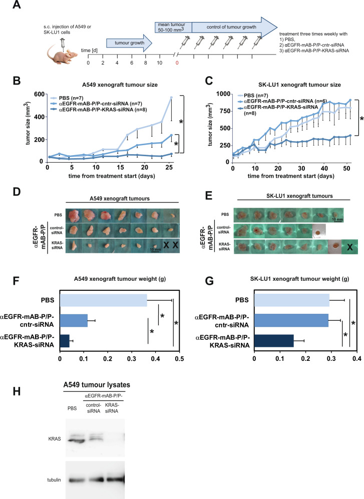
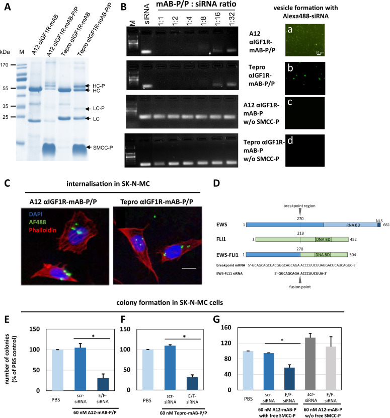
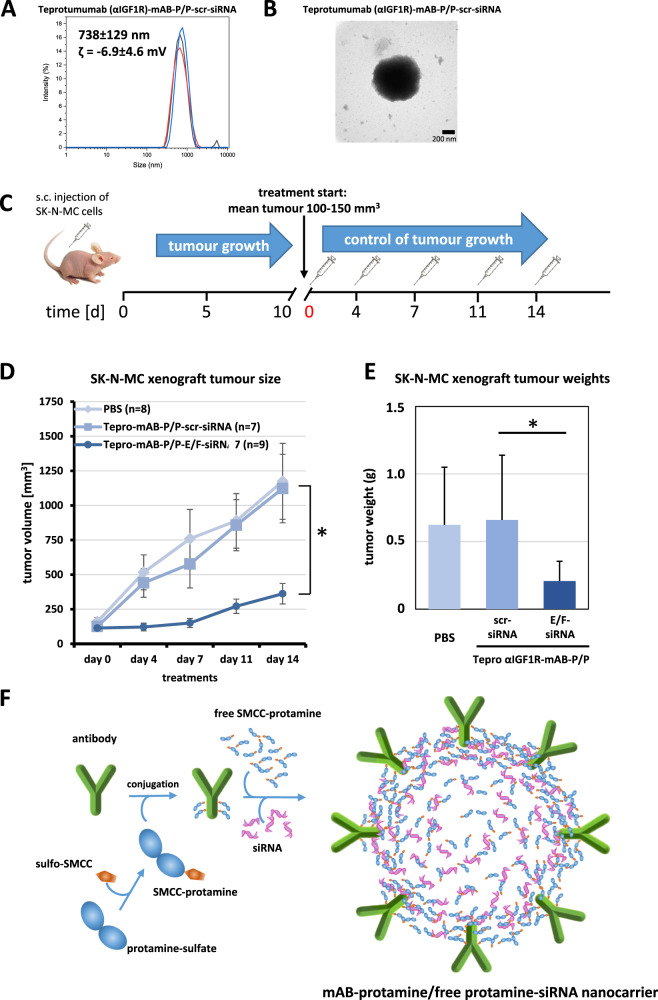
The small arginine-rich protein protamine condenses complete genomic DNA into the sperm head. Here, we applied its high RNA binding capacity for spontaneous electrostatic assembly of therapeutic nanoparticles decorated with tumour-cell-specific antibodies for efficiently targeting siRNA. Fluorescence microscopy and DLS measurements of these nanocarriers revealed the formation of a vesicular architecture that requires presence of antibody-protamine, defined excess of free SMCC-protamine, and anionic siRNA to form. Only these complex nanoparticles were efficient in the treatment of non-small-cell lung cancer (NSCLC) xenograft models, when the oncogene KRAS was targeted via EGFR-mediated delivery. To show general applicability, we used the modular platform for IGF1R-positive Ewing sarcomas. Anti-IGR1R-antibodies were integrated into an antibody-protamine nanoparticle with an siRNA specifically against the oncogenic translocation product EWS/FLI1. Using these nanoparticles, EWS/FLI1 knockdown blocked in vitro and in vivo growth of Ewing sarcoma cells. We conclude that these antibody-protamine-siRNA nanocarriers provide a novel platform technology to specifically target different cell types and yet undruggable targets in cancer therapy by RNAi.


Conflict of interest statement

NB, LW, GL, WEB, and SB have filed two patent applications on electrostatic nanocarrier technology. All other authors disclose no conflict of interest.

Figures

Fig. 1
Fig. 1. Attributes of effective anti-EGFR-mAB-protamine conjugation ratios.
A Concentrations tested and resulting molar ratios of anti-(α)EGFR antibody (αEGFR-mAB) cetuximab to SMCC-protamine for the effective conjugation of both components. B Coomassie-stained SDS-PAGE showing uncoupled αEGFR-mAB cetuximab compared to the conjugation products that were coupled as depicted in A. The formation of a protamine-conjugated heavy chain (HC-P) and light chain (LC-P) showed an optimum at a 1:32 conjugation ratio with no further increase at higher ratios. CH Band-shift assays exhibiting siRNA binding capacity. IN Internalisation of Alexa488-control-siRNA complexed by αEGFR-protamine and free SMCC-protamine (αEGFR-mAB-P/P) in A549 cells. Complexes of αEGFR-mAB-P/P transport Alexa488-siRNA into cells (left panel rectangles), with detailed magnifications (right panels). OT Colony formation assays using the complexes analysed in C-H and I-N in EGFR-positive A549 cells. Significant effect of αEGFR-mAB-P/P transported KRAS siRNA effect in contrast to control scrambled (scr) siRNA is only seen in conjugate preparations with 1:32 molar ratio mAB to protamine (R). Conversely, lower ratios show ineffective binding of siRNA (C, D), do not internalise (I, J) and achieve no sufficient functional effect (OQ), while preparations with higher molar excess of protamine-SMCC show toxicity independent of KRAS knockdown (S, T). A further selection of increments of coupling ratios between 1:20 to 1:40 were presented in Supplementary Fig. 2. Cet:S-P αEGFR-antibody cetuximab conjugated to SMCC-protamine at the indicated ratios, S-P SMCC-protamine. Mean ± SD of three independent experiments. Two-sided t-test, *p < 0.05.
Fig. 2
Fig. 2. Anti-EGFR-mAB-protamine (αEGFR-mAB-P) conjugates do not bind and transport siRNA efficiently after depletion of free SMCC-protamine by HPLC.
A Coomassie-stained SDS-PAGE showing αEGFR-mAB-P, αEGFR-mAB-P coupled with 32x SMCC-protamine and HPLC-fractions 25–31 of anti-EGFR-mAB coupled with 32x SMCC-protamine after depletion of unbound SMCC-protamine; HC heavy chain, LC light chain, -P SMCC-protamine. B Band-shift assays showing that non-SMCC-protamine-depleted αEGFR-mAB-P binds siRNA in a 1:8 to 1:16 molar ratio (left part), whereas the chromatographically depleted αEGFR-mAB-P does not bind siRNA. CE Dynamics of internalisation of Alexa488-siRNA by confocal internalisation studies with depleted vs. non-depleted αEGFR-mAB-P with fluorescence-tagged siRNA on A549 NSCLC cells. Blue fluorescence for nuclei, red for actin, green for internalised Alexa488-siRNA. C Non-depleted αEGFR-mAB-P transports Alexa488-siRNA to A549 intracellular vesicles. Please note the cytoplasmic and perinuclear localisation of Alexa488-positive vesicles (compare also to Supplementary Fig. 5). The purification process leads to abolished internalisation of siRNA (D). SMCC-protamine alone does not work as an unspecific transfection agent (E). Scale bars 10 µm. F Dynamics of Alexa488-siRNA internalisation to A549 cells mediated by non-depleted αEGFR-mAB-P/P vs. SMCC-protamine-depleted αEGFR-mAB-P, controlled by SMCC-protamine only as carrier molecule in flow cytometric analysis (n = 3; error bars indicate SEM). Statistical significance was tested by two-way ANOVA with subsequent Tukey’s multiple comparison test (*p < 0.05, **p < 0.01, ***p < 0.001, ****p < 0.0001). Of note, only the non-SMCC-protamine-depleted conjugate presented a significant siRNA internalisation compared to SMCC-protamine-depleted and SMCC-protamine from 6 h into A549 cells. G, H Functional relevance of free SMCC-protamine in the reaction mixture. Non-depleted αEGFR-mAB-P-transported KRAS siRNA leads to significant reduction of colony formation in NSCLC A549 cells (G) and NSCLC SK-LU1 cells (H) in comparison to control (scrambled, scr) siRNA, whereas the depletion of free SMCC-protamine completely abolishes this effect. SMCC-protamine alone (without αEGFR-mAB) again is not able to efficiently transport and internalise KRAS siRNA seen by no effect in colony assay (H, right bars; Mean ± SD of three independent experiments. Two-sided t-test, *p < 0.05).
Fig. 3
Fig. 3. αEGFR-mAB-protamine (αEGFR-mAB-P) conjugates formed nanoparticles require free SMCC-protamine to form a stable complex with siRNA.
A The 1:32 mAB-protamine conjugate mixture was depleted of excess free SMCC-protamine (free P) by protein G-affinity chromatography. Fractions without excess free SMCC-protamine were compared to fractions still containing excess free SMCC-protamine and the unconjugated SMCC-protamine (without αEGFR-mAB-P) in dynamic light scattering spectroscopy (DLS). Only fractions containing αEGFR-mAB-P, excess unconjugated SMCC-protamine and siRNA exhibited the ability to form larger nanostructures overnight confirmed by dynamic light scattering spectroscopy (black curves, particle size 427 ± 12 nm), but not SMCC-protamine-depleted (red curves, 3.2 nm), or preparations only consisting of free SMCC-protamine and control (scramble, scr) siRNA (blue curves, 5.7 nm) after 2 h of self-assembly. BD Non-purified αEGFR-mAB-P conjugate preparations in complex with Alexa488-siRNA were mounted on glass slides and subjected to fluorescence microscopy. The particles detected in DLS analysis could be verified in microscopy in fluorescence (B and C) as well as bright field microscopy (D, same frame as in C). E αEGFR-mAB-P conjugate depleted from free SMCC-protamine in complex with Alexa488-siRNA were mounted on glass slides and subjected to fluorescence microscopy. No nanoparticles could be observed here. Also, formulations lacking αEGFR-mAB-P, consisting only of SMCC-protamine formed no such particles visible in microscopy (not shown). F αEGFR-mAB-P/P-scrm siRNA nanoparticles were left to form for 2 h and subjected to electron microscopy on copper grids by phospho-Wolfram negative staining. GI αEGFR-mAB-P/P-Alexa488-siRNA nanocarriers formed for 2 h (G, green), were immobilised o/n on treated glass surface, were stained with Alexa647-anti-human-IgG (αhuman-IgG-Alexa647) (H, red). Nanocarrier structures show prominent staining of αhIgG-Alexa647 of the targeting cetuximab antibodies only on surface regions and siRNA within the vesicles (I, overlay).
Fig. 4
Fig. 4. Deciphering preconditions for effective nanoparticle formation between anti-EGFR-mAB-SMCC-protamine conjugate, free SMCC-protamine and siRNA.
AF αEGFR-mAB was conjugated with rising excess of free SMCC-protamine ranging from 1:1 molar ratio to 1:100 excess of SMCC-protamine in chamber slides (see Fig. 1A for reaction details). Resulting conjugates were used to bind siRNA in a cell-free standardised assay. The 1:32 ratio mAB to SMCC-protamine formed a homogeneous population of stable particles in the range up to 0.5–2 µm (D), see detail in G, whereas the other conjugates were incompetent to form stable particles. Stable particles subjected to confocal laser scan (CLS) microscopy optical sections showed a homogeneous distribution of fluorescent Alexa488 signals within the particle (G). HK αEGFR-mAB-P depleted of free SMCC-protamine can form nanoparticles when at 32x free SMCC-protamine (J) is re-added to the conjugate with Alexa488-siRNA. L αEGFR-mAB-P depleted of free SMCC-protamine is effective in the inhibition of A549 cell colony formation when 32x free SMCC-protamine is re-added to the conjugate with anti-KRAS-siRNA in contrast to unspecific control (scrambled, scr) siRNA. Mean ± SD of three independent experiments. Two-sided t-test, *p < 0.05 (two-sided t-test). MS Vesicle formation with 60 nM αEGFR-mAB-P in presence of 32x SMCC-protamine and rising (1:0.6–1:40) molar ratios of Alexa488-control-siRNAs compared to the antibody concentration. Vesicle formation can be observed at 5 to 10x molar excess of siRNA (P, Q). Upper panels: Fluorescence microscopy of Alexa488-siRNA positive vesicle. Lower panels: Phase contrast of the same preparations as in upper panels.
Fig. 5
Fig. 5. The protamine-conjugated EGFR-monoclonal antibody complex binds siRNA, internalises via the endosomal pathway specifically into EGFR-expressing cells, inhibits KRAS expression upon KRAS-siRNA transport, and specifically inhibits cellular colony formation.
A Vesicular tracking in A549 NSCLC cells. Cells treated with αEGFR-mAB-P/Alexa488-siRNA/P were subjected to Lysotracker red staining. The vesicles containing Alexa488 rarely colocalized with Lysotracker staining (merged picture see white arrows for green Alexa-siRNA fluorescence outside red lysosomes). B Western blot for KRAS of two NSCLC cell lines treated with PBS, carrier-control-siRNA and carrier-KRAS siRNA accompanied by blot for actin as loading control (same blot). CE In vitro response of colony growth to treatment with KRAS mutation-specific siRNAs. Cell lines indicated were pre-incubated with antibody-conjugates coupled to siRNAs, resuspended in soft agar and cultivated for ~1 week. Cntr-siRNA, control-siRNA; PBS, vehicle control without active treatment. Mean ± SD of three independent experiments. Two-sided t-test, *p < 0.05. Specific molecular characteristics of the cell lines are given in parenthesis.
Fig. 6
Fig. 6. NSCLC xenograft growth is decreased upon knockdown of KRAS via the anti-EGFR-mAB-protamine (P)/siRNA/P nanostructures.
A Treatment scheme of the in vivo experiments. BF Results of systemic in vivo application of targeted nanocarriers on A549 and SK-LU1 xenograft tumours. B Tumour growth curves for A549 (means–SEM). C Tumour growth curves for SK-LU1 (means–SEM). Both tumour growth curves show significant reduction of tumour growth in anti-KRAS siRNA treatment groups. D, E Size of excised tumours at the end of the respective experiments (X = individual tumours in anti-KRAS siRNA group were treated to extinction). F, G Weight statistics of the excised tumours (g, gram). H Proof of KRAS oncogene knockdown in excised tumour tissue by KRAS western blot ex vivo. Cntr control scrambled siRNA. *denote significant differences between groups (p < 0.05, two-sided t-test).
Fig. 7
Fig. 7. Knockdown of Ewing-specific EWS-FLI1 fusion protein by αIGF1R-mAB-protamine/siRNA/protamine nanoparticles targeting Ewing sarcoma cells.
A IGF1R-targeting mABs A12 (cixutumumab) and Tepro (teprotumumab) were expressed and purified using a GMP-like method and then conjugated to protamine to enable siRNA binding and transport. IgG-protamine conjugates exhibit a decent molecular weight shift (arrows). HC heavy chain, LC light chain, -P SMCC-protamine. B Band-shift assay using αIGF1R-mABs-protamine (-P) and different ratios of siRNA (left panels). Antibody-protamine complexes without free SMCC-protamine do not bind siRNA anymore. Right panels (a–d): αIGF1R-mAB-P in presence of free SMCC-protamine can form nanoparticles with Alexa488-control-siRNA (a: A12, b: Tepro). αIGF1R-mAB-P depleted from free SMCC-protamine cannot form these nanoparticles with Alexa488-control-siRNA (c: A12, d: Tepro). C Anti-IGF1R-mAB-protamine with free SMCC-protamine shuttled Alexa488-marked control-siRNA to SK-N-MC Ewing cells (left panel: A12-mAB-P, right panel: Tepro-mAB-P). Bar represents 10 µm. D Schematic representation of the breakpoint regions in the human EWS protein, the FLI-1 protein and the resulting oncogenic fusion protein EWS-FLI1 along with the breakpoint mRNA sequence and the EWS-FLI1-specific siRNA sequence that was used for this study (see EG and Fig. 8) (BD binding domain, NLS nuclear localisation signal). EG SK-N-MC cells were treated with protamine-conjugated A12 (E) or Tepro (F) formed siRNA nanoparticles as indicated and subjected to colony formation assays. E/F–siRNA is an siRNA interfering with the mRNA of the driving Ewing sarcoma EWS-FLI1 as depicted in D. G SK-N-MC cells treated with αIGF1R (A12)-mAB-protamine/EWS-FLI1-siRNA/P nanoparticles form significantly less colonies in soft agar than cells treated with αIGF1R (A12)-mAB-protamine/contr (scr)-siRNA/P. No differences in colony formation can be observed when SK-N-MC cells were treated with αIGF1R-mAB-protamine conjugates without free SMCC-protamine not resulting in the formation of nanocarriers. Shown here are means of three independent experiments ± SD. Asterisk indicates significant differences (p < 0.05, two-sided t-test).
Fig. 8
Fig. 8. Ewing sarcoma xenograft tumour growth is inhibited upon knockdown of oncogenic EWS-FLI1 translocation product through systemic therapy with αIGF1R-mAB-protamine-siRNA-protamine nanocarriers.
A Dynamic light scattering spectroscopy (DLS) of teprotumumab (αIGF1R)-mAB-protamine-siRNA-protamine nanocarriers reveals a particles size of 738 ± 129 nm with a zeta-potential of −6.9 ± 4.6 mV. B αIGF1R-mAB-P/P-scrm siRNA nanoparticles were left to form for 2 h and subjected to electron microscopy on copper grids by phospho-Wolfram negative staining. C Treatment scheme of the in vivo experiments. Nanoparticles were given intraperitoneally as visualised. D, E Results of systemic in vivo application of targeted nanocarriers on SK-N-MC xenograft tumours. D Tumour growth curves SK-N-MC treated with αIGF1R-mAB teprotumumab (“Tepro”)-protamine-siRNA/P nanoparticles (means ± SEM; two-sided t-test, *p < 0.05). E Weight statistics of the excised tumours at the end of the experiment (mean ± SD. Two-sided t-test, *p < 0.05). F Illustration of a cross section through an idealised nanoparticle structure fulfilling the conditions for an effective antibody-protamine-siRNA-SMCC-protamine nanocarrier complex deduced from our experiments. Electrostatic binding bridges are formed between mAB, with some protamines (cationic) coupled to the targeting antibody, siRNA (anionic), and free SMCC-protamine (cationic). The nanostructures assemble spontaneously into the optimal and most stable electrostatic status and function as nanocarriers for siRNA.

References

    1. Carr JA, Silverman N. The heparin-protamine interaction. A review. J Cardiovasc Surg. 1999;40:659–66. - PubMed
    1. Mochizuki T, Olson PJ, Szlam F, Ramsay JG, Levy JH. Protamine reversal of heparin affects platelet aggregation and activated clotting time after cardiopulmonary bypass. Anesth Analg. 1998;87:781–5. doi: 10.1213/00000539-199810000-00008. - DOI - PubMed
    1. Rossmann P, Matousovic K, Horácek V. Protamine-heparin aggregates. Their fine structure, histochemistry, and renal deposition. Virchows Arch B Cell Pathol Incl Mol Pathol. 1982;40:81–98. doi: 10.1007/BF02932853. - DOI - PubMed
    1. Smull CE, Ludwig EH. Enhancement of the plaque-forming capacity of poliovirus ribonucleic acid with basic proteins. J Bacteriol. 1962;84:1035–40. doi: 10.1128/jb.84.5.1035-1040.1962. - DOI - PMC - PubMed
    1. Pardi N, Hogan MJ, Porter FW, Weissman D. mRNA vaccines—a new era in vaccinology. Nat Rev Drug Discov. 2018;17:261–79. doi: 10.1038/nrd.2017.243. - DOI - PMC - PubMed

Publication types

MeSH terms