Review
. 2022 Apr 8;8(4):667-692.
doi: 10.1021/acsinfecdis.1c00546. Epub 2022 Feb 27.

Aptamers for Viral Detection and Inhibition

Affiliations
Review

Aptamers for Viral Detection and Inhibition

Banani Chakraborty et al. ACS Infect Dis. .

Abstract

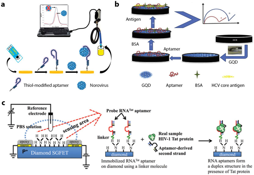
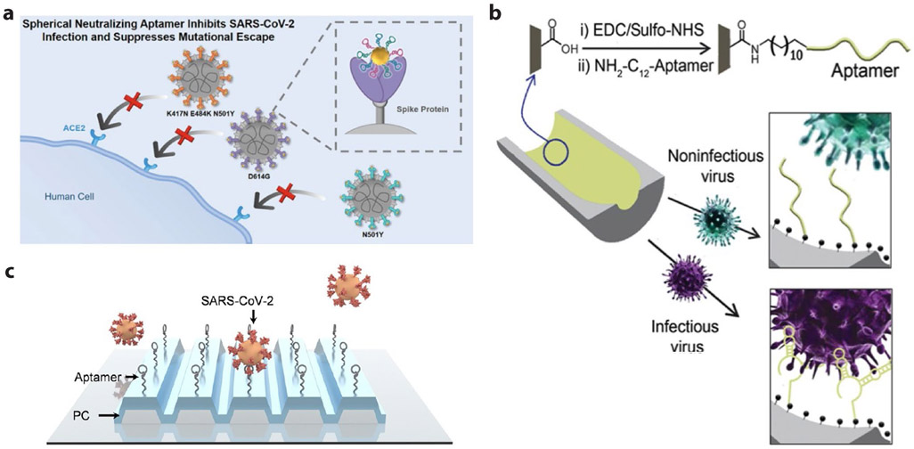
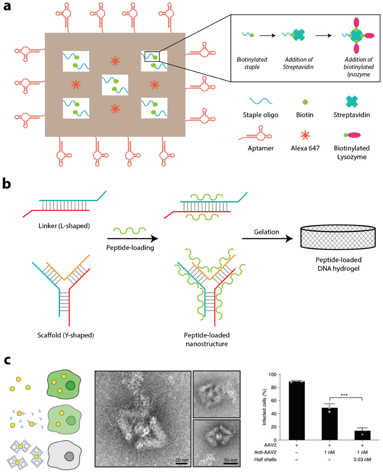
Recent times have experienced more than ever the impact of viral infections in humans. Viral infections are known to cause diseases not only in humans but also in plants and animals. Here, we have compiled the literature review of aptamers selected and used for detection and inhibition of viral infections in all three categories: humans, animals, and plants. This review gives an in-depth introduction to aptamers, different types of aptamer selection (SELEX) methodologies, the benefits of using aptamers over commonly used antibody-based strategies, and the structural and functional mechanism of aptasensors for viral detection and therapy. The review is organized based on the different characterization and read-out tools used to detect virus-aptasensor interactions with a detailed index of existing virus-targeting aptamers. Along with addressing recent developments, we also discuss a way forward with aptamers for DNA nanotechnology-based detection and treatment of viral diseases. Overall, this review will serve as a comprehensive resource for aptamer-based strategies in viral diagnostics and treatment.

Keywords: DNA nanostructures; aptamers; aptasensors; inhibition; sensing; viruses.


Figures

Figure 1.
Figure 1.
Aptamer configurations and targeting. (a) Secondary structures of the aptamers. Mechanism of aptamer binding through molecular recognition and folding for (b) biosensing and (c) drug delivery.
Figure 2.
Figure 2.. Outline of SELEX.
(a) A degenerate nucleic-acid sequence library is incubated with the target molecule under defined solution conditions. (b) Target-bound nucleic acids are partitioned. (c–e) Species with lower binding affinity are removed and the bound species are eluted, allowing preferential amplification of higher affinity species. This enriched pool is then used as the starting point in subsequent cycles. Typically, 10 to 20 cycles are carried out before aptamer characterization. In early rounds, species with no affinity are competed out of the pool. In later rounds, molecules with affinity compete for binding sites on the target. Such competition results in enhancement of the pool binding-affinity in a manner similar to Darwinian evolution. Recent technical developments described in the text are listed alongside each step in brackets. CE, capillary electrophoresis; SELEX, systematic evolution of ligands by exponential enrichment; SPR, surface plasmon resonance. Image reproduced with permission from ref. . Copyright 2006 Springer Nature.
Figure 3.
Figure 3.. Electrochemical aptasensors.
(a) A thiolated norovirus-specific DNA aptamer self-assembled onto a gold nanoparticle-modified screen-printed carbon electrode. Binding of the virus to the immobilized aptamer causes a decrease in the redox current, measured via square wave voltammetry. Reproduced from ref.. (b) Use of glassy carbon electrode (GCE) with graphene quantum dots for HCV core antigen detection. Reproduced with permission from ref. . Copyright 2017 Elsevier. (c) Schematic structure of diamond-FET-based RNA aptamer for HIV-1 Tat protein detection based on changes in the surface charge. Reproduced with permission from ref. . Copyright 2013 Elsevier.
Figure 4.
Figure 4.. Enzyme-linked electrochemical aptasensors.
(a) Enzyme catalysis in ultra-low ion strength media to develop an ion strength increase-based impedance biosensor for H5N1 virus. Reproduced from ref.. (b) Schematic diagram of H5N1 viral protein detection using the enzymatic reaction of the substrate 4-amino phenyl phosphate with the surface formed aptamer/H5N1/antiH5N1-alkaline phosphatase on gold nanoparticle-modified screen-printed carbon electrode. Reproduced with permission from ref. . Copyright 2015 Elsevier. (c) Working principle of the norovirus nanozyme aptasensor. Reproduced with permission from ref. . Copyright 2019 American Chemical Society.
Figure 5.
Figure 5.. Optical aptasensors.
(a) SPR based aptasensor: upon binding the target (virus) the surface plasmon angle of the reflected light changes resulting in a difference in plasmon resonance. (b) LSPR based biosensor for real-time detection: a large array of nanoantennas is incorporated into a microfluidic chamber system that guides analyte solutions precisely over the sensitive area. Optical readout is realized with a spectrometer and spectra are continuously recorded upon chemical reactions; the inset illustrates the investigated biochemical reaction, which is immobilization, backfilling, and hybridization of short DNA sequences. Reproduced from ref.. (c) Schematic illustration of the preparation of aptamer-Ag@SiO2 sensor and the determination of rHA protein of H5N1. Reproduced with permission from ref. . Copyright 2015 Elsevier. (d) Schematic of selective virus sizing and counting by fluorescent nanoparticle tracking. Reproduced from ref.. (e) SERS imaging-based assay using a 3D nano-popcorn plasmonic aptasensor: (i) Detection of DNA using Cy3-labeled aptamer probes (left) or recognition of A/H1N1 virus (right), (ii) resulting in increased Raman signal (left) or decreased Raman signal intensity (right), respectively. Reproduced with permission from ref. . Copyright 2020 Elsevier. (f) A sandwich-like aptasensor for influenza virus detection: 1) primary aptamer is immobilized onto Ag nanoparticles, 2) virus is captured with primary aptamers, 3) secondary aptamers interact with virus, providing the SERS signal. Reproduced from ref., (g) Schematic representation of the construction of a chemiluminescence aptasensor based on magnetic separation and immunoassay. Reproduced from ref., (h) Working principle for the single universal aptamer detection of different kinds of influenza viruses under two different reaction conditions. Reproduced with permission from ref. . Copyright 2016 Elsevier. (i) Molecular beacon aptamer strategy for analyzing the viral protein (Tat). (j) Protein-binding aptamer assisted detection of the H1N1 influenza A virus based on fluorescence polarization. Reproduced with permission from ref. . Copyright 2013 Royal Society of Chemistry.
Figure 6.
Figure 6.. Non-electrochemical aptasensors.
(a) Mechanism for direct and indirect ELONA where the virus is immobilized on the surface or the aptamer is immobilized on surface, respectively. (b) Mechanism for aptamer based lateral flow assay: LFA strip includes positive control line with antibody binding to the target virus and a test line with streptavidin immobilized aptamer. Upon binding of the target virus, the AuNP-Ab complex shows the right signal; in absence of virus no line is visible in test region.
Figure 7.
Figure 7.. DNA aptamers selected for SARS-CoV-2 viral detection and inhibition.
(a) Schematic of SNAP to block the interaction between the RBD of SARS-CoV-2 and host ACE2 with synergetic strategy of multivalent multisite binding and steric hindrance. Reproduced from ref.. (b) Scheme of Infectious virus detection using aptamer-functionalized nanopore sensors. Reproduced from ref. (c).Working principle of label-free optical detection for intact SARS-CoV-2 using surface immobilized DNA aptamers and PRISM system. Reproduced with permission from ref. . Copyright 2022 American Chemical Society.
Figure 8.
Figure 8.. DNA nanostructure based viral detection.
(a) Schematic illustration of construction of DNA-tetrahedra-based electrochemical immunosensor, which senses the pathogen using redox-labelled antibody attached to the top vertex of the tetrahedron. (b) Star-shaped DNA architecture, carrying five molecular beacon-like motifs and five FRET pairs. Binding of DENV reconfigures the structure, resulting in a FRET signal due to the change in distance between the dye pairs. (c) Fluorescently labelled DNA nanobarcodes for detecting a mixture of viral pathogens. (d) Detection of HIV DNA by fluorescent labelled DNA sensors combined with enzyme-based rolling cycle amplification. (e) DNA nanoswitches reconfigure from a linear “off” state to a looped “on” state on detecting viral RNA.
Figure 9.
Figure 9.. DNA nanostructures for viral therapy.
(a) Scheme of aptamer-functionalized DNA origami nanostructure loaded with antibacterial peptide. The aptamers are decorated around the origami to target four bacteria in parallel and treat the disease. (b) Schematic representation of synthesis of peptide-loaded DNA hydrogel via electrostatic crosslinking process for controlled drug delivery. (c) Left: scheme of inhibiting AAV2 infection by DNA origami half shells; Middle: TEM images of AAV2 virus particles captured by DNA origami half shells; Right: Quantification of infected cells for conditions with AAV2 virus only, AAV2 plus antibody at 1 nM (IC50 concentration), and AAV2 plus DNA origami half shells decorated with antibodies inside shells. Reproduced with permission from ref. . Copyright 2021 Springer Nature.

Similar articles

Cited by

References

    1. Assaad F; Borecka I, Nine-year study of WHO virus reports on fatal viral infections. Bull World Health Organ 1977, 55 (4), 445–453. - PMC - PubMed
    1. Tobin NH; Campbell AJP; Zerr DM; Melvin AJ, Life-Threatening Viral Diseases and Their Treatment. In Pediatric Critical Care, 2011, 1324–1335.
    1. Keaney D; Whelan S; Finn K; Lucey B, Misdiagnosis of SARS-CoV-2: A Critical Review of the Influence of Sampling and Clinical Detection Methods. Med. Sci 2021, 9 (2), 36. - PMC - PubMed
    1. Ménard-Moyon C; Bianco A; Kalantar-Zadeh K, Two-Dimensional Material-Based Biosensors for Virus Detection. ACS Sensors 2020, 5 (12), 3739–3769. - PubMed
    1. Kevadiya BD; Machhi J; Herskovitz J; Oleynikov MD; Blomberg WR; Bajwa N; Soni D; Das S; Hasan M; Patel M; Senan AM; Gorantla S; McMillan J; Edagwa B; Eisenberg R; Gurumurthy CB; Reid SPM; Punyadeera C; Chang L; Gendelman HE, Diagnostics for SARS-CoV-2 infections. Nat. Mater 2021, 20 (5), 593–605. - PMC - PubMed

Publication types

Substances