. 2022 Oct;18(10):2397-2408.
doi: 10.1080/15548627.2022.2038501. Epub 2022 Feb 27.

Autophagy deficiency abolishes liver mitochondrial DNA segregation

Affiliations

Autophagy deficiency abolishes liver mitochondrial DNA segregation

Katiane Tostes et al. Autophagy. 2022 Oct.

Abstract

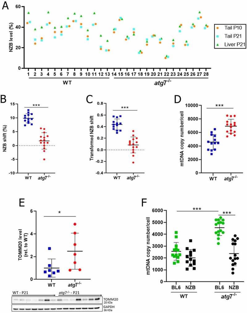
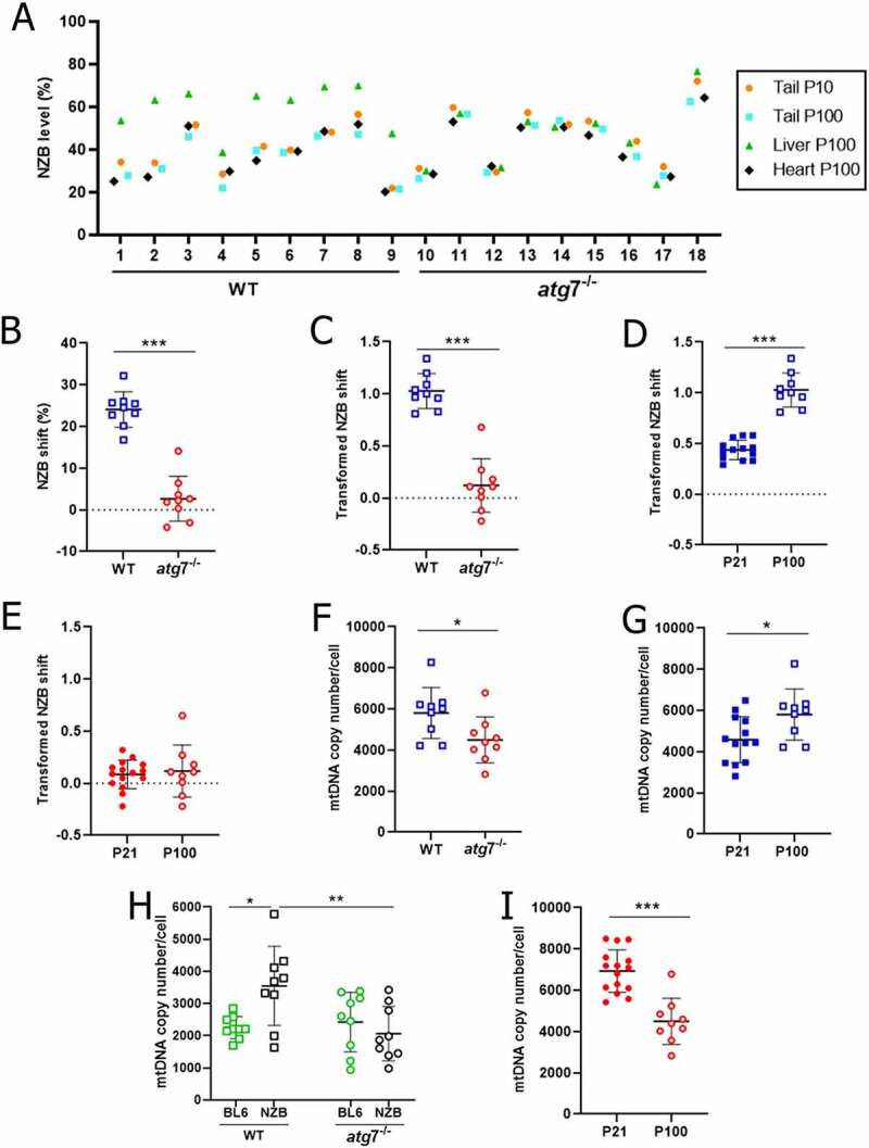
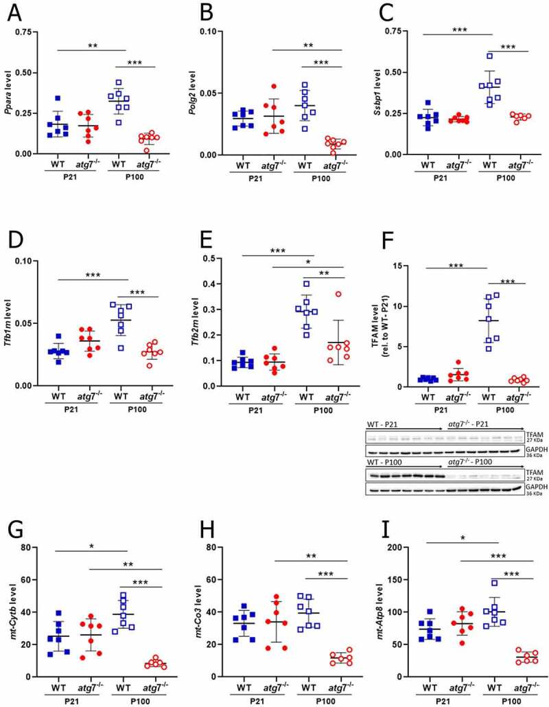
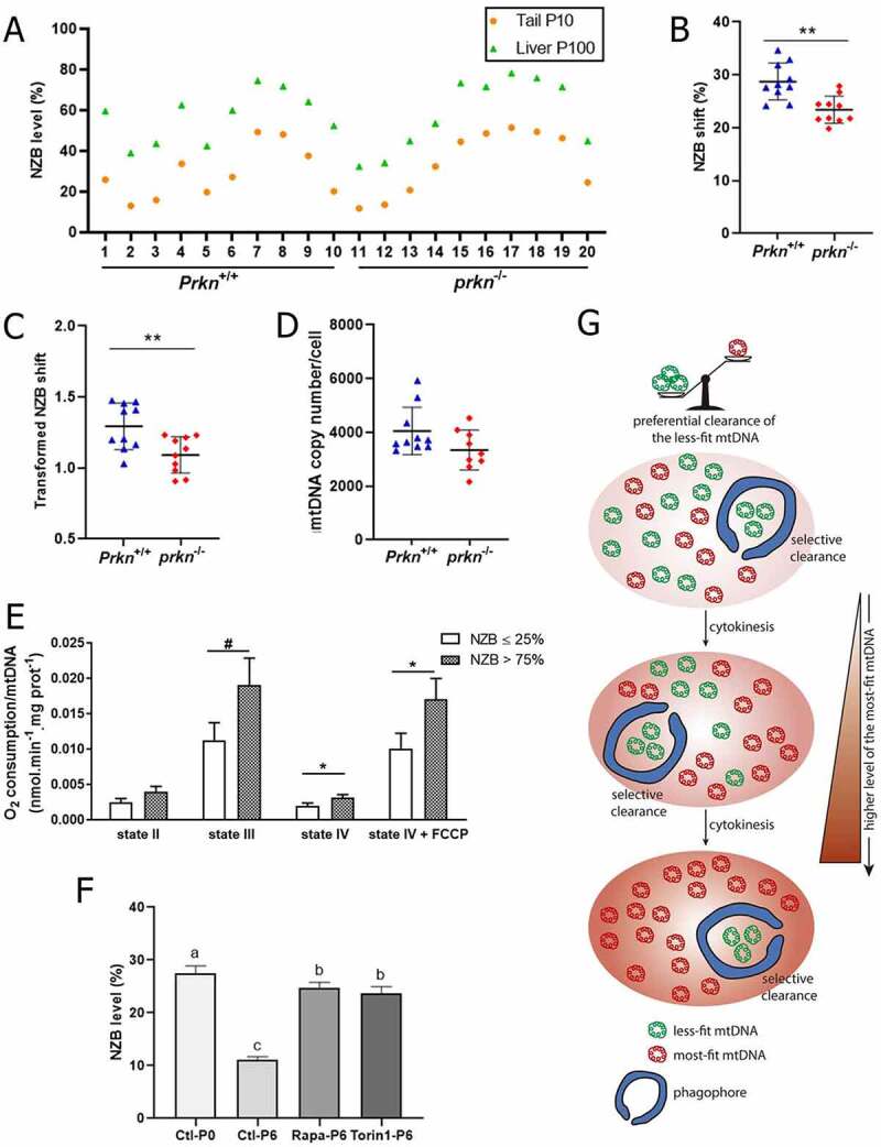
Mutations in the mitochondrial genome (mtDNA) are ubiquitous in humans and can lead to a broad spectrum of disorders. However, due to the presence of multiple mtDNA molecules in the cell, co-existence of mutant and wild-type mtDNAs (termed heteroplasmy) can mask disease phenotype unless a threshold of mutant molecules is reached. Importantly, the mutant mtDNA level can change across lifespan as mtDNA segregates in an allele- and cell-specific fashion, potentially leading to disease. Segregation of mtDNA is mainly evident in hepatic cells, resulting in an age-dependent increase of mtDNA variants, including non-synonymous potentially deleterious mutations. Here we modeled mtDNA segregation using a well-established heteroplasmic mouse line with mtDNA of NZB/BINJ and C57BL/6N origin on a C57BL/6N nuclear background. This mouse line showed a pronounced age-dependent NZB mtDNA accumulation in the liver, thus leading to enhanced respiration capacity per mtDNA molecule. Remarkably, liver-specific atg7 (autophagy related 7) knockout abolished NZB mtDNA accumulat ion, resulting in close-to-neutral mtDNA segregation through development into adulthood. prkn (parkin RBR E3 ubiquitin protein ligase) knockout also partially prevented NZB mtDNA accumulation in the liver, but to a lesser extent. Hence, we propose that age-related liver mtDNA segregation is a consequence of macroautophagic clearance of the less-fit mtDNA. Considering that NZB/BINJ and C57BL/6N mtDNAs have a level of divergence comparable to that between human Eurasian and African mtDNAs, these findings have potential implications for humans, including the safe use of mitochondrial replacement therapy.Abbreviations: Apob: apolipoprotein B; Atg1: autophagy-related 1; Atg7: autophagy related 7; Atp5a1: ATP synthase, H+ transporting, mitochondrial F1 complex, alpha subunit 1; BL6: C57BL/6N mouse strain; BNIP3: BCL2/adenovirus E1B interacting protein 3; FCCP: carbonyl cyanide 4-(trifluoromethoxy)phenylhydrazone; GAPDH: glyceraldehyde-3-phosphate dehydrogenase; MAP1LC3A: microtubule-associated protein 1 light chain 3 alpha; MAP1LC3B: microtubule-associated protein 1 light chain 3 beta; mt-Atp8: mitochondrially encoded ATP synthase 8; MT-CO1: mitochondrially encoded cytochrome c oxidase I; MT-CO2: mitochondrially encoded cytochrome c oxidase II; mt-Co3: mitochondrially encoded cytochrome c oxidase III; mt-Cytb: mitochondrially encoded cytochrome b; mtDNA: mitochondrial DNA; MUL1: mitochondrial ubiquitin ligase activator of NFKB 1; nDNA: nuclear DNA; Ndufa9: NADH:ubiquinone oxireductase subunit A9; NDUFB8: NADH:ubiquinone oxireductase subunit B8; Nnt: nicotinamide nucleotide transhydrogenase; NZB: NZB/BINJ mouse strain; OXPHOS: oxidative phosphorylation; PINK1: PTEN induced putative kinase 1; Polg2: polymerase (DNA directed), gamma 2, accessory subunit; Ppara: peroxisome proliferator activated receptor alpha; Ppia: peptidylprolyl isomerase A; Prkn: parkin RBR E3 ubiquitin protein ligase; P10: post-natal day 10; P21: post-natal day 21; P100: post-natal day 100; qPCR: quantitative polymerase chain reaction; Rpl19: ribosomal protein L19; Rps18: ribosomal protein S18; SD: standard deviation; SEM: standard error of the mean; SDHB: succinate dehydrogenase complex, subunit B, iron sulfur (Ip); SQSTM1: sequestosome 1; Ssbp1: single-stranded DNA binding protein 1; TFAM: transcription factor A, mitochondrial; Tfb1m: transcription factor B1, mitochondrial; Tfb2m: transcription factor B2, mitochondrial; TOMM20: translocase of outer mitochondrial membrane 20; UQCRC2: ubiquinol cytochrome c reductase core protein 2; WT: wild-type.

Keywords: Atg7; NZB; heteroplasmy; mitochondria; mitophagy; parkin.


Conflict of interest statement

No potential conflict of interest was reported by the author(s).

Figures

Figure 1.
Figure 1.
Liver mtDNA segregation relied on Atg7. (A) NZB mtDNA level in the liver (P21) and tail (P10 and P21) for WT (n = 13) and atg7−/− (n = 15). (B) Difference in NZB mtDNA level (NZB shift), within mouse, between liver (P21) and tail (P21) for WT (n = 13) and atg7−/− (n = 15). (C) Transformed NZB shift = ln(h(h0-1)/h0(h-1)). h, liver NZB level (proportion) at P21. h0 = tail NZB level (proportion) at P21 for WT (n = 13) and atg7−/− (n = 15). (D) Liver total mtDNA copy number/cell at P21 for WT (n = 13) and atg7−/− (n = 15). (E) Protein levels of TOMM20 in the liver at P21 for WT (n = 13) and Atg7−/− (n = 15), normalized by GAPDH amount. TOMM20 levels are shown in relation to the WT control. Western blotting membranes of target proteins in the liver are presented in the bottom. (F) Copies of NZB and BL6 mtDNA/cell in the liver at P21 for WT (n = 13) and atg7−/− (n = 15). Data information: In (A), dots are indicative of individual values in the liver (P21) and tail (P10 and P21) of each mouse. In (B-F), dots are indicative of individual values of each mouse, while solid horizontal lines and whiskers represent the mean ± SD. Horizontal lines above dots in (B-F) refer to statistical comparisons of the mean values, where *P < 0.05 and ***P < 0.0001. See also Figure S2.
Figure 2.
Figure 2.
Impaired autophagy abolished liver NZB mtDNA segregation during development into adulthood. (A) NZB mtDNA level in the liver (P100), heart (P100) and tail (P10 and P100) for WT (n = 9) and atg7−/− (n = 9). (B) Difference in NZB mtDNA level (NZB shift), within mouse, between liver (P100) and tail (P100) for WT (n = 9) and atg7−/− (n = 9). (C) Transformed NZB shift = ln(h(h0-1)/h0(h-1)). h, liver NZB level (proportion) at P100. h0 = tail NZB level (proportion) at P100 for WT (n = 9) and atg7−/− (n = 9). (D-E) Transformed NZB shift in WT (D) and atg7−/− (E) livers at P21 (WT, n = 13; atg7−/−, n = 15) and P100 (WT, n = 9; atg7−/−, n = 9). P21 data correspond to Figure 1C. (F) Liver mtDNA copy number/cell at P100 for WT (n = 9) and atg7−/− (n = 9). (G) Liver total mtDNA copy number/cell in WT at P21 (n = 13) and P100 (n = 9). P21 data correspond to Figure 1D. (H) Copies of NZB and BL6 mtDNA/cell in the liver at P100 for WT (n = 9) and atg7−/− (n = 9). (I) Liver total mtDNA copy number/cell in atg7−/− (H) mice at P21 (n = 15) and P100 (n = 9); P21 data correspond to Figure 1D. Data information: in (A), dots are indicative of individual values in the liver (P100), heart (P100) and tail (P10 and P100) of each mouse. In (B-I), dots are indicative of individual values of each mouse, while solid horizontal lines and whiskers represent the mean ± SD. Horizontal lines above dots in (B-I) refer to statistical comparisons of the mean values, where *P < 0.05, **P < 0.01 and ***P < 0.0001. See also Figure S2.
Figure 3.
Figure 3.
Autophagy arrest downregulated in the liver of adult mice factors required for mtDNA replication and transcription, resulting in lower mtDNA-encoded OXPHOS transcripts. (A-H) Transcript levels of Ppara (A), Polg2 (B), Ssbp1 (C), Tfb1m (D) and Tfb2m (E) in the liver at P21 (WT, n = 7; atg7−/−, n = 7) and P100 (WT, n = 7; atg7−/−, n = 7), normalized by Rps18 transcript levels. (F) Protein levels of TFAM in the liver at P21 (WT, n = 7; atg7−/−, n = 7) and P100 (WT, n = 7; atg7−/−, n = 7), normalized by GAPDH amount. TFAM levels are shown in relation to the WT control at P21. Western blotting membranes of target proteins in the liver are presented in the bottom. Transcript levels of mt-Cytb (G), mt-Co3 (H) and mt-Atp8 (I) in the liver at P21 (WT, n = 7; atg7−/−, n = 7) and P100 (WT, n = 7; atg7−/−, n = 7), normalized by the geometric mean of Rpl19 and Ppia transcript levels. Data information: Dots are indicative of individual values of each mouse, while solid horizontal lines and whiskers represent the mean ± SD. Horizontal lines above dots refer to statistical comparisons of the mean values, where *P < 0.05, **P < 0.01 and ***P < 0.0001. See also Figure S3.
Figure 4.
Figure 4.
NZB accumulation in the liver had a minor contribution of Prkn and associated with enhanced respiration capacity per mtDNA molecule. (A) NZB mtDNA level in the liver (P100) and tail (P10) for Prkn+/+ (n = 10) and prkn−/− (n = 10). (B) Difference in NZB mtDNA level (NZB shift), within mouse, between liver (P100) and tail (P10) for Prkn+/+ (n = 10) and prkn−/− (n = 10). (C) Transformed NZB shift = ln(h(h0-1)/h0(h-1)). h = liver NZB level (proportion) at P100. h0 = tail NZB level (proportion) at P10 for Prkn+/+ (n = 10) and prkn−/− (n = 10). (D) Liver mtDNA copy number/cell at P100 for Prkn+/+ (n = 10) and prkn−/− (n = 10). (E) O2 consumption per mtDNA molecule assessed in liver isolated mitochondria under states II, III (ADP), IV (oligomycin) and IV + FCCP. Isolated mitochondria contained ≤ 25% (ranging from 5% to 25%; n = 6) or > 75% (ranging from 76% to 87%; n = 6) NZB mtDNA. (F) Level of NZB mtDNA assessed in cultured fibroblasts at passage 0 (P0) and 6 (P6). Fibroblasts were treated from P0 to P6 with either 1 μM rapamycin (Rapa, n = 3) or 1 μM torin 1 (n = 3); control (Ctl, n = 3). (G) Hypothetical model explaining liver mtDNA segregation. Liver mitochondrial segregation is a consequence of autophagic clearance of the less-fit mtDNA variant. As a result, this mechanism drives a rapid age-dependent accumulation of the most-fit mtDNA, potentially leading to variant fixation. Data information: In (A), dots are indicative of individual values in the liver (P100) and tail (P10) of each mouse. In (B-D), dots are indicative of individual values of each mouse, while solid horizontal lines and whiskers represent the mean ± SD. In (E-F), bars and whiskers represent the mean ± SEM. Horizontal lines above dots or bars in (B-E) refer to statistical comparisons of the mean values, where *P < 0.05, **P < 0.01 and #P = 0.0656. Different letters above bars in (F) indicate significant statistical difference (P < 0.05). See also Figure S4.

References

    1. Craven L, Alston CL, Taylor RW, et al. Recent advances in mitochondrial disease. Annu Rev Genomics Hum Genet. 2017;18(1):257–275. - PubMed
    1. Stewart JB, Chinnery PF.. Extreme heterogeneity of human mitochondrial DNA from organelles to populations. Nat Rev Genet. 2020;22(2):106–118. - PubMed
    1. Gorman GS, Schaefer AM, Ng Y, et al. Prevalence of nuclear and mitochondrial DNA mutations related to adult mitochondrial disease. Ann Neurol. 2015;77(5):753–759. - PMC - PubMed
    1. Wallace DC. Mitochondrial genetic medicine. Nat Genet. 2018;50(12):1642–1649. - PubMed
    1. Payne BAI, Wilson IJ, Yu-Wai-Man P, et al. Universal heteroplasmy of human mitochondrial DNA. Hum Mol Genet. 2013;22(2):384–390. - PMC - PubMed

Publication types

MeSH terms

Substances