

A standardized assay for the quantitative detection of serum HBV RNA in chronic hepatitis B patients
- PMID: 35220917
- PMCID: PMC8920369
- DOI: 10.1080/22221751.2022.2045874
A standardized assay for the quantitative detection of serum HBV RNA in chronic hepatitis B patients
Abstract
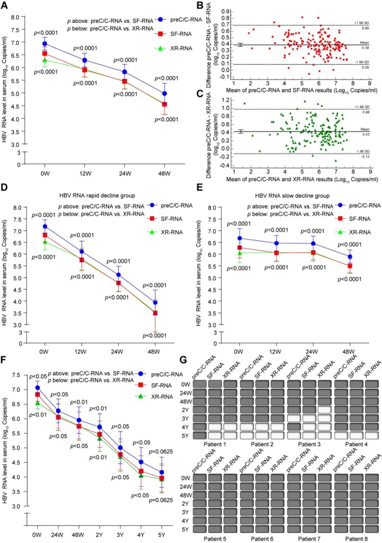
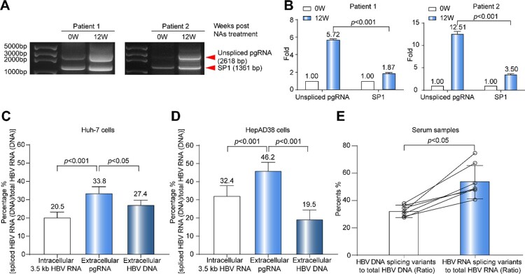
Serum hepatitis B virus (HBV) pregenomic RNA (pgRNA) is a surrogate marker for reflecting the transcriptional activity of covalently closed circular DNA. However, there is still no standardized assay for the quantitative detection of serum HBV RNA in chronic hepatitis B patients. In this study, quantitative polymerase chain reactions for detecting the preC/C-RNA (preC/C region HBV pgRNA), SF-RNA (splicing variants-free pgRNA) and XR-RNA (X region remained pgRNA) regions were set up. The dynamic changes of serum pgRNA splicing variants and 3' terminal truncations were analysed in three retrospective cohorts: 35 treatment-naive chronic HBV-infected patients (cohort A), 52 chronic hepatitis B (CHB) patients who received nucleos(t)ide analogs (NAs) therapy for 48 weeks (cohort B) and eight CHB patients who are under long-term NAs treatment (cohort C). The accuracy and sensitivity of HBV RNA detection were assessed by the National Standard of HBV RNA. We confirmed that high proportions of pgRNA splicing variants and 3' terminal truncations were present and significantly affect the quantitative detection of serum HBV RNA in both treatment-naive and NAs-treated CHB patients. To achieve the higher accuracy and sensitivity on the detection of HBV RNA level, the primers and probes should be designed at the 5' terminal region of HBV genome and outside the mainly spliced sequence of pgRNA, especially for CHB patients under long-term NAs treatment. This study would help to better understand the significance of the pgRNA splicing variants and 3' terminal truncations, and further guide the clinical detection of serum HBV RNA.
Keywords: 3′ terminal truncations; HBV RNA; nucleos(t)ide analogs; quantitative detection; splicing variants.
Conflict of interest statement
One patent which belongs to Peking University of China is relevant to this work. GY, RC, YL, JZ, ZG, JW and FL are employees of Peking University.
Figures


Similar articles
-
Serum hepatitis B virus RNA detectability, composition and clinical significance in patients with ab initio hepatitis B e antigen negative chronic hepatitis B.Virol J. 2022 Jan 29;19(1):22. doi: 10.1186/s12985-022-01749-7. Virol J. 2022. PMID: 35093105 Free PMC article.
-
Factors and virological significance of hepatitis B virus pregenomic RNA status after 5 years of antiviral therapy.Int J Infect Dis. 2021 Apr;105:418-423. doi: 10.1016/j.ijid.2021.02.116. Epub 2021 Mar 3. Int J Infect Dis. 2021. PMID: 33676002
-
Diagnostic Value of Detection of Pregenomic RNA in Sera of Hepatitis B Virus-Infected Patients with Different Clinical Outcomes.J Clin Microbiol. 2020 Jan 28;58(2):e01275-19. doi: 10.1128/JCM.01275-19. Print 2020 Jan 28. J Clin Microbiol. 2020. PMID: 31723011 Free PMC article.
-
Progression and status of antiviral monitoring in patients with chronic hepatitis B: From HBsAg to HBV RNA.World J Hepatol. 2018 Sep 27;10(9):603-611. doi: 10.4254/wjh.v10.i9.603. World J Hepatol. 2018. PMID: 30310538 Free PMC article. Review.
-
Biogenesis of serum HBV RNA and clinical phenomena of serum HBV RNA in chronic hepatitis B patients before and after receiving nucleos(t)ide analogues therapy.J Viral Hepat. 2024 May;31(5):255-265. doi: 10.1111/jvh.13926. Epub 2024 Feb 8. J Viral Hepat. 2024. PMID: 38332479 Review.
Cited by
-
The Levels of Serum HBV Pre-Genomic RNA and Its Associated Factors Among HBV-Infected Patients: A Retrospective Cohort Study in Hangzhou, Zhejiang, China.Int J Gen Med. 2024 Oct 15;17:4669-4680. doi: 10.2147/IJGM.S480283. eCollection 2024. Int J Gen Med. 2024. PMID: 39429953 Free PMC article.
-
Detection technology and clinical applications of serum viral products of hepatitis B virus infection.Front Cell Infect Microbiol. 2024 Jul 5;14:1402001. doi: 10.3389/fcimb.2024.1402001. eCollection 2024. Front Cell Infect Microbiol. 2024. PMID: 39035352 Free PMC article. Review.
-
Diversity of the nucleic acid forms of circulating HBV in chronically infected patients and its impact on viral cycle.Hepatol Int. 2022 Dec;16(6):1259-1272. doi: 10.1007/s12072-022-10389-6. Epub 2022 Aug 4. Hepatol Int. 2022. PMID: 35927368
-
A study on pregenomic RNA and factors in the pregnant and postpartum women with chronic HBV infection based on real world.Front Cell Infect Microbiol. 2025 Apr 4;15:1539356. doi: 10.3389/fcimb.2025.1539356. eCollection 2025. Front Cell Infect Microbiol. 2025. PMID: 40256451 Free PMC article.
-
Hepatitis B Core Antibody Level: A Surrogate Marker for Host Antiviral Immunity in Chronic Hepatitis B Virus Infections.Viruses. 2023 May 3;15(5):1111. doi: 10.3390/v15051111. Viruses. 2023. PMID: 37243197 Free PMC article. Review.
References
-
- Trépo C, Chan HL, Lok A.. Hepatitis B virus infection. Lancet (London, England). 2014 Dec 6;384(9959):2053–2063. - PubMed
-
- Wang J, Shen T, Huang X, et al. . Serum hepatitis B virus RNA is encapsidated pregenome RNA that may be associated with persistence of viral infection and rebound. J Hepatol. 2016 Oct;65(4):700–710. - PubMed
-
- Jansen L, Kootstra NA, van Dort KA, et al. . Hepatitis b virus pregenomic RNA Is present in Virions in plasma and is associated with a response to pegylated interferon alfa-2a and nucleos(t)ide analogues. J Infect Dis. 2016 Jan 15;213(2):224–232. - PubMed
MeSH terms
Substances
LinkOut - more resources
Full Text Sources
Other Literature Sources