Review
. 2022 Jan;35(1):7-17.
doi: 10.1293/tox.2021-0058. Epub 2021 Nov 18.

Luteal toxicity evaluation in rats

Affiliations
Review

Luteal toxicity evaluation in rats

Yoshikazu Taketa. J Toxicol Pathol. 2022 Jan.

Abstract

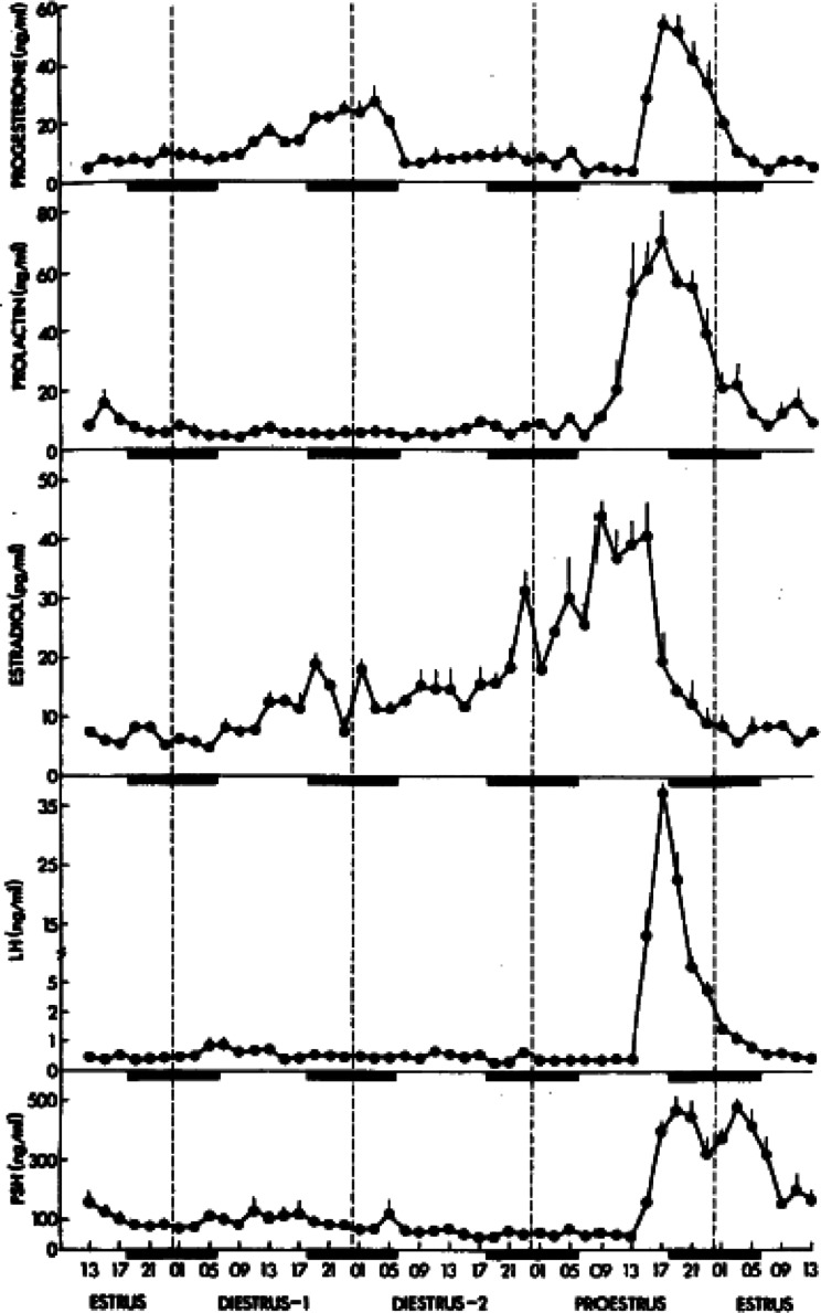
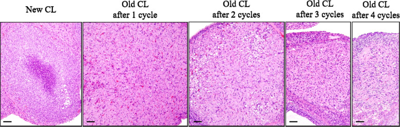
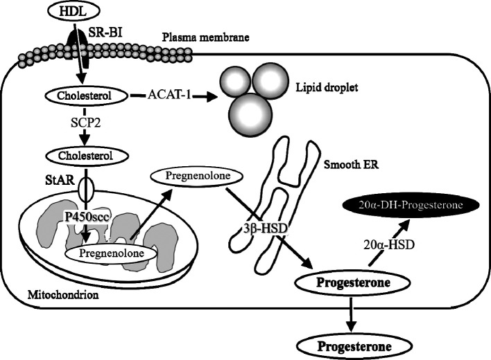
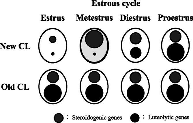
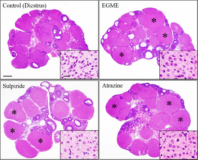
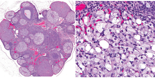
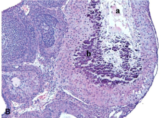
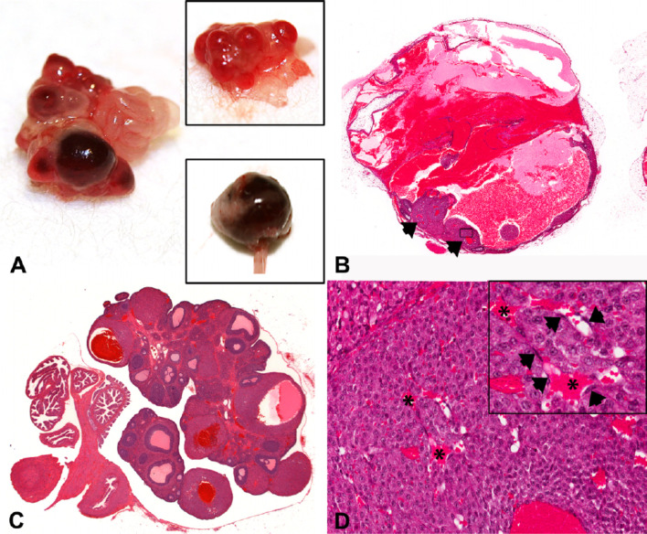
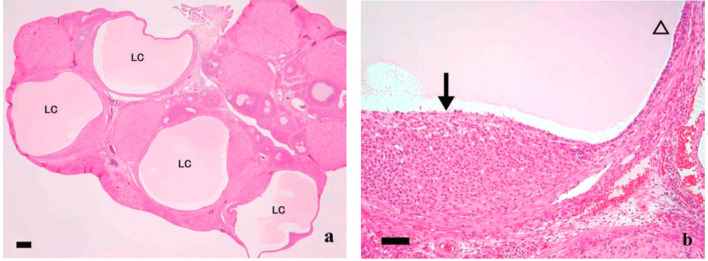
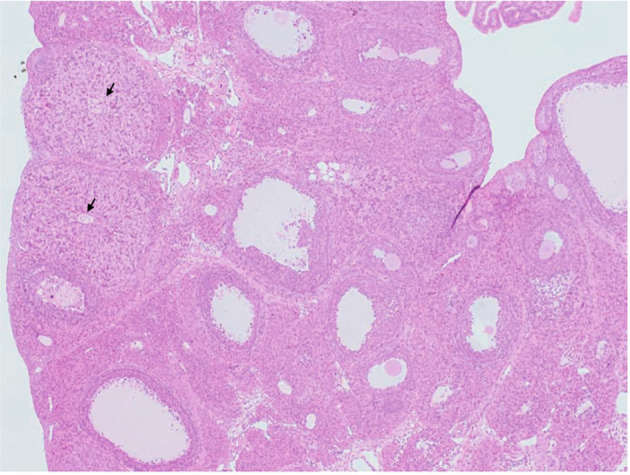
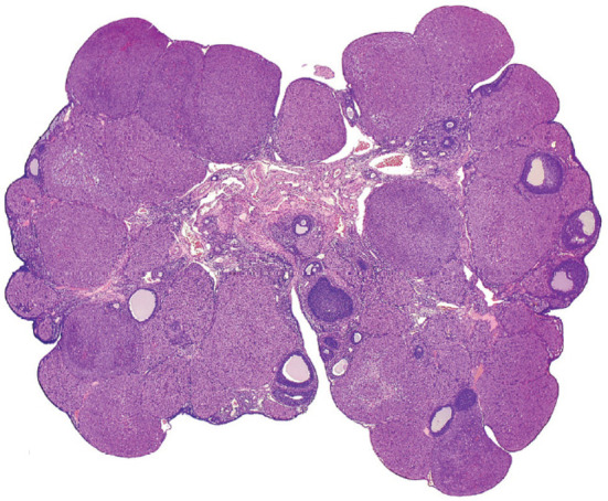
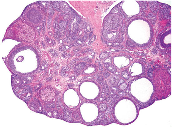
The corpora lutea (CL) are endocrine glands that form in the ovary after ovulation and secrete the steroid hormone, progesterone (P4). P4 plays a critical role in estrous and menstrual cycles, implantation, and pregnancy. The incomplete rodent estrous cycle stably lasts 4-5 days and its morphological features can be distinguished during each estrous cycle stage. In rat ovaries, there are two main types of CL: newly formed ones due to the current ovulation (new CL), and CL remaining from prior estrous cycles (old CL). In the luteal regression process, CL were almost fully regressed after four estrous cycles in Sprague-Dawley rats. P4 secretion from CL in rodents is regulated by the balance between synthesis and catabolism. In general, luteal toxicity should be evaluated by considering antemortem and postmortem data. Daily vaginal smear observations provided useful information on luteal toxicity. In histopathological examinations, not only the ovaries and CL but also other related tissues and organs including the uterus, vagina, mammary gland, and adrenal glands, must be carefully examined for exploring luteal changes. In this review, histological and functional characteristics of CL in rats are summarized, and representative luteal toxicity changes are presented for improved luteal toxicity evaluation in preclinical toxicity research.

Keywords: corpora lutea; luteal toxicity; ovary; progesterone; rat.


Figures

Fig. 1.
Fig. 1.
Time-course changes in reproductive hormone levels; progesterone (P4), prolactin (PRL), estradiol, luteinizing hormone (LH), and follicle-stimulating hormone. The rat estrous cycle is subdivided into four subsequent phases, proestrus, estrus, metestrus/diestrus 1, and diestrus/diestrus 2. Reproduced with permission from the Oxford University Press; taken from Smith et al. Endocrinology. 96: 219–226. 1975.
Fig. 2.
Fig. 2.
Time-course histological changes of luteal regression. New corpora lutea (CL) were composed of luteal cells with a small amount of basophilic cytoplasm. Old CL, after 1 cycle, reached maximum size and were characterized by luteal cells with abundant eosinophilic cytoplasm and distinct cell borders, as well as indistinct interstitial cells. Old CL after 2 cycles were smaller than Old CL after 1 cycle, had conspicuous interstitium, and luteal cell borders were slightly indistinct. Old CL after 3 cycles had more conspicuous interstitium and were smaller than Old CL after 2 cycles. After 4 cycles of new formation, CL almost completely regressed. Bars represent 50 µm. Modified from Taketa et al. Toxicol Pathol. 39: 372–380. 2011.
Fig. 3.
Fig. 3.
A schema of the progesterone (P4) biosynthesis process in the luteal cells. High-density lipoprotein (HDL) is the main source of cholesterol for CL in rodents. Scavenger receptor class B type I (SR-BI) is the authentic HDL receptor mediating the selective uptake of HDL-derived cholesterol esters. After uptake, the storage and turnover of free cholesterol in lipid droplets is processed through acyl coenzyme A-cholesterol acyl transferase (ACAT-1)-catalyzed cholesterol ester formation. Intracellular transport of hydrophobic, free cholesterol appears to be actively directed by proteins including sterol carrier proteins (SCP2). The cholesterol esters are transported to the outer mitochondrial membrane and then to the inner membrane by proteins including steroidogenic acute regulatory protein (StAR). Once the cholesterol reaches the inner mitochondrial membrane, mitochondrial P450 cholesterol side-chain cleavage (P450scc) transforms cholesterol into pregnenolone and 3β-hydroxysteroid dehydrogenase (3β-HSD) in the smooth endoplasmic reticulum (ER), and transforms pregnenolone into P4. In CL with functional regression, 20α-hyroxysteroid dehydrogenase (20α-HSD) catabolizes P4 into the inactive progestin, 20α-dihydroprogesterone (20α-DHP).
Fig. 4.
Fig. 4.
Overview of steroidogenic and luteolytic gene levels in new and old CL across the estrous cycle in rats. The sizes of the circles represent the levels of steroidogenic genes (gray circles) and luteolytic genes (black circles). The new CL at metestrus (bold line), which have the capacity for P4 secretion, showed notably high steroidogenic gene and low luteolytic gene levels. Luteolytic genes in the new CL were remarkably low at estrus and metestrus, and gradually increased thereafter. In the old CL, relatively high steroidogenic and markedly high luteolytic gene levels were consistently retained throughout the estrous cycle. Reproduced with permission from the Elsevier; taken from Taketa et al. Exp Toxicol Pathol. 64: 775–782. 2012.
Fig. 5.
Fig. 5.
Luteal cell hypertrophy induced by 2-week administration of ethylene glycol monomethyl ether (EGME), sulpiride, or atrazine. Luteal cells become hypertrophied with abundant eosinophilic cytoplasm following EGME, sulpiride, or atrazine treatment compared to respective control CL at diestrus stage. Asterisks indicate hypertrophied CL. Bars represent 500 µm, and bars in the inset images represent 20 µm. Reproduced with permission from the Oxford University Press; taken from Taketa et al. Toxicol Sci. 121: 267–278. 2011.
Fig. 6.
Fig. 6.
Representative images of vacuolation in luteal cells. The experimental detail is unknown. Reproduced with permission of the Japanese Society of Toxicologic Pathology from Dixson et al. Nonproliferative and proliferative lesions of the rat and mouse female reproductive system. J Toxicol Pathol. 27: 1S–107S. 2014.
Fig. 7.
Fig. 7.
Degeneration/necrosis of CL in the ovaries of rats treated with sunitinib, a potent inhibitor of vascular endothelial growth factor (VEGF), platelet-derived growth factor (PDGF), stem cell factor receptor (KIT), FMS-like tyrosine kinase-3 (FLT3), and rearranged during transfection (RET) receptors, at 6 mg/kg/day for up to 6 months (×200 magnification). Note necrosis (a) and mineralization (b). Reproduced with permission from the SAGE Publications; taken from Patyna et al. Toxicol Pathol. 36: 905–916. 2008.
Fig. 8.
Fig. 8.
Images of hemorrhagic cystic degeneration of CL. (A) Gross photograph of an ovary from a nude rat treated with platelet-derived growth factor receptor (PDGFR) a and b inhibitors compared to a control ovary (upper inset). The ovary was enlarged, with abnormal nodular areas of red discoloration. The lower inset shows a severely enlarged ovary. (B and C) Sub-gross images from WH rats treated with PDGFR a and b inhibitors showing severe (B) and minimal (C) cystic hemorrhagic dilatation/degeneration of CL. In (B), the ovary was severely dilated with hemorrhage. Residual abnormal CL are present (arrowheads) with areas of hemorrhage in the central lumen. In (C), CL were abnormal, with dilated and hemorrhagic central cavities. (D) Higher power photomicrograph of Fig. 1B (boxed area: original magnification ×20) showing an abnormal CL with several dilated sinusoids (*). Note the absence of interstitial cells (pericytes/endothelium) (arrowheads) throughout CL. Reproduced with permission from the SAGE Publications; taken from Hall et al. Toxicol Pathol. 44: 98–111. 2016.
Fig. 9.
Fig. 9.
Luteal cyst in rats treated with mifepristone, which is the synthetic steroid with antiprogesterone and antiglucocorticoid activities. (a) Multiple fluid-filled luteal cysts (LC) were observed. Bars show 200 µm. (b) Large cyst lined by thin (open arrowhead) and massive (arrows) luteinized cell layers. Bars show 50 µm. From Tamura et al. J Toxicol Sci. 34: SP31–42. 2009.
Fig. 10.
Fig. 10.
Image of unovulated CL. CL-retained oocytes (arrows) were observed in the ovaries of rats treated with peroxisome proliferator-activated receptor α/γ (PPARα/γ) dual agonist for 4 weeks. From Sato et al. J Toxicol Sci. 34: SP137–SP146. 2009.
Fig. 11.
Fig. 11.
Representative image of increased number of CL. The experimental detail is unknown. Reproduced with permission of the Japanese Society of Toxicologic Pathology from Dixson et al. Nonproliferative and proliferative lesions of the rat and mouse female reproductive system. J Toxicol Pathol. 27: 1S–107S. 2014.
Fig. 12.
Fig. 12.
Representative image of decreased number or absent CL. The experimental detail is unknown. Follicular cysts were also observed in the ovary. Reproduced with permission of the Japanese Society of Toxicologic Pathology from Dixson et al. Nonproliferative and proliferative lesions of the rat and mouse female reproductive systems. J Toxicol Pathol. 27: 1S–107S. 2014.

Similar articles

Cited by

References

    1. Sanbuissho A, Yoshida M, Hisada S, Sagami F, Kudo S, Kumazawa T, Ube M, Komatsu S, and Ohno Y. Collaborative work on evaluation of ovarian toxicity by repeated-dose and fertility studies in female rats. J Toxicol Sci. 34(Suppl 1): SP1–SP22. 2009. - PubMed
    1. Stouffer RL. Structure, function, and regulation of the corpus luteum. In: Knobil and Neill’s Physiology of Reproduction, 3rd ed. JD Neill (ed). Elsevier Academic Press, San Diego. 475–526. 2006.
    1. Rothchild I. The regulation of the mammalian corpus luteum. Recent Prog Horm Res. 37: 183–298. 1981. - PubMed
    1. Yoshida M, Sanbuissyo A, Hisada S, Takahashi M, Ohno Y, and Nishikawa A. Morphological characterization of the ovary under normal cycling in rats and its viewpoints of ovarian toxicity detection. J Toxicol Sci. 34(Suppl 1): SP189–SP197. 2009. - PubMed
    1. Kaneko S, Sato N, Sato K, and Hashimoto I. Changes in plasma progesterone, estradiol, follicle-stimulating hormone and luteinizing hormone during diestrus and ovulation in rats with 5-day estrous cycles: effect of antibody against progesterone. Biol Reprod. 34: 488–494. 1986. - PubMed