Case Reports
. 2022 Jan;35(1):107-111.
doi: 10.1293/tox.2021-0039. Epub 2021 Oct 25.

Porencephaly with an optic organ abnormality in a beagle dog

Affiliations
Case Reports

Porencephaly with an optic organ abnormality in a beagle dog

Osamu Hashiguchi et al. J Toxicol Pathol. 2022 Jan.

Abstract

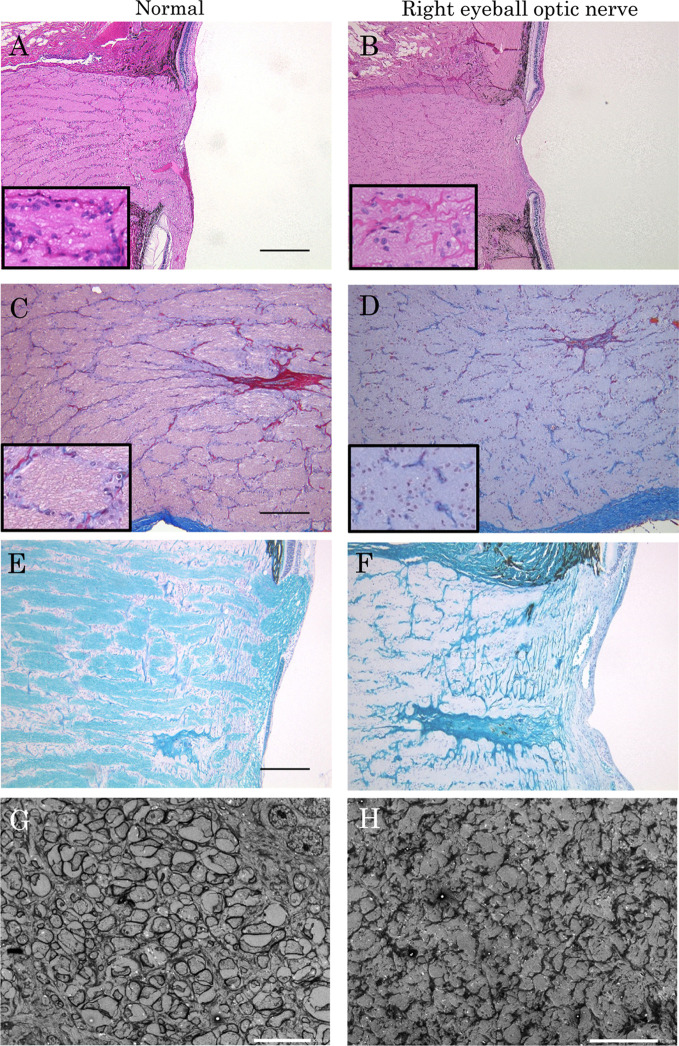
A female TOYO beagle dog showed porencephaly and visual organ abnormalities. At necropsy, there was a cavity filled with cerebrospinal fluid in the right cerebral hemisphere and an adhesion area between the cerebral cortex and the skull, which was partially thickened. Additionally, the right optic nerve showed a slight decrease in diameter. Histopathological examination revealed increased glial fibers and collagen fibers, hemosiderin deposition, and an increased number of microglia in the adhesion area, along with a marked reduction of the cerebral parenchyma. In the right eyeball, the retina and optic nerve showed focal atrophy in the nerve fiber layer and inner granular layer to full retinal atrophy and hypoplasia of the myelinated nerve fibers, respectively. Electron microscopic examination revealed hypoplasia of the myelin sheath of nerve fibers in the right optic nerve. This is an extremely rare case of porencephaly and congenital optic nerve hypoplasia, along with independent retinal thinning.

Keywords: TOYO beagle; optic nerve hypoplasia; porencephaly.


Figures

Fig. 1.
Fig. 1.
Microscopic photographs of a cystic lesion of the cerebrum. (A) Low magnification of the brain. A large cystic structure (asterisk) forms in the cerebrum. The area within the black circle indicates an adhesion between the cystic lesion and the dura of the skull. HE stain. (B) Adhesion site between the cerebral parenchyma and the dura of the skull. The skull of the adhesion area shows thickening. HE stain. (C) and (D) High magnification of photograph (B). There is an increase in glial fibers (C, HE stain; bar=100 μm) and collagen fibers (D, MT stain; bar=100 μm) in the cerebral parenchyma. (E) Microglia-like cells containing brown pigment were observed in the cerebral parenchyma near the adhesion site (arrowhead). The insert shows high magnification of the brown pigment. HE stain; bar=50 μm. (F) The brown pigments were positive for BB, suggesting hemosiderin. BB stain; bar=50 µm. (G) Cells with brown pigment were positive for the Iba1 antibody, suggesting microglia. Iba1 immunostaining; bar=50 μm.
Fig. 2.
Fig. 2.
Microscopic photographs of retinal thinning in the right eyeball. The central area of the retina shows a normal structure. Toward the peripheral area of the retina, the thickness of the retina is reduced, ranging from focal lesions consisting of atrophy of the nerve fiber layer and the inner nuclear layer to entire atrophy involving all layers of the retina. HE stains; bar=50 μm.
Fig. 3.
Fig. 3.
Microscopic and electron microscopic photographs of a normal optic nerve (A, C, and E: light microscopy, G: electron microscopy) and a slight decrease in diameter of the right optic nerve (B, D, and F: light microscopy, H: electron microscopy). A and B: The diameter of the right optic disc (B) is smaller than that in the normal optic nerve (A). Each insert shows high magnifications of A or B. HE stain; bar=500 µm. C and D: An abnormal arrangement of fibrous connective tissue is observed in the right optic nerve (D). Each insert shows high magnifications of C or D. MT stain; bar=200 µm. E and F: The myelin sheath of the right optic nerve (F) is less stained with LFB compared to that of the normal optic nerve (E). LFB stain, bar=200 μm. G and H: The myelin sheath in the myelinated nerve fibers is unclear in the right optic nerve (H), bar=2 μm.

Similar articles

Cited by

References

    1. Russell SA. Cranial abnormalities. In: Textbook of Fetal Abnormalities, 2nd ed. S Bower, P Twining, JM McHugo, and DW Pilling (eds). Churchill Livingstone, London. 95–141. 2006.
    1. Kirkland PD. Akabane virus infection. Rev Sci Tech. 34: 403–410. 2015. - PubMed
    1. Tonni G, Ferrari B, Defelice C, and Centini G. Neonatal porencephaly in very low birth weight infants: ultrasound timing of asphyxial injury and neurodevelopmental outcome at two years of age. J Matern Fetal Neonatal Med. 18: 361–365. 2005. - PubMed
    1. Verbeek E, Meuwissen ME, Verheijen FW, Govaert PP, Licht DJ, Kuo DS, Poulton CJ, Schot R, Lequin MH, Dudink J, Halley DJ, de Coo RI, den Hollander JC, Oegema R, Gould DB, and Mancini GM. COL4A2 mutation associated with familial porencephaly and small-vessel disease. Eur J Hum Genet. 20: 844–851. 2012. - PMC - PubMed
    1. Garigliany MM, Hoffmann B, Dive M, Sartelet A, Bayrou C, Cassart D, Beer M, and Desmecht D. Schmallenberg virus in calf born at term with porencephaly, Belgium. Emerg Infect Dis. 18: 1005–1006. 2012. - PMC - PubMed

Publication types