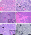
Malignant pinealoma observed in the deep cerebral parenchyma of a male Wistar rat
- PMID: 35221505
- PMCID: PMC8828600
- DOI: 10.1293/tox.2021-0047
Malignant pinealoma observed in the deep cerebral parenchyma of a male Wistar rat
Abstract
This report describes a case of spontaneous malignant pinealoma in a 90-week-old male Wistar rat. The tumor mass occurred in the deep cerebral parenchyma and no intact pineal gland was observed in the area between the posterior-dorsal median line of the cerebrum and the cerebellum. The tumor was characterized by a large nodular proliferation occupying the central area of the brain, extending from the dorsal surface to the base of the brain, corresponding to the thalamus. The tumor cells had round to irregular oblong nuclei approximately 5-17 μm in diameter and showed faintly or moderately eosinophilic cytoplasm and indistinct cell boundaries. Immunohistochemically, the tumor cells were positive for synaptophysin and partially positive for neuron-specific enolase (NSE). The tumor showed malignant features including cellular pleomorphism, high mitotic index, necrotic foci, and invasive and extensive growth and was, therefore, diagnosed as an extremely rare malignant pinealoma in the deep cerebral parenchyma.
Keywords: Wistar; pineal gland; pinealoma; rat; synaptophysin.
©2022 The Japanese Society of Toxicologic Pathology.
Figures


Similar articles
-
Occurrence of Pineal Gland Tumors in Combined Chronic Toxicity/Carcinogenicity Studies in Wistar Rats.Toxicol Pathol. 2015 Aug;43(6):838-43. doi: 10.1177/0192623315572700. Epub 2015 Mar 9. Toxicol Pathol. 2015. PMID: 25755100
-
Cytologic feature by squash preparation of pineal parenchyma tumor of intermediate differentiation.Diagn Cytopathol. 2008 Oct;36(10):749-53. doi: 10.1002/dc.20884. Diagn Cytopathol. 2008. PMID: 18773448
-
Papillary tumor of the pineal region.Neuropathology. 2008 Feb;28(1):87-92. doi: 10.1111/j.1440-1789.2007.00832.x. Epub 2007 Dec 5. Neuropathology. 2008. PMID: 18069972
-
Morphologic characterization of spontaneous nervous system tumors in mice and rats.Toxicol Pathol. 2000 Jan-Feb;28(1):178-92. doi: 10.1177/019262330002800123. Toxicol Pathol. 2000. PMID: 10669006 Review.
-
Primary pineal malignant melanoma with B-Raf V600E mutation: a case report and brief review of the literature.Acta Neurochir (Wien). 2015 Jul;157(7):1267-70. doi: 10.1007/s00701-015-2427-3. Epub 2015 May 15. Acta Neurochir (Wien). 2015. PMID: 25976339 Review.
References
-
- Maitra A. The endocrine system. In: Robbins and Cotran Pathologic Basis of Disease, 9th ed. V Kumar, AK Abbas, and JC Aster (eds). Elsevier, Philadelphia. 1073–1139. 2015.
-
- La Perle KMD. Endocrine system. In: Pathologic Basis of Veterinary Disease, 5th ed. JF Zachary, and MD McGavin (eds). Mosby, St Louis. 660–697. 2012.
-
- Higgins RJ, Bollen AW, Dickinson PJ, and Siso-Llonch S. Tumors of the nervous system. In: Tumors in Domestic Animals, 5th ed. Meuten DJ (ed). Wiley-Blackwell, Raleigh. 834–891. 2017.
-
- Weber K, Garman RH, Germann PG, Hardisty JF, Krinke G, Millar P, and Pardo ID. Classification of neural tumors in laboratory rodents, emphasizing the rat. Toxicol Pathol. 39: 129–151. 2011. - PubMed
-
- Louis DN, Perry A, Reifenberger G, von Deimling A, Figarella-Branger D, Cavenee WK, Ohgaki H, Wiestler OD, Kleihues P, and Ellison DW. The 2016 World Health Organization Classification of Tumors of the Central Nervous System: a summary. Acta Neuropathol. 131: 803–820. 2016. - PubMed

