

Case Report: Moving Tumor-Like Foci Behind Refractory Epilepsy-Cerebral Sparganosis Successfully Treated by Surgery After Failure of Praziquantel Treatment
- PMID: 35222259
- PMCID: PMC8866191
- DOI: 10.3389/fneur.2022.838849
Case Report: Moving Tumor-Like Foci Behind Refractory Epilepsy-Cerebral Sparganosis Successfully Treated by Surgery After Failure of Praziquantel Treatment
Abstract
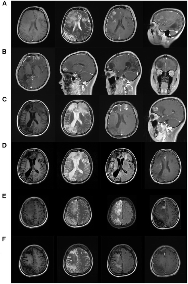
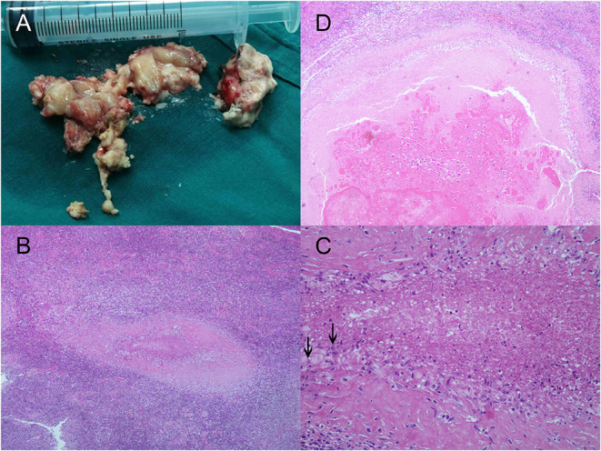
Cerebral sparganosis is clinically non-specific and easily misdiagnosed, exposing patients to the risk of severe brain damage and neurological dysfunction caused by actively migrating larvae. Diagnostic biomarkers from typical cases can help to establish an early diagnosis and proper treatment. We present a 25-year-old woman who suffered from 9 years of refractory epilepsy and was misdiagnosed with glioma and subjected to surgery. The postoperative pathology confirmed granuloma, and the tumor-like foci reappeared 3 months later. Along with the "tunnel sign" on MRI, cerebral sparganosis was suspected and confirmed by positive serum and cerebrospinal fluid antibodies against Spirometra mansoni. The patient visited us after a failure of four cycles of praziquantel treatment, recurrent seizures and hemiplegia with basal ganglia foci. Craniotomy was not carried out until the larva moved to the superficial lobe on follow-up MRIs, and pathology revealed sparganosis granuloma. The patient became seizure-free and recovered myodynamia but had long-lasting cognitive dysfunction due to severe brain damage. This case indicated the importance of tunnel signs and moving tumor-like foci on MRI as diagnostic clues of cerebral sparganosis. An early diagnosis is vitally important to avoid severe neural dysfunction by the long-living and moving larvae. Surgical removal of the larva is a critical remedy for cases failed by praziquantel treatment.
Keywords: Spirometra mansoni; cerebral sparganosis; craniotomy; refractory epilepsy; tunnel sign.
Copyright © 2022 Chen, Chen and Kang.
Conflict of interest statement
The authors declare that the research was conducted in the absence of any commercial or financial relationships that could be construed as a potential conflict of interest.
Figures
Similar articles
-
A case report: 1-year follow-up of cerebral sparganosis mansoni with a stroke-like onset.BMC Neurol. 2019 May 29;19(1):105. doi: 10.1186/s12883-019-1335-1. BMC Neurol. 2019. PMID: 31142276 Free PMC article.
-
Cerebral sparganosis presenting with atypical postcontrast magnetic resonance imaging findings: a case report and literature review.BMC Infect Dis. 2019 Aug 27;19(1):748. doi: 10.1186/s12879-019-4396-2. BMC Infect Dis. 2019. PMID: 31455261 Free PMC article. Review.
-
Cerebral sparganosis: case report and review of the European cases.Acta Neurochir (Wien). 2015 Sep;157(8):1339-43; discussion 1343. doi: 10.1007/s00701-015-2466-9. Epub 2015 Jun 18. Acta Neurochir (Wien). 2015. PMID: 26085111 Review.
-
Cerebral sparganosis in mainland Chinese patients.J Clin Neurosci. 2013 Nov;20(11):1514-9. doi: 10.1016/j.jocn.2012.12.018. Epub 2013 Jul 30. J Clin Neurosci. 2013. PMID: 23911107
-
Surgical treatment of a patient with live intracranial sparganosis for 17 years.BMC Infect Dis. 2022 Apr 9;22(1):353. doi: 10.1186/s12879-022-07293-7. BMC Infect Dis. 2022. PMID: 35397512 Free PMC article.
Cited by
-
Recent update on cerebral sparganosis: A bibliometric analysis and scientific mapping.Narra J. 2024 Aug;4(2):e982. doi: 10.52225/narra.v4i2.982. Epub 2024 Aug 15. Narra J. 2024. PMID: 39280299 Free PMC article.
-
Natural variation in the binding pocket of a parasitic flatworm TRPM channel resolves the basis for praziquantel sensitivity.Proc Natl Acad Sci U S A. 2023 Jan 3;120(1):e2217732120. doi: 10.1073/pnas.2217732120. Epub 2022 Dec 27. Proc Natl Acad Sci U S A. 2023. PMID: 36574686 Free PMC article.
-
Cerebral sparganosis in a child with corpus callosum invasion: a case report.BMC Infect Dis. 2023 May 25;23(1):350. doi: 10.1186/s12879-023-08322-9. BMC Infect Dis. 2023. PMID: 37231358 Free PMC article.
References
-
- Lei W, Fei W. Analysis of clinical characteristics in 24 cases of cerebral sparganosis. China Trop Med. (2016) 16:698–701. 10.13604/j.cnki.46-1064/r.2016.07.19 - DOI
-
- Feng C, Jie W, Yuqin Z. Clinical and radiological analyses of 27 cases of brain parasitic diseases. China Modern Doctor. (2014) 52:48–50.
Publication types
LinkOut - more resources
Full Text Sources