Review
. 2022 Feb 11:12:814635.
doi: 10.3389/fmicb.2021.814635. eCollection 2021.

The Regulation of Integrated Stress Response Signaling Pathway on Viral Infection and Viral Antagonism

Affiliations
Review

The Regulation of Integrated Stress Response Signaling Pathway on Viral Infection and Viral Antagonism

Yongshu Wu et al. Front Microbiol. .

Abstract

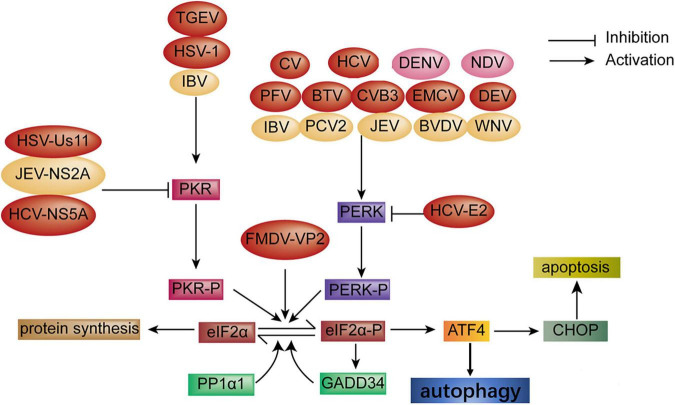
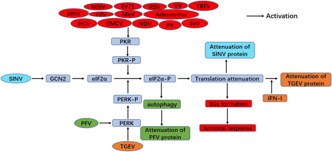
The integrated stress response (ISR) is an adaptational signaling pathway induced in response to different stimuli, such as accumulation of unfolded and misfolded proteins, hypoxia, amino acid deprivation, viral infection, and ultraviolet light. It has been known that viral infection can activate the ISR, but the role of the ISR during viral infection is still unclear. In some cases, the ISR is a protective mechanism of host cells against viral infection, while viruses may hijack the ISR for facilitating their replication. This review highlighted recent advances on the induction of the ISR upon viral infection and the downstream responses, such as autophagy, apoptosis, formation of stress granules, and innate immunity response. We then discussed the molecular mechanism of the ISR regulating viral replication and how viruses antagonize this cellular stress response resulting from the ISR.

Keywords: eIF2α phosphorylation; host; integrated stress response; unfolded protein response; viral replication.


Conflict of interest statement

The authors declare that the research was conducted in the absence of any commercial or financial relationships that could be construed as a potential conflict of interest.

Figures

FIGURE 1
FIGURE 1
Schematic diagram of the domain organization of the four mammalian eIF2α kinases. Polypeptides are boxes running from N- to C-terminal domains from left to right. Length in amino acids is of proteins. The abbreviations of domains are listed: SP, signal peptide; TM, transmembrane domain; KD, kinase domain; DS RBD, double-stranded RNA binding domain; PKD, pseudokinase domain; His/Rs, histidyl-tRNA synthetase-related domain; RB, ribosome binding. Domains involved in sensing stress signals/activation are in green and black. Kinase domains are yellow and brown, other domains are colored blue, and domains are drawn to scale.
FIGURE 2
FIGURE 2
Diagram of the activation of autophagy and apoptosis via the ISR signaling pathway during viral infection. Autophagy: Autophagy is activated through the PKR-eIF2α pathway upon infection with transmissible gastroenteritis virus (TGEV) and HSV-1 infection; CV, HCV, PFV, BTV, CVB3, encephalomyocarditis virus (EMCV) and DEV infection, respectively, induces autophagy through the PERK-eIF2α pathway. FMDV-VP2 induces autophagy through interaction with HSPB1 and activation of the eIF2α-ATF4 pathway. In turn, HSV-Us11, HCV-NS5A, and HCV-E2 protein block autophagy (red color). Apoptosis: Apoptosis is induced via the PERK and PKR-eIF2α pathway under infection with IBV, PCV2, BVDV, JEV, and WNV, respectively. Oppositely, the JEV-NS2A protein inhibits apoptosis (yellow color). In addition, autophagy and apoptosis simultaneously are induced through the PERK-eIF2α pathway during DENV and NDV infection, respectively (pink color).
FIGURE 3
FIGURE 3
Diagram of SGs formation under viral infection. SGs formation: EV71, VV, HIV, HCV, SeV, EMCV, RSV, TBEV, MeV, TMEV, Adenovirus, NDV, and SINV infection induce SGs formation through the PKR-eIF2α-P signaling pathway. MeV, Reovirus, SFV, rotavirus (RV), HSV, VSV, MHV, and MRV benefit SGs formation via direct eIF2α phosphorylation (blue color). However, MNV, nervous necrosis virus (NNV) infection, and the expression of IAV-NS1, FMDV-Lpro, PV-3Cpro, TMEMV-Lpro, and EMCV-3C inhibit SGs formation through blocking eIF2α phosphorylation (red color). SGs formation is increased via the PERK-eIF2α-P signaling during human cytomegalovirus (HCMV) infection. SINV infection enhances SGs formation through the GCN2-eIF2α-P signaling pathway.
FIGURE 4
FIGURE 4
Antiviral response of the ISR during viral infection. PERK-eIF2α signaling suppresses viral replication through inducing IFN-I production and eIF2α phosphorylation-mediated translation attenuation with TGEV infection (orange color). PFV infection inhibits viral replication through PERK-mediated autophagy (green color). In addition, viral infection induces SGs formation through PKR-eIF2α phosphorylation and plays antiviral response, such as MNV, EV71, RSV, VV, TBEV, SINV, HIV, MeV, Adenovirus, HCV, EMCV, NDV, PV and SeV (red color). GCN2-eIF2α signaling inhibit viral replication upon SINV infection (blue color).

Similar articles

Cited by

References

    1. Abrahamyan L. G., Chatel-Chaix L., Ajamian L., Milev M. P., Monette A., Clement J. F., et al. (2010). Novel staufen1 ribonucleoproteins prevent formation of stress granules but favour encapsidation of HIV-1 genomic RNA. J. Cell Sci. 123 369–383. 10.1242/jcs.055897 - DOI - PubMed
    1. Aitken C. E., Lorsch J. R. (2012). A mechanistic overview of translation initiation in eukaryotes. Nat. Struct. Mol. Biol. 19 568–576. 10.1038/nsmb.2303 - DOI - PubMed
    1. Albornoz A., Carletti T., Corazza G., Marcello A. (2014). The stress granule component TIA-1 binds tick-borne encephalitis virus RNA and is recruited to perinuclear sites of viral replication to inhibit viral translation. J. Virol. 88 6611–6622. 10.1128/JVI.03736-13 - DOI - PMC - PubMed
    1. Alexander D. E., Ward S. L., Mizushima N., Levine B., Leib D. A. (2007). Analysis of the role of autophagy in replication of herpes simplex virus in cell culture. J. Virol. 81 12128–12134. 10.1128/JVI.01356-07 - DOI - PMC - PubMed
    1. Ambrose R. L., Mackenzie J. M. (2013a). ATF6 signaling is required for efficient west nile virus replication by promoting cell survival and inhibition of innate immune responses. J. Virol. 87 2206–2214. 10.1128/JVI.02097-12 - DOI - PMC - PubMed

LinkOut - more resources