. 2022 Feb 11:13:834033.
doi: 10.3389/fimmu.2022.834033. eCollection 2022.

Human CD4 T Cells From Thymus and Cord Blood Are Convertible Into CD8 T Cells by IL-4

Affiliations

Human CD4 T Cells From Thymus and Cord Blood Are Convertible Into CD8 T Cells by IL-4

Helena Nunes-Cabaço et al. Front Immunol. .

Abstract

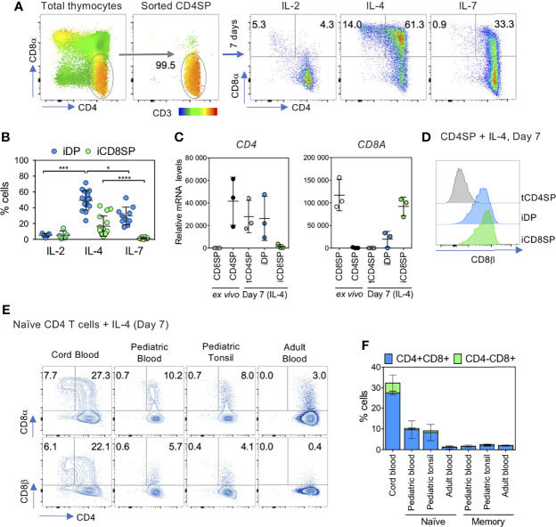
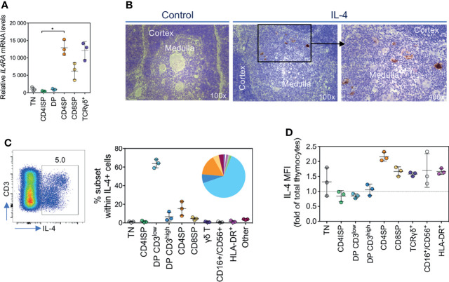
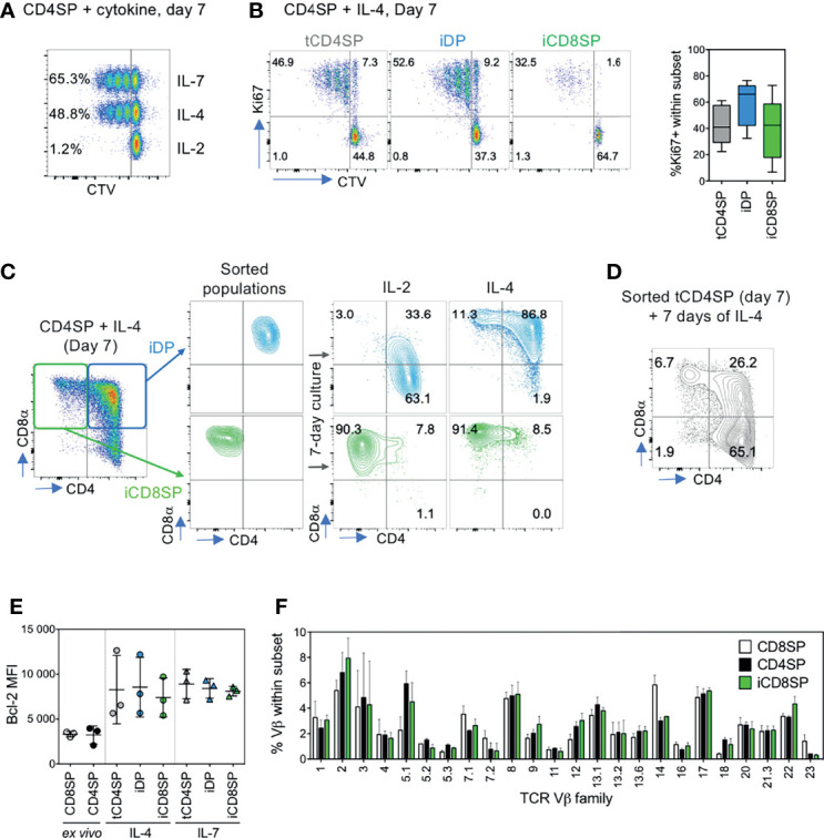
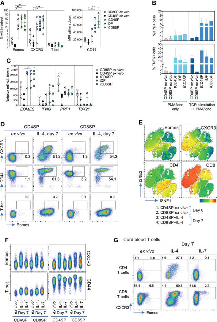
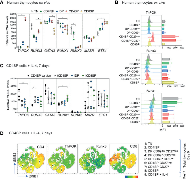
Commitment to the CD4+ or CD8+ T cell lineages is linked to the acquisition of a functional program broadly defined by helper and cytotoxic properties, respectively. The mechanisms underlying these processes in the human thymus remain largely unclear. Moreover, recent thymic emigrants are thought to have some degree of plasticity, which may be important for the shaping of the immune system and adjustment to specific peripheral needs. We show here that IL-4 induces proliferation-independent de novo synthesis of CD8αβ in human CD4 single-positive (SP) thymocytes, generating a stable CD8SP population that features a diverse TCRαβ repertoire, CD4 expression shut-down and ThPOK downregulation. IL-4 also promotes an innate-like program in both CD4SP and CD8SP thymocytes, characterized by Eomes upregulation in the absence of T-bet, in line with its recognized role in the generation of thymic innate-like CD8+ T cells. The clinical relevance of these findings is further supported by the profile of IL-4 production and IL-4 receptor expression that we identified in the human thymus. Importantly, human cord blood CD4+ T cells preserve the ability to generate Eomes+ CD8+ T cells in the presence of IL-4, with implications in neonatal immunity. Our results support a role for IL-4 in the dynamic regulation of human thymocyte plasticity and identify novel strategies to modulate immune responses.

Keywords: CD4+ and CD8+ T cell lineage commitment; IL-4; cord blood; human thymus; innate-like T cells.


Conflict of interest statement

The authors declare that the research was conducted in the absence of any commercial or financial relationships that could be construed as a potential conflict of interest.

Figures

Figure 1
Figure 1
IL-4 induces the upregulation of CD8αβ on human CD4SP thymocytes and in peripheral CD4+ T cells. (A) CD4SP thymocytes, sorted as CD3highCD4+CD8negCD25neg TCRγδneg, were incubated with IL-2, IL-4 or IL-7 for 7 days and CD8α expression was assessed at the end of the culture. CD3 expression is presented as an heatmap overlay on initial populations. (B) Frequency of cytokine-induced double-positive (iDP) and CD8 single-positive (iCD8SP) thymocytes after 7 days of culture with IL-2, IL-4 or IL-7. Each dot represents a thymus. Statistics was only performed within the same subset (iDP or iCD8SP). (C) CD4 and CD8A mRNA expression in CD4SP and CD8SP thymocytes ex vivo and in sorted populations 7 days after IL-4 culture. tCD4SP: cytokine-treated CD4 single-positive thymocytes. (D) Expression of CD8β on tCD4SP, iDP and iCD8SP generated from incubation of CD4SP thymocytes with IL-4 for 7 days. (E) Expression of CD8α and CD8β on sorted naïve CD4+ T cells from cord, pediatric or adult blood and pediatric tonsil after a 7-day culture with IL-4 (left dot plots) and (F) quantification of iDP and iCD8SP cells generated from naïve and memory T cells (right panel; n=2/3). Results in graphs are presented as mean ± SD. *p < 0.05, ***p < 0.001, ****p < 0.0001.
Figure 2
Figure 2
CD4SP thymocytes express IL-4Rα and produce high levels of IL-4. (A) IL4RA mRNA levels in developing T cells. TN: triple-negative; CD4ISP: CD4 immature single-positive; DP: double-positive; CD4SP and CD8SP: CD4+ or CD8+ single-positive. (B) Analysis of IL-4 expression in the human thymus by immunohistochemistry. (C) Flow cytometric analysis of IL-4-producing thymic populations, as frequency of total IL-4 production. (D) Median fluorescence intensity (MFI) of IL-4+ cells within each subset analyzed. Results in graphs are presented as mean ± SD. *p < 0.05.
Figure 3
Figure 3
iCD8SP cells generated from CD4SP thymocytes in the presence of IL-4 are stable and diverse. (A) Proliferation of CD4SP thymocytes, sorted as CD3highCD4+CD8negCD25neg TCRγδneg cells, in response to IL-2, IL-4 and IL-7, as measured by Cell Trace Violet (CTV) dilution. Frequency of CTVneg cells is presented in each population, and data are representative of 3 experiments. (B) Proliferation of tCD4SP, iDP and iCD8SP thymocytes in response to IL-4, as measured by CTV dilution or Ki67 frequency (graph: n=5). (C) Stability of IL-4-induced CD8 expression, as assessed by sorting iDP and iCD8SP populations on day 7 of IL-4 culture and either maintaining cells in IL-4 or switching them to IL-2 for 7 more days. (D) CD8 induction in CD4+CD8neg (tCD4SP) cells sorted 7 days after IL-4 culture of CD4SP thymocytes and cultured in the presence of IL-4 for 7 additional days. (E) Bcl-2 median fluorescence intensity (MFI) of different populations before and after exposure to IL-4 or IL-7. (F) TCR Vβ repertoire of CD8SP, CD4SP and IL-4-induced iCD8SP thymocytes (n=3). Results in graphs are presented as mean ± SD.
Figure 4
Figure 4
IL-4 induces an innate phenotype on CD4SP thymocytes and cord blood CD4+ T cells. (A) Frequency of Eomes, CXCR3 and T-bet and MFI of CD44 within CD4SP or CD8SP thymocytes ex vivo, or tCD4SP, iDP and iCD8SP cells 7 days after exposure to IL-4. (B) Frequency of IFN-γ+ and TNF-α+ cells within ex vivo CD4SP and CD8SP thymocytes or cells treated with IL-4 upon stimulation with PMA/ionomycin, both with or without TCR stimulation for 5 days (n=3-5). (C) Transcriptional expression of EOMES, IFNG, PRF1 and TBX21 in CD4SP and CD8SP cells ex vivo or after IL-4 culture. (D) Representative dot plots of Eomes, CXCR3, CD44 and T-bet expression in CD4SP and CD8SP thymocytes ex vivo and after 7 days of culture with IL-4. (E) tSNE clustering analysis of the concatenated flow cytometry data of ex vivo and IL-4-exposed CD4SP and CD8SP cells, based on the following markers: CD3, CD4, CD8α, CD8β, Eomes and CXCR3. Eomes, CXCR3, CD4 and CD8 expression are presented as heatmap overlays over the populations analyzed. (F) Representative dot plots of innate-related markers in CD4SP and CD8SP thymocytes ex vivo and after 7 days of culture with IL-4 or IL-7. (G) Eomes and CXCR3 expression in cord blood CD4+ or CD8+ T cells ex vivo or cultured with IL-4 or IL-7 for 7 days. Results in graphs are presented as mean ± SD. *p < 0.05, **p < 0.01, ***p < 0.001, ***p<0.0001.
Figure 5
Figure 5
IL-4-induced iCD8SP thymocytes have downregulated ThPOK mRNA and protein. (A) Expression of ThPOK (ZBTB7B), RUNX3, GATA3, RUNX1, RUNX2, MAZR and ETS1 mRNA levels in sorted ex vivo human thymocyte populations. TN: triple-negative; CD4ISP: CD4 immature single-positive; DP: double-positive; CD4SP and CD8SP: CD4+ or CD8+ single-positive. (B) Median fluorescence intensity (MFI) of ThPOK, Runx3 and Runx1 expression during human T cell development (n=3). (C) ThPOK (ZBTB7B), RUNX3, GATA3, RUNX1, RUNX2, MAZR and ETS1 mRNA levels in CD4SP thymocytes ex vivo and in sorted tCD4SP, iDP and iCD8SP populations after IL-4 treatment. (D) tSNE dimensional reduction analysis of concatenated flow cytometry results (total thymocytes and IL-4-treated CD4SP thymocytes), based on the following markers: CD3, CD4, CD8α, CD69, CD27, CD45RA, ThPOK and Runx3. The expression of CD4, ThPOK, Runx3 and CD8 are presented as overlays over the populations analyzed. Results in graphs are presented as mean ± SD. *p < 0.05, **p < 0.01.

References

    1. Spits H. Development of αβ T Cells in the Human Thymus. Nat Rev Immunol (2002) 2:760–72. doi: 10.1038/nri913 - DOI - PubMed
    1. Vanhecke D, Verhasselt B, Smedt MD, Leclercq G, Plum J, Vandekerckhove B. Human Thymocytes Become Lineage Committed at an Early Postselection CD69+ Stage, Before the Onset of Functional Maturation. J Immunol (1997) 159:5973–83. - PubMed
    1. Vanhecke D, Leclercq G, Plum J, Vandekerckhove B. Characterization of Distinct Stages During the Differentiation of Human CD69+CD3+ Thymocytes and Identification of Thymic Emigrants. J Immunol (1995) 155:1862–72. - PubMed
    1. Egawa T. Regulation of CD4 and CD8 Coreceptor Expression and CD4 Versus CD8 Lineage Decisions. Adv Immunol (2015) 125:1–40. doi: 10.1016/bs.ai.2014.09.001 - DOI - PubMed
    1. Taniuchi I. CD4 Helper and CD8 Cytotoxic T Cell Differentiation. Annu Rev Immunol (2018) 36:579–601. doi: 10.1146/annurev-immunol-042617-053411 - DOI - PubMed

Publication types