. 2022 Mar;23(3):222.
doi: 10.3892/etm.2022.11146. Epub 2022 Jan 17.

Metformin reduces chondrocyte pyroptosis in an osteoarthritis mouse model by inhibiting NLRP3 inflammasome activation

Affiliations

Metformin reduces chondrocyte pyroptosis in an osteoarthritis mouse model by inhibiting NLRP3 inflammasome activation

Jiangbo Yan et al. Exp Ther Med. 2022 Mar.

Abstract

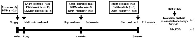
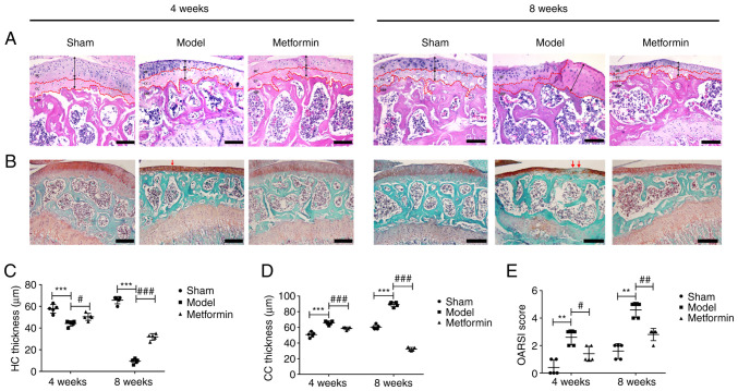
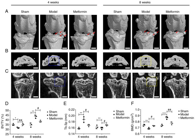
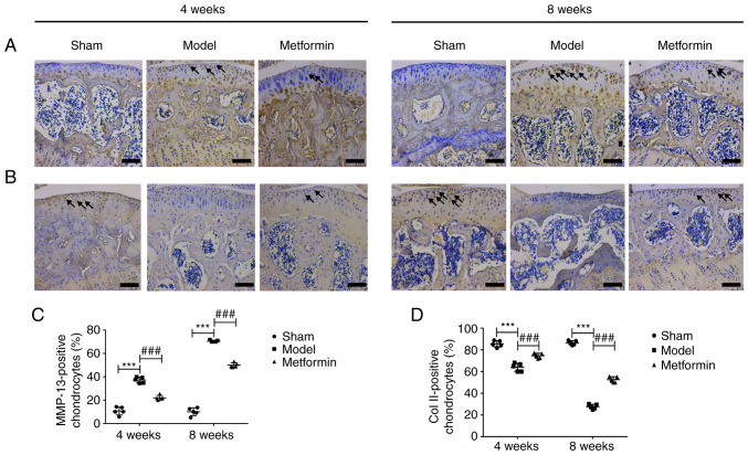
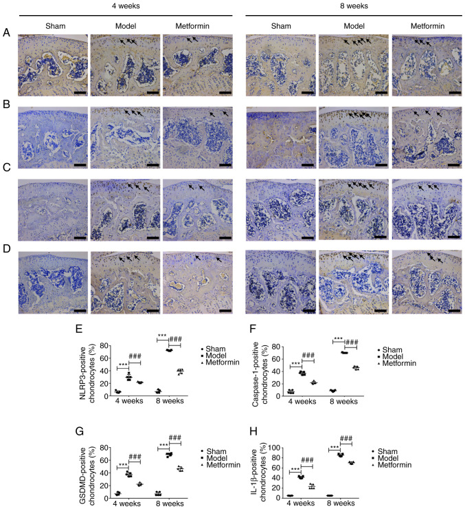
Osteoarthritis (OA) is an age-related degenerative disease, and its incidence is increasing with the ageing of the population. Metformin, as the first-line medication for the treatment of diabetes, has received increasing attention for its role in OA. The purpose of the present study was to confirm the therapeutic effect of metformin in a mouse model of OA and to determine the mechanism underlying the resultant delay in OA progression. The right knees of 8-week-old C57BL/6 male mice were subjected to destabilization of the medial meniscus (DMM). Metformin (200 mg/kg) was then administered daily for 4 or 8 weeks. Safranin O-fast green staining, H&E staining and micro-CT were used to analyse the structure and morphological changes. Immunohistochemical staining was used to detect type II collagen (Col II), matrix metalloproteinase 13 (MMP-13), NOD-like receptor protein 3 (NLRP3), caspase-1, gasdermin D (GSDMD) and IL-1β protein expression. Reverse transcription-quantitative PCR was used to detect the mRNA expression of NLRP3, caspase-1, GSDMD and IL-1β. Histomorphological staining showed that metformin delayed the progression of OA in the DMM model. With respect to cartilage, metformin decreased the Osteoarthritis Research Society International score, increased the thickness of hyaline cartilage and decreased the thickness of calcified cartilage. Regarding the mechanism, in cartilage, metformin increased the expression of Col II and decreased the expression of MMP-13, NLRP3, caspase-1, GSDMD and IL-1β. In addition, in subchondral bone, metformin inhibited osteophyte formation, increased the bone volume fraction (%) and the bone mineral density (g/cm3), decreased the trabecular separation (mm) in early stage of osteoarthritis (4 weeks) but the opposite in an advanced stage of osteoarthritis (8 weeks). Overall, metformin inhibited the activation of NLRP3 inflammasome, decreased cartilage degradation, reversed subchondral bone remodelling and inhibited chondrocyte pyroptosis.

Keywords: NOD-like receptor protein 3; chondrocytes; metformin; osteoarthritis; pyroptosis.


Conflict of interest statement

The authors declare that they have no competing interests.

Figures

Figure 1
Figure 1
Schematic diagram of the animal experiment. DMM, destabilization of the medial meniscus; RT-qPCR, reverse transcription-quantitative PCR.
Figure 2
Figure 2
Metformin can decrease cartilage degradation in a destabilization of the medial meniscus model. (A) Mouse H&E staining results (magnification, x100). HC and CC thickness are marked by dotted lines. Black arrows indicated the variation in HC and CC thickness. Scale bar=200 µm. (B) Mouse safranin O-fast green staining results (magnification, x100). Red arrows indicated the cartilage degradation. Scale bar=200 µm. (C) HC and (D) CC thickness. (E) OARSI scores of cartilage structural damage in mice. One-way ANOVA followed by Tukey's multiple comparison test was used to compare data among groups after testing the data for homogeneity of variance. Non-parametric data (OARSI scores) were analysed using the Kruskal-Wallis H test followed by Dunn's test. **P<0.01; ***P<0.001; #P<0.05; ##P<0.01; ###P<0.001. HC, hyaline cartilage; CC, calcified cartilage; OARSI, Osteoarthritis Research Society International.
Figure 3
Figure 3
Metformin can improve subchondral bone remodelling in a destabilization of the medial meniscus model. (A) Representative micro-CT images. Osteophyte and cartilage degeneration are marked by red arrows. Scale bar=2,000 µm. (B) Micro-CT two-dimensional scan of tibial subchondral bone. Scale bar=2,000 µm. (C) Micro-CT two-dimensional reconstructions of tibial subchondral bone. Scale bar=2,000 µm. Subchondral bone loss in the tibia is highlighted using a blue box. Increases in bone mass and bone sclerosis are highlighted using a yellow box. Quantification of bone morphological parameters (D) BV/TV, (E) Tb.Sp and (F) BMD. One-way ANOVA followed by Tukey's multiple comparison test was used to compare data among groups after testing the data for homogeneity of variance. *P<0.05; **P<0.01 and ***P<0.001; #P<0.05; ##P<0.01. BV/TV, bone volume fraction; Tb.Sp, trabecular separation; BMD, bone mineral density.
Figure 4
Figure 4
Metformin can enhance cartilage matrix anabolism and inhibit its catabolism. (A) MMP-13 and (B) Col II immunohistochemistry test results (magnification, x400). Scale bar=100 µm. Black arrows indicate the positive cells. The ratios of positive cells immunoreactive for (C) MMP-13 and (D) Col II. One-way ANOVA followed by Tukey's multiple comparison test was used to compare data among groups after testing the data for homogeneity of variance. ***P<0.001; ###P<0.001. MMP-13, matrix metalloproteinase 13; Col II, type II collagen.
Figure 5
Figure 5
Metformin can decrease chondrocyte pyroptosis in a destabilization of the medial meniscus model at the protein level. (A) NLRP3, (B) caspase-1, (C) GSDMD and (D) IL-1β immunohistochemistry test results (magnification, x400). Scale bar=100 µm. Black arrows indicate the positive cells. The ratio of positive cells immunoreactive for (E) NLRP3, (F) caspase-1, (G) GSDMD and (H) IL-1β. One-way ANOVA followed by Tukey's multiple comparison test was used to compare data among groups after testing the data for homogeneity of variance. ***P<0.001; ###P<0.001. NLRP3, NOD-like receptor protein 3; GSDMD, gasdermin D.
Figure 6
Figure 6
Metformin can decrease chondrocyte pyroptosis in a destabilization of the medial meniscus model at gene level. Relative expression levels of (A) NLRP3, (B) caspase-1, (C) GSDMD and (D) IL-1β mRNA. One-way ANOVA followed by Tukey's multiple comparison test was used to compare data among groups after testing the data for homogeneity of variance. *P<0.05 and ***P<0.001; #P<0.05; ##P<0.01; ###P<0.001. NLRP3, NOD-like receptor protein 3; GSDMD, gasdermin D.

References

    1. Li Z, Huang Z, Bai L. The P2X7 receptor in osteoarthritis. Front Cell Dev Biol. 2021;9(628330) doi: 10.3389/fcell.2021.628330. - DOI - PMC - PubMed
    1. Yu D, Jordan KP, Bedson J, Englund M, Blyth F, Turkiewicz A, Prieto-Alhambra D, Peat G. Population trends in the incidence and initial management of osteoarthritis: Age-period-cohort analysis of the clinical practice research datalink, 1992-2013. Rheumatology (Oxford) 2017;56:1902–1917. doi: 10.1093/rheumatology/kex270. - DOI - PMC - PubMed
    1. Fu C, Zheng C, Lin J, Ye J, Mei Y, Pan C, Wu G, Li X, Ye H, Liu X. Cibotium barometz polysaccharides stimulate chondrocyte proliferation in vitro by promoting G1/S cell cycle transition. Mol Med Rep. 2017;15:3027–3034. doi: 10.3892/mmr.2017.6412. - DOI - PMC - PubMed
    1. Fischer H, Koenig U, Eckhart L, Tschachler E. Human caspase 12 has acquired deleterious mutations. Biochem Biophys Res Commun. 2002;293:722–726. doi: 10.1016/S0006-291X(02)00289-9. - DOI - PubMed
    1. Charlier E, Relic B, Deroyer C, Malaise O, Neuville S, Collée J, Malaise MG, De Seny D. Insights on molecular mechanisms of chondrocytes death in osteoarthritis. Int J Mol Sci. 2016;17(2146) doi: 10.3390/ijms17122146. - DOI - PMC - PubMed