. 2022 Mar;23(3):232.
doi: 10.3892/etm.2022.11156. Epub 2022 Jan 20.

Combination treatment with sorafenib and wh-4 additively suppresses the proliferation of liver cancer cells

Affiliations

Combination treatment with sorafenib and wh-4 additively suppresses the proliferation of liver cancer cells

Su-Hong Chen et al. Exp Ther Med. 2022 Mar.

Abstract

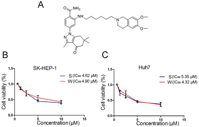
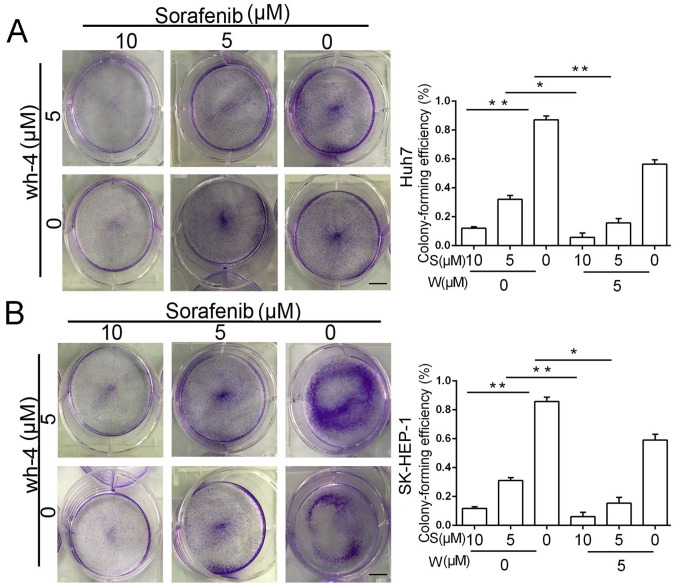
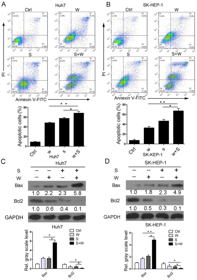
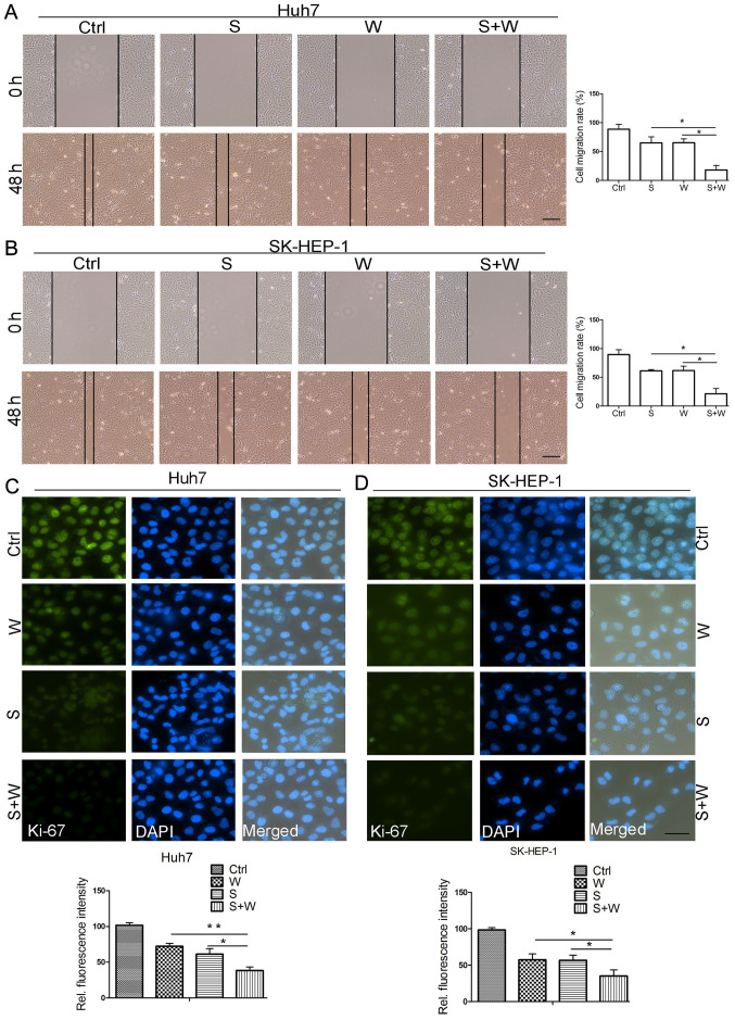
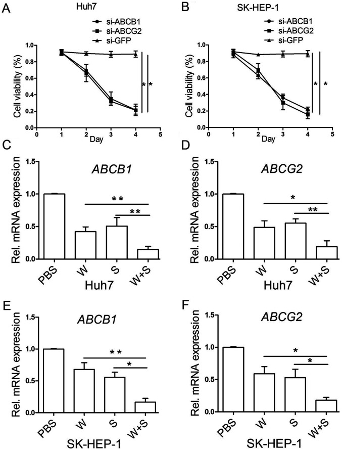
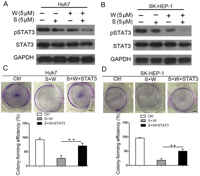
Sorafenib is currently used to treat hepatocellular carcinoma (HCC). However, the development of chemoresistance to sorafenib is a major limitation for sorafenib-based therapy in patients with HCC. In the present study, the effect of the combination therapy of sorafenib and wh-4 on the proliferation of liver cancer cells was investigated. The results showed that sorafenib with wh-4 additively suppressed the proliferation of liver cancer cells. The colony formation of liver cancer cells decreased significantly in response to the combination treatment of sorafenib with wh-4, and it also induced the apoptosis of liver cancer cells. Western blot analysis demonstrated decreased expression of Bcl2, and increased expression of Bax in liver cancer cells treated with a combination of sorafenib and wh-4. Moreover, the migration of liver cancer cells was inhibited. The combination treatment of sorafenib with wh-4 reduced the expression levels of ABCB1 and ABCG2 which are responsible for resistance. Finally, STAT3 overexpression abolished the proliferation inhibition effect of sorafenib with wh-4 on liver cancer cells, and sorafenib and wh-4 suppressed the proliferation of liver cancer cells by STAT3 pathway. Together, these results suggest that sorafenib-wh4 combination treatment is a potential novel therapeutic approach to suppress the proliferation of liver cancer cells.

Keywords: combination treatment; hepatocellular carcinoma cells; proliferation; sorafenib; wh-4.


Conflict of interest statement

The authors declare that they have no competing interests.

Figures

Figure 1
Figure 1
Cell viability of liver cancer cells analyzed by MTT assay. (A) Formal chemical structure of wh-4. (B and C) Three thousand cells were seeded in a 96-well dish and incubated for ~12 h. Cell were then treated with the indicated doses of sorafenib and wh-4 for 48 h. The effect of sorafenib, wh-4 and combination treatment on the cell viability of Huh7, SK-HEP-1 cells was evaluated by the MTT assay. IC50, half-maximal inhibitory concentration; S, sorafenib; W, wh-4.
Figure 2
Figure 2
Sorafenib combined with wh-4 additively decreases the viability of liver cancer cells. (A and B) Fifty thousand cells were seeded in a 6-well dish, and the cells were treated with sorafenib, wh-4 or their combination for 48 h. The plates were incubated at 37˚C in a humidified incubator for 21 days. Culture media was replenished every 3 days. The colonies of more than 40 cells was visualized as positive and stained with 0.5% crystal violet for 30 min. Scale bar, 10 mm. *P<0.05 and **P<0.01. S, sorafenib; W, wh-4.
Figure 3
Figure 3
Flow cytometry and western blot assays demonstrates that combination treatment with sorafenib and wh-4 additively induces liver cancer cell apoptosis. (A and B) Analysis of liver cancer cells treated with sorafenib (5 µM), wh-4 (5 µM) and their combination by flow cytometry. The combination contained 5 µM each of sorafenib and wh-4. After 24 h, cells were collected and the effects were analyzed by flow cytometry. (C and D) Analysis of liver cancer cells treated with 5 µM sorafenib, 5 µM wh-4 and their combination (5 µM concentration of each drug) by western blotting. GAPDH was considered as the loading control. *P<0.05 and **P<0.01. S, sorafenib; W, wh-4; Ctrl, control; Rel., relative.
Figure 4
Figure 4
Sorafenib with wh-4 suppresses cell migration. (A and B) Cells were subjected to 6-well plates. The cells were treated with 5 µM sorafenib, 5 µM wh-4 and their combination (5 µM each of sorafenib and wh-4) for 24 h. A 200-µl sterile pipette tip was used to scratch the dish bottom. After 48 h, the width was recorded using captured images. The combination contained sorafenib (5 µM) and wh-4 (5 µM). Scale bar, 50 µm. (C and D) Ki-67 analysis of cell proliferation. Cells were treated with sorafenib (5 µM), wh-4 (5 µM) and their combination (5 µM concentration of each drug) for 24 h. Scale bar, 100 µm. *P<0.05 and **P<0.01. S, sorafenib; W, wh-4; Ctrl, control; Rel., relative.
Figure 5
Figure 5
Sorafenib with wh-4 decrease the levels of chemoresistant genes. (A and B) Cell proliferation analysis of liver cancer cells after ABCB1 and ABCG2 knockdown. (C and D) Cell viability analysis of liver cancer cells by MTT assay. The liver cancer cells were knockdown with siRNA. (E and F) Reverse transcription-quantitative PCR analysis of ABCB1 and ABCG2. Tumor cells were treated with sorafenib (5 µM), wh-4 (5 µM) and their combination (5 µM concentration of each drug) for 24 h. *P<0.05 and **P<0.01. S, sorafenib; W, wh-4; siRNA/si-, small interfering RNA; Rel., relative.
Figure 6
Figure 6
STAT3 signaling pathway mediates liver cancer cell apoptosis induced by sorafenib with wh-4. (A and B) Analysis of STAT3 levels in cells treated with sorafenib (5 µM), wh-4 (5 µM) and their combination (5 µM sorafenib and 5 µM wh-4) for 24 h. GAPDH served as the loading control. (C and D) A colony-formation assay was used to analyze whether STAT3 was key to apoptosis induced by sorafenib with wh-4. Colony formation was detected in cells treated with sorafenib (5 µM), wh-4 (5 µM) and their combination (5 µM sorafenib and 5 µM wh-4) for 48 h. Colonies were stained with 0.5% crystal violet. Scale bar, 10 mm. **P<0.01. S, sorafenib; W, wh-4; Ctrl, control; pSTAT3, phosphorylated STAT3.

Similar articles

References

    1. Torre LA, Bray F, Siegel RL, Ferlay J, Lortet-Tieulent J, Jemal A. Global cancer statistics, 2012. CA Cancer J Clin. 2015;65:87–108. doi: 10.3322/caac.21262. - DOI - PubMed
    1. Villanueva A, Minguez B, Forner A, Reig M, Llovet JM. Hepatocellular carcinoma: Novel molecular approaches for diagnosis, prognosis, and therapy. Annu Rev Med. 2010;61:317–328. doi: 10.1146/annurev.med.080608.100623. - DOI - PMC - PubMed
    1. Vitale A, Volk ML, Pastorelli D, Lonardi S, Farinati F, Burra P, Angeli P, Cillo U. Use of sorafenib in patients with hepatocellular carcinoma before liver transplantation: A cost-benefit analysis while awaiting data on sorafenib safety. Hepatology. 2010;51:165–173. doi: 10.1002/hep.23260. - DOI - PubMed
    1. Reyes R, Wani NA, Ghoshal K, Jacob ST, Motiwala T. Sorafenib and 2-Deoxyglucose synergistically inhibit proliferation of both Sorafenib-Sensitive and -Resistant HCC cells by inhibiting ATP production. Gene Expr. 2017;17:129–140. doi: 10.3727/105221616X693855. - DOI - PMC - PubMed
    1. Shen YC, Hsu C, Cheng AL. Molecular targeted therapy for advanced hepatocellular carcinoma: Current status and future perspectives. J Gastroenterol. 2010;45:794–807. doi: 10.1007/s00535-010-0270-0. - DOI - PubMed