Case Reports
. 2022 Feb 9:9:820956.
doi: 10.3389/fvets.2022.820956. eCollection 2022.

Mediastinal Fibrosarcoma in a Dog-Case Report

Affiliations
Case Reports

Mediastinal Fibrosarcoma in a Dog-Case Report

Alysha M McGrath et al. Front Vet Sci. .

Abstract

This represents the first published case report of mediastinal fibrosarcoma in a dog. An 8-year-old male neutered mixed breed dog was presented for evaluation of lethargy and increased panting. Thoracic focused assessment with sonography for trauma revealed moderate pleural effusion. Thoracic radiograph findings were suggestive of a cranial mediastinal mass. Computed tomography revealed a mass within the right ventral aspect of the cranial mediastinum. On surgical exploration, a cranial mediastinal mass with an adhesion to the right cranial lung lobe was identified and removed en-bloc using a vessel sealant device and requiring a partial lung lobectomy. Histopathology results described the cranial mediastinal mass as fibrosarcoma with reactive mesothelial cells identified within the sternal lymph node. The patient was treated with systemic chemotherapy following surgical removal. To date, the dog has survived 223 days following diagnosis with recurrence noted 161 days following diagnosis and radiation therapy was initiated. Primary cranial mediastinal fibrosarcoma while a seemingly rare cause of thoracic pathology in dogs, should be considered in the differential diagnosis for a cranial mediastinal mass.

Keywords: blunt dissection; case report; chemotherapy; dog; doxorubicin; fibrosarcoma; mediastinum; surgery.


Conflict of interest statement

The authors declare that the research was conducted in the absence of any commercial or financial relationships that could be construed as a potential conflict of interest.

Figures
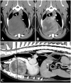
Figure 1
Figure 1
Transverse (A,B) and sagittal (C) plane CT images of the cranial mediastinal mass with a black line representing the location on the sagittal image. The cranial vena cava (arrowheads) is compressed ventrodorsally by the large mediastinal mass, being bordered dorsally by the brachiocephalic trunk. The mass is soft tissue attenuating, with heterogeneous contrast enhancement, and a well-defined margin. Note the pleural (*) and mediastinal (#) fluid that is not contrast enhancing. Window width and window level were adjusted to 300/40 (soft tissue window), the slice thickness was 2 mm, and the image was acquired with 120 kVp.
Figure 2
Figure 2
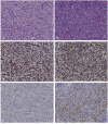
Mediastinal mass (A–C) and sternal lymph node (D–F), canine. (A) The mediastinal mass is composed of densely packed, disorganized, interwoven bundles and streams of neoplastic spindle cells on scant fibrous stroma (40X). (B) Immunohistochemistry for vimentin of the mediastinal mass shows strong intracytoplasmic labeling within neoplastic spindle cells. (C) Immunohistochemistry for cytokeratin of the mediastinal mass shows no labeling among the neoplastic spindle cells. (D) The subcapsular and medullary sinuses of the sternal lymph node are infiltrated by sheets of pleomorphic round cells exhibiting marked anisocytosis and anisokaryosis, frequent binucleation in which the nuclei are squished together creating a flattened appearance of their coalescing borders, and emperiopolesis. (E) Immunohistochemistry for vimentin of the sternal lymph node shows strong intracytoplasmic labeling within pleomorphic round cells. (F) Immunohistochemistry for cytokeratin of the sternal lymph node shows moderate to strong intracytoplasmic labeling within pleomorphic round cells.

References

    1. Yoon J, Feeney DA, Cronk DE, Anderson KL, Ziegler LE. Computed tomographic evaluation of canine and feline mediastinal masses in 14 patients. Vet Radiol Ultrasound. (2004) 45:542–6. 10.1111/j.1740-8261.2004.04093.x - DOI - PubMed
    1. Pintore L, Bertazzolo W, Bonfanti U, Gelain ME, Bottero E. Cytological and histological correlation in diagnosis feline and canine mediastinal masses. J Small Anim Pract. (2014) 55:28–32. 10.1111/jsap.12161 - DOI - PubMed
    1. Reeve EJ, Mapletoft EK, Schiborra F, Maddox TW, Lamb CR, Warren-Smith CMR. Mediastinal lymphoma in dogs is homogenous compared to thymic epithelial neoplasia and is more likely to envelop the cranial vena cava in CT images. Vet Radiol Ultrasound. (2020) 61:25–32. 10.1111/vru.12812 - DOI - PubMed
    1. Kuntz CA. Thoracic surgical oncology. Clin Tech Small Anim Pract. (1998) 13:47–52. 10.1016/S1096-2867(98)80027-3 - DOI - PubMed
    1. Liptak JM, Kamstock DA, Dernell WS, Ehrhart EJ, Rizzo SA, Withrow SJ. Cranial mediastinal carcinomas in nine dogs. Vet Comp Oncol. (2008) 6:19–30. 10.1111/j.1476-5829.2007.00133.x - DOI - PubMed

Publication types