The Expression and Clinical Significance of PCNAP1 in Hepatocellular Carcinoma Patients
- PMID: 35224110
- PMCID: PMC8881134
- DOI: 10.1155/2022/1817694
The Expression and Clinical Significance of PCNAP1 in Hepatocellular Carcinoma Patients
Abstract
Background: Long noncoding RNAs (lncRNAs) play an important role in many cancer progression. The aim of this study was to evaluate the expression level and clinical significance of the lncRNA, proliferating cell nuclear antigen pseudogene 1 (PCNAP1), in cancer tissue and the plasma of patients with hepatocellular carcinoma (HCC).
Methods: Quantitative real-time polymerase chain reaction was used to detect the expression of PCNAP1 in HCC tissue, adjacent tissue, and plasma. Spearman's rank correlation analysis was performed to assess relationships among cancer tissue, plasma PCNAP1, and plasma AFP. Kaplan-Meier analysis was used to assess survival of HCC patient with high and low expression of PCNAP1. The survival difference was compared by the log-rank test. The use of plasma levels PCNAP1 for diagnosing HCC was evaluated by receiver operating characteristic curve analysis.
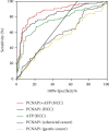
Results: The expression of PCNAP1 in HCC tissue was significantly higher than in adjacent tissue (P < 0.01). The PCNAP1 levels were related to the TNM stage, lymph node metastasis, and tumor maximum diameter (P < 0.05) but were not related to gender and age (P = 0.459 and 0.656). Patients with greater levels of PCNAP1 had poorer survival than patients with lower levels of expression (P < 0.01). Compared to the healthy control group, a gastric cancer group, and a colorectal cancer group, HCC patient plasma levels of PCNAP1 were significantly greater (P < 0.01). The area under the curve (AUC) of plasma PCNAP1 in HCC patients was 0.83 (95% CI: 0.78-0.88). With a cut-off value of plasma PCNAP1 at 1.27, an HCC diagnostic sensitivity of 70.08%, and a specificity of 85.04%, was the maximum diagnostic efficiency achieved.
Conclusion: This study demonstrates PCNAP1 levels to be increased in HCC patients. As such, PCNAP1 may be a new tool useful in disease diagnosis and prognosis.
Copyright © 2022 Yanhong Chen et al.
Conflict of interest statement
The authors declare that there is no conflict of interest regarding the publication of this paper.
Figures


Similar articles
-
Diagnostic and prognostic value of lncRNA cancer susceptibility candidate 9 in hepatocellular carcinoma.World J Gastroenterol. 2019 Dec 28;25(48):6902-6915. doi: 10.3748/wjg.v25.i48.6902. World J Gastroenterol. 2019. PMID: 31908394 Free PMC article.
-
Down-regulation of long non-coding RNA GAS5-AS1 and its prognostic and diagnostic significance in hepatocellular carcinoma.Cancer Biomark. 2018;22(2):227-236. doi: 10.3233/CBM-170781. Cancer Biomark. 2018. PMID: 29660898
-
Low expression of long noncoding RNA CTC-297N7.9 predicts poor prognosis in patients with hepatocellular carcinoma.Cancer Med. 2019 Dec;8(18):7679-7692. doi: 10.1002/cam4.2618. Epub 2019 Nov 1. Cancer Med. 2019. PMID: 31674731 Free PMC article.
-
LncRNA PCNAP1 modulates hepatitis B virus replication and enhances tumor growth of liver cancer.Theranostics. 2019 Jul 9;9(18):5227-5245. doi: 10.7150/thno.34273. eCollection 2019. Theranostics. 2019. PMID: 31410212 Free PMC article.
-
Diagnostic Value of lncRNAs as Biomarker in Hepatocellular Carcinoma: An Updated Meta-Analysis.Can J Gastroenterol Hepatol. 2018 Oct 15;2018:8410195. doi: 10.1155/2018/8410195. eCollection 2018. Can J Gastroenterol Hepatol. 2018. PMID: 30410873 Free PMC article.
Cited by
-
Predictive value of cuproptosis and disulfidptosis-related lncRNA in head and neck squamous cell carcinoma prognosis and treatment.Heliyon. 2024 Sep 17;10(18):e37996. doi: 10.1016/j.heliyon.2024.e37996. eCollection 2024 Sep 30. Heliyon. 2024. PMID: 39323825 Free PMC article.
-
Pseudogenes as Potential Diagnostic, Prognostic and Therapeutic Biomarkers in Colorectal Cancer: A Systematic Review.Cancer Rep (Hoboken). 2025 Jun;8(6):e70263. doi: 10.1002/cnr2.70263. Cancer Rep (Hoboken). 2025. PMID: 40556338 Free PMC article.
-
Combinatorial Gene Expression Profiling of Serum HULC, HOTAIR, and UCA1 lncRNAs to Differentiate Hepatocellular Carcinoma from Liver Diseases: A Systematic Review and Meta-Analysis.Int J Mol Sci. 2024 Jan 19;25(2):1258. doi: 10.3390/ijms25021258. Int J Mol Sci. 2024. PMID: 38279264 Free PMC article.
References
MeSH terms
Substances
LinkOut - more resources
Full Text Sources
Medical
Research Materials

