Case Reports
. 2022 Feb 16;6(2):ytac067.
doi: 10.1093/ehjcr/ytac067. eCollection 2022 Feb.

Case report of the broad spectrum of late complications in an adult patient with univentricular physiology palliated by the Fontan circulation

Affiliations
Case Reports

Case report of the broad spectrum of late complications in an adult patient with univentricular physiology palliated by the Fontan circulation

Marieke Nederend et al. Eur Heart J Case Rep. .

Abstract

Background: At the most severe end of the spectrum of congenital heart disease are patients with an univentricular physiology. They comprise a heterogeneous group of congenital heart malformations that have the common characteristic that the cardiac morphology is not equipped for sustaining a biventricular circulation.

Case summary: Here, we present a case of an adult patient after Fontan palliation, illustrative of the complex clinical course and the broad spectrum of complications that can be encountered during follow-up, highlighting the need for a multidisciplinary approach in the clinical care for these patients.

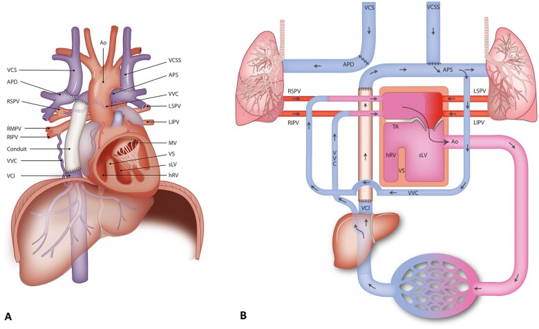
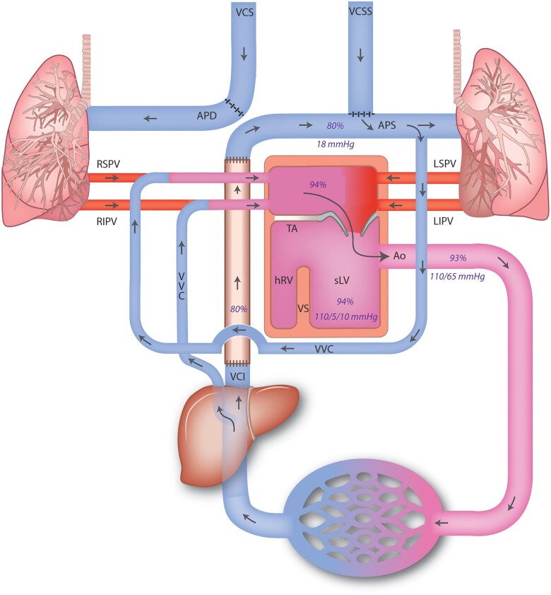
Discussion: During the surgical Fontan procedure, the inferior vena cava is connected to the pulmonary circulation, after prior connection of the superior vena cava to the pulmonary arterial circulation. The resulting cavopulmonary connection, thus lacking a subpulmonic ventricle, provides non-pulsatile passive flow of oxygen-poor blood from the systemic venous circulation into the lungs, and the functional monoventricle pumps the oxygen-rich pulmonary venous return blood into the aorta. With an operative mortality of <5% and current 30-year survival rates up to 85%, the adult population of patients with a Fontan circulation is growing. This increase in survival is, however, inevitably accompanied by long-term complications affecting multiple organ systems, resulting in decline in cardiovascular performance.

Conclusion: For optimal treatment, the evaluation in a multidisciplinary team is mandatory, using the specific expertise of the team members to timely detect and address late complications and to support quality of life.

Keywords: Case report; Congenital heart disease; Fontan circulation; Fontan failure; Fontan-related liver disease; Long-term complications; Univentricular heart.


Figures

Figure 1
Figure 1
(A) Graphic depiction of the anatomy of the patient. (B) Schematic depiction of the anatomy and the circulation of the patient. Ao, aorta; APD, arteria pumonalis dextra/right; APS, arteria pulmonalis sinistra/left; hRV, hypoplastic right ventricle; LIPV, left inferior pulmonary vein; LSPV, left superior pulmonary vein; MV, mitral valve; RIPV, right inferior pulmonary vein; RMPV, right middle pulmonary vein; RSPV, right superior pulmonary vein; sLV, systemic (single) left ventricle; TA, tricuspid atresia; VCI, vena cava inferior; VCS, vena cava superior; VCSS, persisting vena cava superior sinistra; VS, ventricular septum; VVC, veno-venous collaterals.
Figure 2
Figure 2
(A) Computed tomography angiography showing the anatomical connections of the mildly dilated left (functionally) single ventricle connected to the aorta, the hypoplastic right ventricle and the extracardiac tunnel. In addition, the subaortic ventricular septal defect is shown. (B) Computed tomography angiography during contrast showing extensive collateral opacification paraesophageal (right), opacification of right inferior pulmonary vein and a segmental right superior pulmonary vein from the right upper lobe. These findings suggested a shunt circulation from the abdominal systemic veins to the left atrium. (C) Computed tomography angiography showing dilated left (mono) atrium and dilated coronary sinus, as well as relatively dilated right pulmonary veins. Congestion with interstitial thickening of the interlobar septa and bronchial cuffing with pleural fluid on the right side, no signs of congestion in the left lung and slim pulmonary veins. Ao, aorta; Ao desc, aorta descendens; CS, coronary sinus; LA, left atrium; LV, left ventricle; PV, pulmonary vein; RV, right ventricle (hypoplastic); VSD, ventricular septal defect.
Figure 3
Figure 3
Schematic depiction of the circulation of the patient with pressures and saturations as measured during cardiac catheterization. Ao, aorta; APD, arteria pumonalis dextra/right; APS, ateria pulmonalis sinistra/left; hRV, hypoplastic right ventricle; LIPV, left inferior pulmonary vein; LSPV, left superior pulmonary vein; MV, mitral valve; RIPV, right inferior pulmonary vein; RSPV, right superior pulmonary vein; sLV, systemic (single) left ventricle; TA, tricuspid atresia; VCI, vena cava inferior; VCS, vena cava superior; VCSS, persisting vena cava superior sinistra; VS, ventricular septum; VVC, veno-venous collaterals.
Figure 4
Figure 4
(A) Angiographic projection (AP 2.2°) showing collateral flow from the inferior vena cava to the right pulmonary vein. Epicardial pacemaker leads indicated by yellow arrows. (B) Angiographic projection (AP 8.4°) showing collateral flow immediately after coiling with four coils within the red circles (VortX pushable coil Boston Scientific 6 mm × 6.5 mm). Note the reduced distal contrast opacification in the collateral vessel. (C) Angiographic projection (AP 8.4°) showing a second small collateral from the inferior vena (subdiaphragmatic) cava to the right pulmonary vein. Previously placed coils indicated by the red circle. (D) Angiographic projection (RIO 27°) showing veno-venous collaterals after additional coiling of smaller collateral with three coils indicated by the green circle (VortX pushable coil Boston Scientific 6 mm × 6.5 mm) Previously placed coils indicated by the red circle.
Figure 5
Figure 5
Computed tomography showing one of the focal lesions (blue arrows) suspicious of hepatocellular carcinoma with characteristic hypervascularity in arterial phase (A). The lesion is iso-attenuating in the portal venous phase (B). There is washout in the delayed phase (C).
Figure 6
Figure 6
Angiographic visualization showing transarterial chemoembolization through the posterior division of the right hepatic artery (black arrows) towards segment VIII (projected over segment VII). Note the previously placed coils in the veno-venous collaterals (red circle).

Similar articles

Cited by

References

    1. Baumgartner H, De Backer J, Babu-Narayan SV, Budts W, Chessa M, Diller G-P. et al. 2020 ESC Guidelines for the management of adult congenital heart disease: The Task Force for the management of adult congenital heart disease of the European Society of Cardiology (ESC). Eur Heart J 2020;41:4153–4154. - PubMed
    1. Rychik J, Atz AM, Celermajer DS, Deal BJ, Gatzoulis MA, Gewillig MH. et al.; On behalf of the American Heart Association Council on Cardiovascular Disease in the Young and Council on Cardiovascular and Stroke Nursing. Evaluation and management of the child and adult with fontan circulation: a scientific statement from the American Heart Association. Circulation 2019;140:e234–e284. - PubMed
    1. van Melle JP, Wolff D, Horer J, Belli E, Meyns B, Padalino M. et al. Surgical options after Fontan failure. Heart 2016;102:1127–1133. - PubMed
    1. Schuuring MJ, Vis JC, van Dijk AP, van Melle JP, Vliegen HW, Pieper PG. et al. Impact of bosentan on exercise capacity in adults after the Fontan procedure: a randomized controlled trial. Eur J Heart Fail 2013;15:690–698. - PubMed
    1. Sumal AS, Kyriacou H, Mostafa A.. Tricuspid atresia: where are we now? J Card Surg 2020;35:1609–1617. - PubMed

Publication types

LinkOut - more resources