

Biomimetic Alveolus-on-a-Chip for SARS-CoV-2 Infection Recapitulation
- PMID: 35224503
- PMCID: PMC8841031
- DOI: 10.34133/2022/9819154
Biomimetic Alveolus-on-a-Chip for SARS-CoV-2 Infection Recapitulation
Abstract
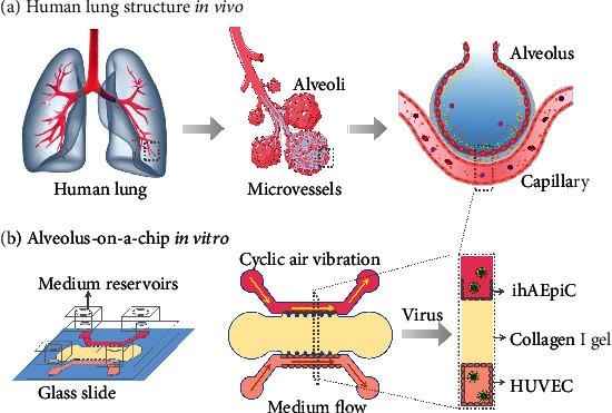
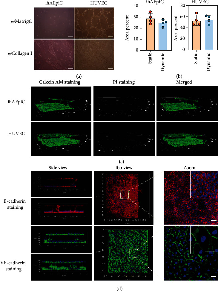
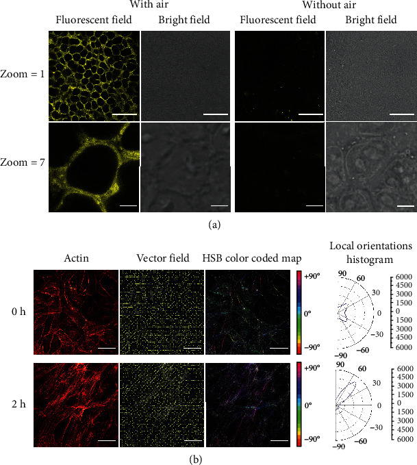
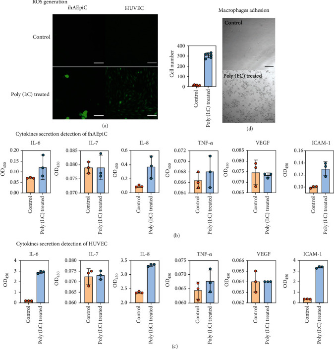
SARS-CoV-2 has caused a severe pneumonia pandemic worldwide with high morbidity and mortality. How to develop a preclinical model for recapitulating SARS-CoV-2 pathogenesis is still urgent and essential for the control of the pandemic. Here, we have established a 3D biomimetic alveolus-on-a-chip with mechanical strain and extracellular matrix taken into consideration. We have validated that the alveolus-on-a-chip is capable of recapitulating key physiological characteristics of human alveolar units, which lays a fundamental basis for viral infection studies at the organ level. Using virus-analogous chemicals and pseudovirus, we have explored virus pathogenesis and blocking ability of antibodies during viral infection. This work provides a favorable platform for SARS-CoV-2-related researches and has a great potential for physiology and pathophysiology studies of the human lung at the organ level in vitro.
Copyright © 2022 Ting Cao et al.
Conflict of interest statement
The authors declare no conflict of interest.
Figures

Similar articles
-
SARS-CoV-2 induced intestinal responses with a biomimetic human gut-on-chip.Sci Bull (Beijing). 2021 Apr 30;66(8):783-793. doi: 10.1016/j.scib.2020.11.015. Epub 2020 Dec 1. Sci Bull (Beijing). 2021. PMID: 33282445 Free PMC article.
-
Biomimetic Human Disease Model of SARS-CoV-2-Induced Lung Injury and Immune Responses on Organ Chip System.Adv Sci (Weinh). 2020 Dec 21;8(3):2002928. doi: 10.1002/advs.202002928. eCollection 2021 Feb. Adv Sci (Weinh). 2020. PMID: 33173719 Free PMC article.
-
Modelling SARS-CoV-2 infection in a human alveolus microphysiological system.Access Microbiol. 2024 Sep 11;6(9):000814.v3. doi: 10.1099/acmi.0.000814.v3. eCollection 2024. Access Microbiol. 2024. PMID: 39697363 Free PMC article.
-
Insights into COVID-19 Vaccine Development Based on Immunogenic Structural Proteins of SARS-CoV-2, Host Immune Responses, and Herd Immunity.Cells. 2021 Oct 29;10(11):2949. doi: 10.3390/cells10112949. Cells. 2021. PMID: 34831172 Free PMC article. Review.
-
Surgical Infection Society Guidance for Operative and Peri-Operative Care of Adult Patients Infected by the Severe Acute Respiratory Syndrome Coronavirus-2 (SARS-CoV-2).Surg Infect (Larchmt). 2020 May;21(4):301-308. doi: 10.1089/sur.2020.101. Epub 2020 Apr 20. Surg Infect (Larchmt). 2020. PMID: 32310715 Review.
Cited by
-
Fighting the SARS-CoV-2 Pandemic: Focusing a New Lens on COVID-19.Research (Wash D C). 2022 Jul 21;2022:9879646. doi: 10.34133/2022/9879646. eCollection 2022. Research (Wash D C). 2022. PMID: 35966758 Free PMC article. No abstract available.
-
Biomimetic Gland Models with Engineered Stratagems.Research (Wash D C). 2023 Sep 15;6:0232. doi: 10.34133/research.0232. eCollection 2023. Research (Wash D C). 2023. PMID: 37719047 Free PMC article. Review.
-
Cancer-on-chip: a 3D model for the study of the tumor microenvironment.J Biol Eng. 2023 Aug 17;17(1):53. doi: 10.1186/s13036-023-00372-6. J Biol Eng. 2023. PMID: 37592292 Free PMC article. Review.
-
Advanced lung organoids and lung-on-a-chip for cancer research and drug evaluation: a review.Front Bioeng Biotechnol. 2023 Nov 7;11:1299033. doi: 10.3389/fbioe.2023.1299033. eCollection 2023. Front Bioeng Biotechnol. 2023. PMID: 38026900 Free PMC article. Review.
-
Microfluidic strategies for biomimetic lung chip establishment and SARS-CoV2 study.Mater Today Bio. 2023 Dec 7;24:100905. doi: 10.1016/j.mtbio.2023.100905. eCollection 2024 Feb. Mater Today Bio. 2023. PMID: 38094656 Free PMC article. Review.
References
-
- Coronavirus disease (COVID-19) pandemic. World Health Organization . https://www.who.int/emergencies/diseases/novel-coronavirus-2019 .
LinkOut - more resources
Full Text Sources
Miscellaneous
