. 2022 Aug;241(2):552-564.
doi: 10.1111/joa.13647. Epub 2022 Feb 27.

True-color 3D rendering of human anatomy using surface-guided color sampling from cadaver cryosection image data: A practical approach

Affiliations

True-color 3D rendering of human anatomy using surface-guided color sampling from cadaver cryosection image data: A practical approach

Jon Jatsu Azkue. J Anat. 2022 Aug.

Abstract

Three-dimensional computer graphics are increasingly used for scientific visualization and for communicating anatomical knowledge and data. This study presents a practical method to produce true-color 3D surface renditions of anatomical structures. The procedure involves extracting the surface geometry of the structure of interest from a stack of cadaver cryosection images, using the extracted surface as a probe to retrieve color information from cryosection data, and mapping sampled colors back onto the surface model to produce a true-color rendition. Organs and body parts can be rendered separately or in combination to create custom anatomical scenes. By editing the surface probe, structures of interest can be rendered as if they had been previously dissected or prepared for anatomical demonstration. The procedure is highly flexible and nondestructive, offering new opportunities to present and communicate anatomical information and knowledge in a visually realistic manner. The technical procedure is described, including freely available open-source software tools involved in the production process, and examples of color surface renderings of anatomical structures are provided.

Keywords: 3D graphics; anatomy education; medical visualization; surface rendering.


Figures

FIGURE 1
FIGURE 1
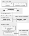
Flow diagram for the surface‐guided color sampling procedure. Cryosection images are converted into a volumetric data file, which can then be resliced in any orthogonal plane for segmentation. Segmentation of the submandibular gland (sb, delimited in red color) from the Visible Human Male is shown. The marching cubes algorithm generates a 3D polygon mesh representing the external surface of the segmented target object. At this stage, a surface mesh can be edited to produce a modified version of the target structure that will be rendered as having been dissected or prepared. The color sampling operation retrieves color information (shades of brown) of those voxels representing the external boundaries of the target structure, using the surface mesh outputted by the previous step as a probe (red), and then assigns color attributes to the corresponding polygon vertices. A schematic representation of this operation in a subgroup of voxels is shown. Finally, the VTK renderer creates color on polygon faces by interpolation of vertex colors and renders the surface. b: Body of mandible; c: Carotid arteries; d: Digastric muscle; f: Facial vein; h: Hyoglossus muscle; hb: Hyoid bone; j: Internal jugular vein; m: Masseter muscle; n: Deep cervical lymph nodes; p: Palatopharyngeus and pharyngeal constrictor muscles; pl: Platysma; pt: Medial pterygoid muscle; sb: Submandibular gland; sg: Styloglossus muscle; sm: Submental vein; st: Sternocleidomastoid muscle; t: Tongue
FIGURE 2
FIGURE 2
Implementation of the color sampling operation using VTK. Input data importation, color sampling, rendering, and file saving steps are shown, also indicating the involved filters or functions in the VTK processing pipeline. The vtkProbeFilter, core of the sampling operation, takes the source volumetric data and the polygon mesh extracted by segmentation as inputs. The filter outputs a copy of the input mesh and appends the extracted color data from scalar values to polygon vertices. A separate step converts scalar values into color for visualization. A mapper converts the polygon mesh into a virtual model for surface rendering. The resulting model can be stored as a file. A python script that performs this operation and renders the resulting surface is provided in Appendix A
FIGURE 3
FIGURE 3
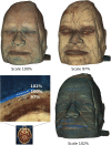
Successful color sampling is dependent on the accuracy of the surface probe. A surface rendition of the Visible Human Female head is shown using correctly sized (100% scale) and distorted probes. Increasing or decreasing probe size by 2%–3% results in sampling colors from voxels outside the body (embedding blue gel) or representing subcutaneous tissues. A cross‐sectional image shows the relative positions of the three probes for reference
FIGURE 4
FIGURE 4
The level of detail of rendered colors and textures depends on the resolution of the source cryosection images. Reconstructions of the cervical spinal cord from the Visible Head from the Visible Korean dataset (left) and the Visible Human Male from the NLM (right) are shown, and the maximum width of the spinal cord in the corresponding original images is indicated. Dorsal and ventral horns are readily identifiable in the higher resolution model, and relatively blurred in the lower resolution one
FIGURE 5
FIGURE 5
Kidney in entirety and after editing the surface probe. Surface rendition of the whole left kidney and attached ureter from the Visible Human Male are shown, as well as virtually dissected versions of the kidney, ureter, and vascular supply after sectioning in the axial and coronal planes. The morphology and internal organization of the renal pyramids are clearly discernible. Note an accessory renal artery entering the inferior pole
FIGURE 6
FIGURE 6
The brain as a whole and after editing the surface probe. Surface renditions of the brain from the isolated head dataset in the Visible Human image collection are shown, both as a whole (top left) and following sectioning in the axial and coronal planes or exposure of the left insular cortex (bottom right) after removal of the overlying frontal, parietal, and temporal cortices. Note whitish discoloration on the posterior short gyrus and long gyrus of the insula due to abrasion during cadaver sectioning
FIGURE 7
FIGURE 7
Stripping layers off the model. A surface rendition of the head of the Visible Male from the Visible Korean dataset is shown, using a surface probe from which two pieces representing portions of the skin and skull overlying the brain had been removed. Note the tenuous purple impregnation of the skin due to the embedding gel
FIGURE 8
FIGURE 8
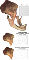
Reducing mesh complexity without losing visual realism. The right temporalis from the Visible Human Female is shown attached to the coronoid process, before texture baking (top). Two versions of the muscle model alone are shown after texture baking and reducing the vertex count by ca. 86% or by ca. 97% (bottom). The vertex count could be reduced dramatically (figures provided in Table 2) without noticeably affecting the overlying bitmap texture. The magnification boxes show the structure of the polygon mesh from approximately the same region at the various levels of simplification

Similar articles

References

    1. Ackerman, M.J. (1998) The Visible Human Project: a resource for anatomical visualization. Studies in Health Technology and Informatics, 52, 1030–1032. - PubMed
    1. Ackerman, M.J. , Yoo, T. & Jenkins, D. (2001) From data to knowledge—the Visible Human Project continues. Studies in Health Technology and Informatics, 84, 887–890. - PubMed
    1. Assaf, Y. & Pasternak, O. (2008) Diffusion tensor imaging (DTI)‐based white matter mapping in brain research: a review. Journal of Molecular Neuroscience, 34, 51–61. - PubMed
    1. Azkue, J.J. (2021a) Embedding interactive, three‐dimensional content in portable document format to deliver gross anatomy information and knowledge. Clinical Anatomy, 34, 919–933. - PubMed
    1. Azkue, J.J. (2021b) External surface anatomy of the postfolding human embryo: computer‐aided, three‐dimensional reconstruction of printable digital specimens. Journal of Anatomy, 239, 1438–1451. 10.1111/joa.13514 - DOI - PMC - PubMed