. 2022 May 2;61(19):e202200951.
doi: 10.1002/anie.202200951. Epub 2022 Mar 11.

Cystine Knot Peptides with Tuneable Activity and Mechanism

Affiliations

Cystine Knot Peptides with Tuneable Activity and Mechanism

Choi Yi Li et al. Angew Chem Int Ed Engl. .

Abstract

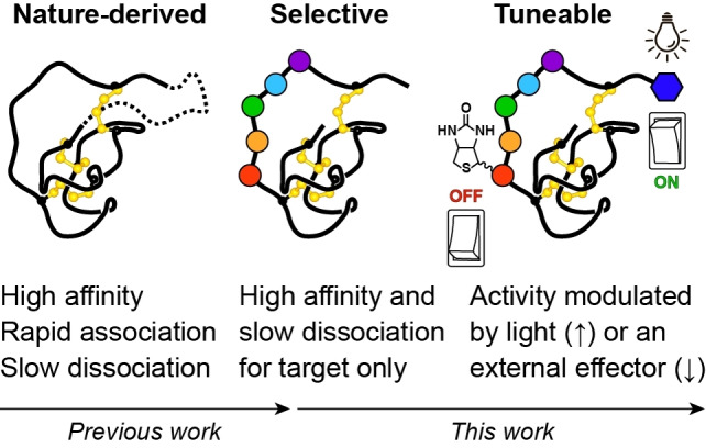
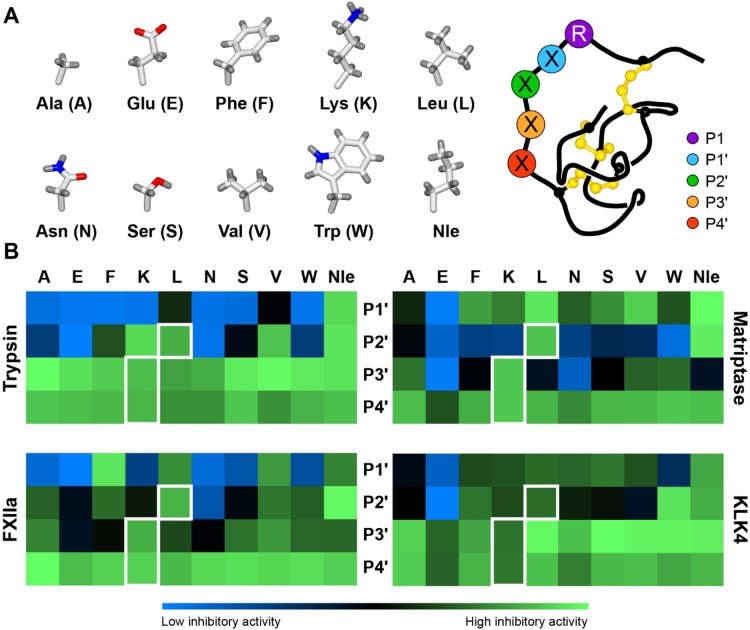
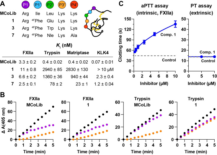
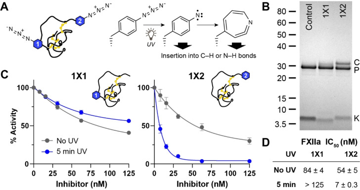
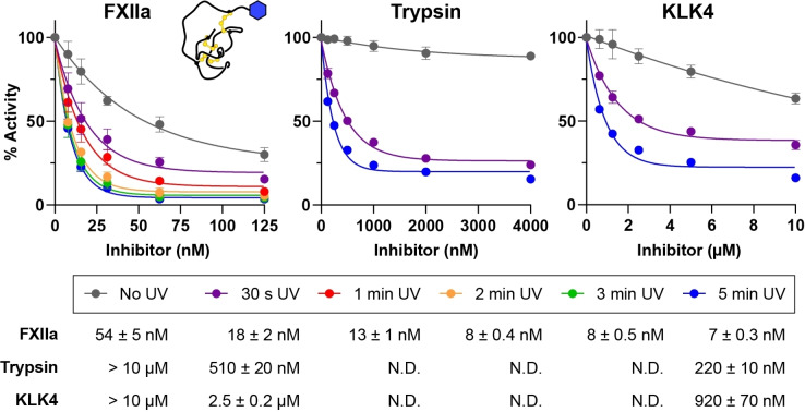
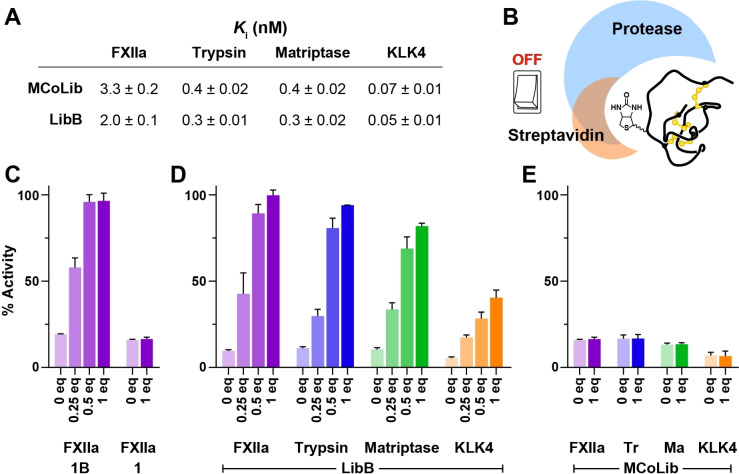
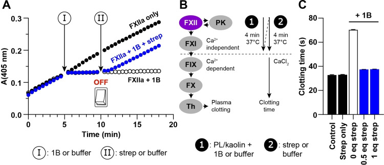
Knottins are topologically complex peptides that are stabilised by a cystine knot and have exceptionally diverse functions, including protease inhibition. However, approaches for tuning their activity in situ are limited. Here, we demonstrate separate approaches for tuning the activity of knottin protease inhibitors using light or streptavidin. We show that the inhibitory activity and selectivity of an engineered knottin can be controlled with light by activating a second mode of action that switches the inhibitor ON against new targets. Guided by a knottin library screen, we also identify a position in the inhibitor's binding loop that permits insertion of a biotin tag without impairing activity. Using streptavidin, biotinylated knottins with nanomolar affinity can be switched OFF in activity assays, and the anticoagulant activity of a factor XIIa inhibitor can be rapidly switched OFF in human plasma. Our findings expand the scope of engineered knottins for precisely controlling protein function.

Keywords: Activity Switch; Enzymes; Inhibitors; Knottins; Photoactivation.


Conflict of interest statement

The authors declare no conflict of interest.

Figures

Scheme 1
Scheme 1
Nature‐derived knottin PIs are acyclic (solid line) or cyclic (dashed line) peptides that bind with high affinity to their targets and show fast association‐slow dissociation rates. In this work, we engineer selective knottin PIs with minimal modifications, then modulate their activity with light or an external effector.
Figure 1
Figure 1
Synthetic knottin libraries for profiling protease specificity. A) Schematic representation of the knottin libraries, where each of the ten amino acids shown was substituted into the P1′ (blue), P2′ (green), P3′ (orange) or P4′ (red) positions. P1 (purple) was fixed as Arg (R). B) Heat maps illustrating the inhibitory activity (relative to enzyme activity in the absence of inhibitor) for knottin variants with selected amino acids (column titles) at a given position (row titles, centre). Inhibitory activity is shown as a gradient from low (blue) to high (green) as indicated in the key below the heat maps. Residues present in MCoTI‐II are highlighted with a white border (P1′ is Ile). Data are from three experiments (mean) and are shown as bar graphs (mean±SD) in Figure S10. Nle denotes norleucine.
Figure 2
Figure 2
Design of potent and selective FXIIa inhibitors via two substitutions. A) Sequence modifications in the binding loop for selected knottin variants (4FPhe denotes 4‐fluoro‐L‐Phe) and K i values (±standard error) against FXIIa, trypsin, matriptase, or KLK4. Data are from three experiments performed in triplicate, and >10 μM indicates less than 50 % inhibition at 10 μM. B) Reaction progress curves showing enzyme activity (y‐axis, mOD) after addition of substrate to FXIIa or trypsin pre‐incubated with inhibitor (orange) or simultaneously with inhibitor (purple). Enzyme activity in the absence of inhibitor (black) illustrates the control rate. Progress curves used to calculate k off for each enzyme‐inhibitor pair (except trypsin‐1) are shown in Figure S15. C) Inhibitory activity of compound (comp.) 1 in coagulation assays using human plasma. Activated partial thromboplastin time (aPTT) assays measure the FXIIa‐initiated intrinsic pathway, and prothrombin time (PT) assays measure the FVIIa/tissue factor‐initiated extrinsic pathway. Data show mean±SD from n=3, and control indicates clotting time in the absence of inhibitor.
Figure 3
Figure 3
A photoreactive knottin with light‐controlled activity. A) Location of the AzF residue in 1X1 or 1X2, and reaction scheme for photoactivation of AzF via formation of a reactive nitrene that can directly insert into C−H or N−H bonds, or undergo ring expansion to form a dehydroazepine intermediate. B) Crosslinking of 1X1 or 1X2 to FXIIa (5 min UV exposure) analysed by SDS‐PAGE (C=complex, P=protease, K=knottin). Control is a 1 variant that lacks a photoreactive amino acid. C) Data from competitive inhibition assays comparing the activity of 1X1 or 1X2 against FXIIa before (grey) or after (blue) UV crosslinking. Data (mean±SD) for each condition are normalised to controls (FXIIa without inhibitor) with or without UV exposure. D) Apparent IC50 values for 1X1 or 1X2 against FXIIa before or after UV crosslinking.
Figure 4
Figure 4
A photoreactive knottin with light‐controlled selectivity. Graphs show data (mean±SD, three experiments in duplicate) from competitive inhibition assays comparing the activity of 1X2 against FXIIa, trypsin or KLK4 with or without exposure to UV light. Exposure times are indicated in the key below the graphs. Data for each exposure time are normalised to controls (protease without inhibitor) exposed to UV light for the same time. IC50 values calculated for each exposure time are shown below the key. For trypsin and KLK4, exposure times of 1–3 min were not performed (N.D.=not determined).
Figure 5
Figure 5
Switching OFF biotin‐labelled knottins using an external effector. A) K i values (±standard error) for the biotin‐labelled inhibitor LibB compared with its non‐labelled counterpart MCoLib. Data are from three experiments performed in triplicate. B) Schematic illustrating the concept of an affinity tag OFF switch based on knottin labelling at P4′ that introduces overlapping binding sites for streptavidin and the target protease. C) Recovery of enzyme activity (y‐axis) after adding 0.25–1 equiv streptavidin (x‐axis) to FXIIa incubated with 1B (≈1 : 20 ratio FXIIa:1B). Activity data are expressed as a % relative to controls with FXIIa and substrate only (mean±SD from three experiments). No recovery of activity was observed for the unlabelled inhibitor 1. D) Recovery of enzyme activity after adding streptavidin to FXIIa (purple), trypsin (blue), matriptase (green) or KLK4 (orange) incubated with LibB. E) No recovery of activity was observed for the non‐labelled inhibitor MCoLib.
Figure 6
Figure 6
Streptavidin rapidly switches OFF a biotin‐labelled knottin in kinetic assays and in human plasma. A) Progress curves showing substrate cleavage by FXIIa (y‐axis, OD). Reactions were initiated by adding FXIIa (5 nM) to substrate (150 μM). At 5 min (I), 1B (500 nM) was added to stop the indicated reactions (blue circles or open circles). At 10 min (II), streptavidin (500 nM, blue circles) or an equivalent volume of buffer (open circles) was added. Recovery of FXIIa activity after adding streptavidin was ≈95 % relative to controls without 1B (black circles). Data show representative curves from one of three experiments. B) Modified aPTT assay for switching OFF 1B in human plasma. Adding phospholipid (PL) and kaolin (1) to citrated plasma initiates activation of factor XII (FXII), which subsequently activates plasma kallikrein (PK) and factor XI (FXI). CaCl2 is required for activation of downstream enzymes: factor IX (FIX), factor X (FX), and thrombin (Th). Adding 1B during the first incubation (1) allows inhibition of FXIIa, which can be switched OFF in the second incubation (2) by adding streptavidin. Clotting times are shown in C). For controls (black bars), buffer was added in the first incubation and buffer or streptavidin in the second. 10 μM 1B was added in the first incubation for the remaining conditions, followed by buffer (open bar) or 0.5–1 equiv streptavidin (blue bars) in the second. Data show the mean±SD from n=4.

Similar articles

Cited by

References

    1. Wang J., Liu Y., Liu Y., Zheng S., Wang X., Zhao J., Yang F., Zhang G., Wang C., Chen P. R., Nature 2019, 569, 509–513. - PubMed
    1. Bridge T., Shaikh S. A., Thomas P., Botta J., McCormick P. J., Sachdeva A., Angew. Chem. Int. Ed. 2019, 58, 17986–17993; - PMC - PubMed
    2. Angew. Chem. 2019, 131, 18154–18161.
    1. Nguyen D. P., Mahesh M., Elsasser S. J., Hancock S. M., Uttamapinant C., Chin J. W., J. Am. Chem. Soc. 2014, 136, 2240–2243. - PMC - PubMed
    1. Petersen S., Alonso J. M., Specht A., Duodu P., Goeldner M., del Campo A., Angew. Chem. Int. Ed. 2008, 47, 3192–3195; - PubMed
    2. Angew. Chem. 2008, 120, 3236–3239.
    1. Salveson P. J., Haerianardakani S., Thuy-Boun A., Kreutzer A. G., Nowick J. S., J. Am. Chem. Soc. 2018, 140, 5842–5852. - PMC - PubMed

Publication types

LinkOut - more resources