

Gene deletion of γ-actin impairs insulin-stimulated skeletal muscle glucose uptake in growing mice but not in mature adult mice
- PMID: 35224890
- PMCID: PMC8882697
- DOI: 10.14814/phy2.15183
Gene deletion of γ-actin impairs insulin-stimulated skeletal muscle glucose uptake in growing mice but not in mature adult mice
Abstract
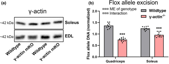
The cortical cytoskeleton, consisting of the cytoplasmic actin isoforms β and/or γ-actin, has been implicated in insulin-stimulated GLUT4 translocation and glucose uptake in muscle and adipose cell culture. Furthermore, transgenic inhibition of multiple actin-regulating proteins in muscle inhibits insulin-stimulated muscle glucose uptake. The current study tested if γ-actin was required for insulin-stimulated glucose uptake in mouse skeletal muscle. Based on our previously reported age-dependent phenotype in muscle-specific β-actin gene deletion (-/- ) mice, we included cohorts of growing 8-14 weeks old and mature 18-32 weeks old muscle-specific γ-actin-/- mice or wild-type littermates. In growing mice, insulin significantly increased the glucose uptake in slow-twitch oxidative soleus and fast-twitch glycolytic EDL muscles from wild-type mice, but not γ-actin-/- . In relative values, the maximal insulin-stimulated glucose uptake was reduced by ~50% in soleus and by ~70% in EDL muscles from growing γ-actin-/- mice compared to growing wild-type mice. In contrast, the insulin-stimulated glucose uptake responses in mature adult γ-actin-/- soleus and EDL muscles were indistinguishable from the responses in wild-type muscles. Mature adult insulin-stimulated phosphorylations on Akt, p70S6K, and ULK1 were not significantly affected by genotype. Hence, insulin-stimulated muscle glucose uptake shows an age-dependent impairment in young growing but not in fully grown γ-actin-/- mice, bearing phenotypic resemblance to β-actin-/- mice. Overall, γ-actin does not appear required for insulin-stimulated muscle glucose uptake in adulthood. Furthermore, our data emphasize the need to consider the rapid growth of young mice as a potential confounder in transgenic mouse phenotyping studies.
Keywords: glucose uptake; skeletal muscle; γ-actin.
© 2022 The Authors. Physiological Reports published by Wiley Periodicals LLC on behalf of The Physiological Society and the American Physiological Society.
Conflict of interest statement
Nothing to declare.
Figures


Similar articles
-
β-Actin shows limited mobility and is required only for supraphysiological insulin-stimulated glucose transport in young adult soleus muscle.Am J Physiol Endocrinol Metab. 2018 Jul 1;315(1):E110-E125. doi: 10.1152/ajpendo.00392.2017. Epub 2018 Mar 13. Am J Physiol Endocrinol Metab. 2018. PMID: 29533739 Free PMC article.
-
Oligomeric resistin impairs insulin and AICAR-stimulated glucose uptake in mouse skeletal muscle by inhibiting GLUT4 translocation.Am J Physiol Endocrinol Metab. 2009 Jul;297(1):E57-66. doi: 10.1152/ajpendo.90744.2008. Epub 2009 May 12. Am J Physiol Endocrinol Metab. 2009. PMID: 19435854
-
Insulin Resistance Is Not Sustained Following Denervation in Glycolytic Skeletal Muscle.Int J Mol Sci. 2021 May 6;22(9):4913. doi: 10.3390/ijms22094913. Int J Mol Sci. 2021. PMID: 34066429 Free PMC article.
-
Restoration of hypoxia-stimulated glucose uptake in GLUT4-deficient muscles by muscle-specific GLUT4 transgenic complementation.J Biol Chem. 1998 Aug 14;273(33):20910-5. doi: 10.1074/jbc.273.33.20910. J Biol Chem. 1998. PMID: 9694838
-
Rac1 signalling towards GLUT4/glucose uptake in skeletal muscle.Cell Signal. 2011 Oct;23(10):1546-54. doi: 10.1016/j.cellsig.2011.05.022. Epub 2011 Jun 13. Cell Signal. 2011. PMID: 21683139 Review.
Cited by
-
Muscle-specific AXIN1 and AXIN2 double knockout does not alter AMPK/mTORC1 signalling or glucose metabolism.J Physiol. 2025 Jul;603(14):3961-3971. doi: 10.1113/JP288854. Epub 2025 Jun 30. J Physiol. 2025. PMID: 40587294 Free PMC article.
-
Insulin signalling and GLUT4 trafficking in insulin resistance.Biochem Soc Trans. 2023 Jun 28;51(3):1057-1069. doi: 10.1042/BST20221066. Biochem Soc Trans. 2023. PMID: 37248992 Free PMC article. Review.
-
SLC7 transporters at the crossroads of amino acid metabolism and diabetes pathophysiology: insights and therapeutic perspectives.Front Nutr. 2025 May 21;12:1467057. doi: 10.3389/fnut.2025.1467057. eCollection 2025. Front Nutr. 2025. PMID: 40469683 Free PMC article. Review.
-
Gain-of-Function Dynamin-2 Mutations Linked to Centronuclear Myopathy Impair Ca2+-Induced Exocytosis in Human Myoblasts.Int J Mol Sci. 2022 Sep 8;23(18):10363. doi: 10.3390/ijms231810363. Int J Mol Sci. 2022. PMID: 36142275 Free PMC article.
-
GLUT4 Trafficking and Storage Vesicles: Molecular Architecture, Regulatory Networks, and Their Disruption in Insulin Resistance.Int J Mol Sci. 2025 Aug 5;26(15):7568. doi: 10.3390/ijms26157568. Int J Mol Sci. 2025. PMID: 40806697 Free PMC article. Review.
References
-
- Brozinick, J. T. , Hawkins, E. D. , Strawbridge, A. B. , & Elmendorf, J. S. (2004). Disruption of cortical actin in skeletal muscle demonstrates an essential role of the cytoskeleton in glucose transporter 4 translocation in insulin‐sensitive tissues. Journal of Biological Chemistry, 279(39), 40699–40706. 10.1074/jbc.M402697200 - DOI - PMC - PubMed
-
- DeFronzo, R. A. , Ferrannini, E. , Groop, L. , Henry, R. R. , Herman, W. H. , Holst, J. J. , Hu, F. B. , Kahn, C. R. , Raz, I. , Shulman, G. I. , Simonson, D. C. , Testa, M. A. , & Weiss, R. (2015). Type 2 diabetes mellitus. Nature Reviews Disease Primers, 1, 1–22. 10.1038/nrdp.2015.19 - DOI - PubMed
-
- DeFronzo, R. A. , & Tripathy, D. (2009). Skeletal muscle insulin resistance is the primary defect in type 2 diabetes. Diabetes Care. 10.2337/dc09-s302. https://pubmed.ncbi.nlm.nih.gov/19875544/ - DOI - PMC - PubMed
Publication types
MeSH terms
Substances
LinkOut - more resources
Full Text Sources
Medical
