

Rescue of photoreceptor with human mesenchyme stem cell and human mesenchyme stem cell expressing erythropoietin in total degeneration of retina animal model
- PMID: 35225544
- PMCID: PMC9114553
- DOI: 10.4103/ijo.IJO_472_21
Rescue of photoreceptor with human mesenchyme stem cell and human mesenchyme stem cell expressing erythropoietin in total degeneration of retina animal model
Abstract
Purpose: This study aimed to investigate the efficacy of human-derived umbilical cord mesenchymal stem cells (HDUMSC) and human-derived umbilical cord mesenchymal stem cells expressing erythropoietin (HDUMSC-EPO) to rescue total degenerated retina in a rat model.
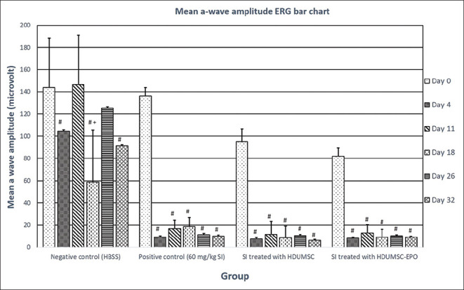
Methods: The study included four treatment groups, namely negative control using normal saline (HBSS) injection, positive control using sodium iodide 60 mg/kg (SI), SI treated with HDUMSC, and SI treated with HDUMSC-EPO given via subretinal and intravenous routes, to test the efficacy of retinal regeneration following SI-induced retinal degeneration. Retinal function in both phases was tested via electroretinography (ERG) and histological staining examining the outer nuclear layer (ONL).
Results: There was a statistically significant result (P < 0.05) in the SI treated with HDUMSC-EPO only when comparing day 11 (mean = 23.6 μv), day 18 (mean = 25.2 μv), day 26 (mean = 26.3 μv), and day 32 (mean = 28.2 μv) to the b-wave ERG on day 4 rescue injection day (mean = 12.5 μv). The SI treated with HDUMSC-EPO showed significant improvement in b-wave ERG readings in the Sprague-Dawley (SD) rat but did not restore baseline readings prior to degeneration (day 0). Both treated groups' ONL thicknesses did not show significant changes compared to the negative control group (HBSS) following rescue therapy.
Conclusion: Total retinal degeneration following intravenous SI injection was observed at 60 mg/kg. SI treated with HDUMSC and HDUMSC-EPO showed no regenerative potential compared to baseline in SI-induced total retina degeneration on ERG or histology, whereas SI treated with HDUMSC-EPO group showed a substantial increase in b-wave ERG amplitude over time.
Keywords: Electroretinography; HDUMSC; HDUMSC-EPO; ONL; Sprague–Dawley rat; retinal degenerative disease.
Conflict of interest statement
None
Figures


References
-
- Guan Y, Cui L, Qu Z, Lu L, Wang F, Wu Y, et al. Subretinal transplantation of rat MSCs and erythropoietin gene modified rat MSCs for protecting and rescuing degenerative retina in rats. Curr Mol Med. 2013;13:1419–31. - PubMed
-
- Kiuchi K, Yoshizawa K, Shikata N, Moriguchi K, Tsubura A. Morphologic characteristics of retinal degeneration induced by sodium iodate in mice. Curr Eye Res. 2002;25:373–9. - PubMed
MeSH terms
Substances
LinkOut - more resources
Full Text Sources
Research Materials
